R E S E A R C H A R T I C L E
Open Access
Algal production of extra and intra-cellular
polysaccharides as an adaptive response to the
toxin crude extract of
Microcystis aeruginosa
Mostafa Mohamed El-Sheekh
1*, Hanan Mohamed Khairy
2and Rania El-Shenody
1Abstract
This is an investigation concerned with studying the possible adaptive response of four different unicellular algae,
AnabaenaPCC 7120,Oscillatoria angustissima,Scendesmus obliquus and Chlorella vulgaris, to the toxin ofMicrocystis aeruginosa(Kützing). The effects of four different concentrations, 25, 50, 100 and 200μg mL-1of microcystins crude extract ofM.aeruginosa, on both intra and extra-cellular polysaccharide levels, in log phase, of the four tested algae were studied. The obtained results showed differential increase in the production levels for both intra and
extra-cellular polysaccharides by the tested algae, compared with the control.S.obliquusandC.vulgarisshowed a resistance to crude toxin higher thanAnabaenaPCC 7120 andO.angustissima. The highly production of
polysaccharides by green algal species under this toxic stress indicated the involvement of these polysaccharides in protecting the algal cells against toxic species and, reflect the biological behavior of particular algal species to the environmental stresses.
Keywords:Allelopathy,Microcystis aeruginosa, Crude extracts, Intra and extra- cellular polysaccharides, Cyanotoxins
Introduction
Microcystis aeruginosa is a common hepatotoxic cyano-bacterium living in eutrophic freshwaters [1]. The inhib-ition of competitors by the release of compounds, a process known as allelopathy, may be important in plank tonic systems [2]. Allelopathy has been hypothesized to play a role in species succession [3], the formation of harmful algal blooms in water resources [4], and the es-tablishment of invasive species [5]. Allelopathy is defined as any process involving secondary metabolites produced by plants and microorganisms that influence the growth and development of biological systems, including posi-tive and negaposi-tive effects [6]. These secondary metabolites are called allelochemicals and play a major role on growth and development in both natural and agro-ecosystems [7]. Most allelopathic are compounds biodegradable and at the same time natural toxins [8].
The production of biologically active substances which promotes the growth of algae and other plant organisms has been reported [9].
The algal cells can release extra-cellular polysacchar-ides (EPS) into the environment; these EPS are ecologic-ally important through their influence on carbon cycle and microbial diversity [10]. They may enhance the bac-terial growth and activity leading to the release of in-organic substances useful for microalgae in the same environment [11]. In addition, these polysaccharides can also make complexes with inorganic ions and thus redu-cing their toxicity to aquatic organisms [12].
Some green algae can develop a defense system by the production of polysaccharides to cope with oxidative
stress induced by microcystin. The results of in vitro
assay of antioxidant activity revealed that these polysac-charides had different activities, depending on their sul-fate contents [13]. On the other hand, toxic blooms of Nodularia spumigenacan provide a potential food source for the heterotrophic food chain [14]. Furthermore, bac-teria can efficiently degrade microcystins in natural waters with previous cyanobacterial history, and heterotrophic nanoflagellates respond quickly to the bacterial growth [15]. However, some species of green algae e.g. Scenedes-mussp. coexist and even flourish in the presence of either toxic cyanobacteria or their toxins [16]. Mass-occurrences
* Correspondence:mostafaelsheekh@yahoo.com
1Botany Department, Faculty of Science, Tanta University, Tanta, Egypt Full list of author information is available at the end of the article
of cyanobacteria have been reported to increase in fre-quency, as well as in intensity, due to eutrophication [17]. It is therefore important to investigate how cyano-bacteria influence on the structure and functioning of other surrounding organisms in the aquatic ecosystem since River water quality zoning could provide essential information for developing river water quality manage-ment policies [18].
Therefore, the aim of this study was to investigate the response of several unicellular algae to the stress of
crude toxin of Microcystis aeruginosa through the
pro-duction of intra and extra-cellular polysaccharides.
Materials and methods Test organisms
Two unicellular blue green algae (AnabaenaPCC. 7120
and Oscillatoria angustissima) and two green algae (Scendesmus obliquusand Chlorella vulgaris) were kind-ly provided from Phycology Laboratory, Faculty of Science, Tanta University.
Culture and crude extract of cyanobacteriumMicrocystis aeruginosa
Microcystis aeruginosawas isolated from fresh water sam-ples from Nile river channel near Tanta city, Egypt, and spread with an inoculating needle on the surface of steri-lized Petri dishes containing solidified, sterile media (Allen’s,
[19]). The cultures of M. aeruginosa purified and
pre-pared in axenic unialgal cultures by Venkataraman [20]. M. aeruginosa was cultured in 1 liter conical flasks
containing 400 mL medium (Allen’s, [19]) and kept in
controlled conditions of continuous light (45 μmol/ms)
and temperature (25±2°C). Algal cells were harvested at
the end of log growth phase, lyophilized and kept in deep freeze (−20°C).
The crude extracts were prepared by suspending 100 mg lyophilized cells in 10 mL of 75% methanol ac-cording to [21] with ultrasonication (ultrasonic probe with characteristics of ~60 W and ~20 KHZ) for 2 min followed by intermittent shaking for 1 h. Debris was re-moved by centrifugation for 10 min at 4000 rpm. The pellet was reextracted and the combined supernatants were evaporated to dryness at 30°C using a rotary evap-orator (Perfit, India).
Treatments
All experiments were carried out in 250 mL conical
flasks, containing 100 mL Allen’s and Stanier medium
which was adjusted to contain 0 (control), 25, 50, 100 and 200 μg/mL of lyophilized cell extract of previously
mentioned cyanobacteria species. Culture conditions of the tested algae were the same as previously mentioned. The experimental cultures were harvested at 3, 6, 9, 12 and 15 days for the determination of intra and extra-cellular polysaccharides. Cultures were grown in tripli-cate for statistical analysis.
Measurement of intra-cellular polysaccharides (IPS)
During the growth, 100 mL of the tested algae cultures were pipette out and centrifuged at 3000 rpm for 10 min. The filtrate was used to estimate extra-cellular poly-saccharides and the pellets were dried and then used to estimate intra-cellular polysaccharide as described by [22]. Intra-cellular polysaccharide (IPS) was extracted by homogenizing the derided pellet in distilled water (50 mL). The homogenates were then heated in water
Table 1 Effect of different concentrations of microcystins crude extract (25, 50, 100 and 200μg/mL) ofMicrocystis
aeruginosain log phase on intra (IPS) and extra-cellular polysaccharides content (EPS) inμg/mL ofAnabaenaPCC.
7120
Microcystin crude extract concentrations (μg/mL)
Polysaccharide Days Control 25 50 100 200 F value P value
0 105±18 105±18 105±18 105±18 105±18
3 182±19 182±19(ns) 182±19(ns) 182±19(ns) 182±19(ns)
IPS 6 275±22 393±34*** 453±32*** 529±28*** 577±23*** 1062.1 0.001
9 523±50 620±23*** 858±52*** 1009±40*** 1153±50***
12 767±34 948±26*** 1239±39*** 1533±35*** 1667±40***
15 1087±78 1307±40*** 1659±36*** 1968±42*** 2531±36***
0 81±20 81±20(ns) 81±20(ns) 81±20(ns) 81±20(ns)
3 150±21 150±21(ns) 150±21(ns) 150±21(ns) 150±21(ns)
EPS 6 234±27 329±21*** 427±24*** 473±37*** 543±27*** 255.86 0.001
9 334±31 426±28*** 516±41*** 618±48*** 716±24***
12 397±25 481±19*** 585±44*** 702±40*** 801±25***
15 476±58 557±27*** 748±37*** 909±25*** 1012±45***
***Highly significant at P≤0.001 using one way analysis of variance (ANOVA). (ns)
bath at 95°C for 6 hours. The extracts were filtrated through Whatman No.2 filter paper, then precipitated with four volumes of 95% ethanol, stirred vigorously and left overnight at 4°C. The precipitated IPS was recovered by centrifugation at 10.000 rpm for 15 min and the supernatant was discarded.
Measurement of extra-cellular polysaccharide (EPS)
The extra-cellular polysaccharides were estimated ac-cording to [23]. To precipitate proteins from the algal culture, trichloroacetic acid (TCA) was added in a final concentration of 4% and the algal filtrate was stirred for 2 h. Precipitated proteins were removed by centrifugation. The clear supernatant was collected which contains EPS. Extra-cellular polysaccharides was precipitated by ethanol and determined as described in IPS.
Statistical analysis
Results are presented as mean ±SD (standard deviation) for three replicates. Data obtained were analyzed statisti-cally to determine the degree of significance between treatments using one, two and three way analysis of
vari-ance (ANOVA) at P ≤ 0.001. The statistical analyses
were carried out using SAS program [24] version 6.12.
Results
Data in Table 1 show the effect of various concentra-tions of microcystins crude extract ofM.aeruginosa(25,
50, 100 and 200 μg/mL), in log phase, on both
extra-cellular polysaccharides (EPS) and intra-extra-cellular
polysac-charides (IPS) ofAnabaenaPCC.7120. Results showed a
significant increase of both EPS and IPS as compared with control. Thus, the crude microcystins extract could induce the production of polysaccharides and their Table 2 Effect of different concentrations of microcystins crude extract (25, 50, 100 and 200μg/mL) ofMicrocystis
aeruginosain log phase on intra (IPS) and extra-cellular polysaccharides content (EPS) inμg/mL ofOscillatoria
angustissima
Microcystin crude extract concentrations (μg/mL)
Polysaccharide Days Control 25 50 100 200 F value P value
0 5±18 95±18(ns) 95±18(ns) 95±18(ns) 95±18(ns)
3 177±37 177±37(ns) 177±37(ns) 177±37(ns) 177±37(ns)
IPS 6 355±38 387±33(ns) 425±29*** 476±35*** 530±34*** 171.1 0.001
9 403±34 440±21(ns) 512±20*** 559±46*** 712±38***
12 461±28 522±29(ns) 582±28*** 728±30*** 863±53***
15 550±45 598±23(ns) 758±62*** 867±58*** 1079±45***
0 156±24 156±24(ns) 156±24(ns) 156±24(ns) 156±24(ns)
3 285±31 285±31(ns) 285±31(ns) 285±31(ns) 285±31(ns)
EPS 6 311±37 377±23(ns) 502±18*** 525±28*** 633±46*** 1111.17 0.001
9 631±28 830±69*** 980±24*** 1051±50*** 1198±33***
12 1045±47 1155±32*** 1327±31*** 1571±35*** 1939±44***
15 1630±34 1963±40*** 2232±85*** 2499±40*** 3043±83***
***Highly significant at P≤0.001 using one way analysis of variance (ANOVA). (ns)Non significant at P≤0. 001 using one way analysis of variance.
Table 4 Analysis of variance of extra and intra-cellular polysaccharides ofOscillatoria angustissimaat different concentrations (25, 50, 100 and 200μg/mL) of
microcystins crude extract ofMicrocystis aeruginosa
Extra-cellular polysaccharides
Treatment F-value P-value Adjusted R2
Concentrations 442.77 0.001 99.81%
Days 5767.13 0.001
Concentrations* days 80.87 0.001
Intra-cellular polysaccharides
Concentrations 128.44 0.001 98.8%
Days 819.17 0.001
Table 3 Analysis of variance of extra and intra-cellular polysaccharides ofAnabaenaPCC.7120 at different concentrations (25, 50, 100 and 200μg/mL) of
microcystins crude extract ofMicrocystis aeruginosa
Extra-cellular polysaccharides
Treatment F- value P- value Adjusted R2
Concentrations 733.68 0.001 99.88%
Days 5044.92 0.001
Concentrations*days 132.10 0.001
Intra-cellular polysaccharides
Concentrations 222.96 0.001 99.8%
amounts increased with increasing the microcystins crude extract concentration. At the end of the
experi-ment (day15), the increase in EPS and IPS forAnabaena
PCC.7120 amounted by (20%, 52.6%, 81.0% and 132.8%) and (17%, 57%, 91% and 112.6%) over control level for EPS and IPS at different concentrations, respectively. One way analysis of variance revealed a high significant increase in extra and intra-cellular polysaccharides of AnabaenaPCC. 7120 as compared with control at (P ≤ 0. 001). On the other hand, inOscillatoria angustissima, EPS and IPS increased, and amounted by 20%, 37%, 53% and 86.7% and 8.7%, 37.8%, 57.6% and 96%, respectively (Table 2). High significant increase in extra and
intra-cellular polysaccharides at (P ≤ 0. 001) compared with
the control at all concentrations of the microcystins
crude extract except the first concentration 25 μg/mL,
the increase in IPS at (all days of experiments) and EPS at 6 days were insignificant.
The suggested model (concentrations, days and inter-ception between them) indicated that the deviation in
EPS and IPS content ofAnabaenaPCC.7120 were 99.88%
and 98.8% for EPS and IPS, respectively (Table 3) and for O. angustissima were 99.81% and 98.8%, respectively (Table 4).
Results revealed that, EPS and IPS content ofScendesmus obliquus and Chlorella vulgaris showed increasing under Table 5 Effect of different concentrations of microcystins crude extract (25, 50, 100 and 200μg/mL) ofMicrocystis
aeruginosain log phase on intra (IPS) and extra-cellular polysaccharides content (EPS) inμg/mL ofScendesmus
obliquus
Microcystin crude extract concentrations (μg/mL)
Polysaccharide Days Control 25 50 100 200 F value P value
0 123±25 123±25(ns) 123±25(ns) 123±25(ns) 123±25(ns)
3 167±30 167±30(ns) 167±30(ns) 167±30(ns) 167±30(ns)
IPS 6 177±35 213±25(ns) 258±33** 368±29*** 446±42*** 654.47 0.001
9 230±37 251±35(ns) 376±30*** 759±36*** 920±40***
12 332±45 397±46(ns) 651±43*** 1168±61*** 1665±33***
15 488±49 652±47*** 1014±41*** 1526±45*** 2460±60***
0 27±8 27±8(ns) 27±8(ns) 27±8(ns) 27±8(ns)
3 64.7±28 64.7±28(ns) 64.7±28(ns) 64.7±28(ns) 64.7±28(ns)
EPS 6 169±29 198.7±11(ns) 220.6±39(ns) 286±14*** 453±19*** 185.94 0.001
9 220±26 317.7±29(ns) 398±25*** 416±20*** 634±52***
12 279±25 434±32*** 518±42*** 636±47*** 948±45***
15 378±26 534±36*** 662±56*** 803±53*** 1068±114***
***Highly significant at P≤0.001 using one way analysis of variance (ANOVA). (ns)Non significant at P≤0. 001 using one way analysis of variance.
Table 6 Effect of different concentrations of microcystins crude extract (25, 50, 100 and 200μg/mL) ofMicrocystis
aeruginosain log phase on intra (IPS) and extra-cellular polysaccharides content (EPS) inμg/mL ofChlorella vulgaris
Microcystin crude extract concentrations (μg/mL)
Polysaccharide Days Control 25 50 100 200 F value P value
0 72±20 72±20(ns) 72±20(ns) 72±20(ns) 72±20(ns)
3 122±20 122±20(ns) 122±20(ns) 122±20(ns) 122±20(ns)
IPS 6 162±27 207±27(ns) 236±38(ns) 256±40*** 325±42*** 397.39 0.001
9 227±30 260±29(ns) 352±36*** 724±53*** 984±78***
12 274±25 367±29*** 435±37*** 998±89*** 1585±78***
15 369±30 463±38*** 591±46*** 1509±64*** 2133±108***
0 54±17 54±17(ns) 54±17(ns) 54±17(ns) 54±17(ns)
3 116±18 116±18(ns) 116±18(ns) 116±18(ns) 116±18(ns)
EPS 6 130±19 146±25(ns) 179±24** 322±24*** 381±22*** 302.29 0.001
9 157±16 183±21(ns) 250±21*** 408±24*** 550±20***
12 203±15 230±21(ns) 324±33*** 474±23*** 781±20***
15 232±16 363±33*** 541±21*** 669±32*** 948±51***
***Highly significant at P≤0.001 using one way analysis of variance (ANOVA). (ns)
different concentrations (25, 50, 100 and 200μg/mL) of
microcystins crude extract ofM.aeruginosa. The increasing in EPS and IPS ofSc.obliquus were (41%, 75%, 112% and 182.5%) and (33.6%, 117.8%, 212.7% and 404%) over the control value for each concentration for EPS and IPS, re-spectively (Table 5). On the other hand, increasing in EPS and IPS ofC.vulgarisamounted by (56%, 133%, 188% and 308.6%) and (25%, 60%, 309% and 478%), respectively over than control (Table 6).
Analysis of variance revealed high significant increase in extra and intra-cellular polysaccharides ofSc.obliquus andChlorella vulgarisat (P≤0. 001) except at 25μg/ml
the increases in IPS ofSc.obliquusand EPS ofChlorella vulgaris(Tables 5 and 6, respectively) were insignificant at the days 6, 9 and 12, and the IPS ofC.vulgariswas in-significant at days 6 and 9 as EPS ofSc.obliquus.
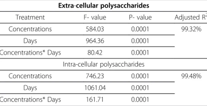
From the suggested statistical model (concentrations, days and interception between them), it could be ob-served that, the deviation for EPS and IPS content ofSc. obliquus was 98.89% and 99.68%, respectively (Table 7) and for C. vulgariswas 99.32% and 99.48%, respectively (Table 8).
Discussion
Microcystis aeruginosa blooms are hazardous to fresh-water flora and fauna due to the production of toxins [25]. M. aeruginosa produces a wide range of toxic meta-bolites. The physiological and ecological role of these compounds remains largely unknown. Some studies have suggested that these compounds may have allelochemical roles, such as compounds that may inhibit competing sympatric macrophytes, algae and microbes [26,27]. The adverse effects of crude microcystins (MCs) could be due to the synergistic interactions among MCs variants or to the presence of unidentified toxic other than MCs in the crude extracts [28]. Leflaive and Ten-Hage [29] reported that cellular extracts containing toxins are often more ac-tive than purified toxin, they suggests that cellular extracts contain a mix of active toxin that may act synergistically.
The possible adaptive response of four unicellular algae (AnabaenaPCC. 7120 andO.angustissimaas blue green algae and S. obliquus; C. vulgaris as green algae) to M. aeruginosa producing toxin were studied. The results indicated that the crude microcystins extract induces the production of extra and intra-cellular polysaccharides of the tested algae and their amounts increased with in-creasing the microcystins crude extract concentration. The production of polysaccharide by green algal species under toxic stress indicated the involvement of this polysaccharide in protecting the algal cells against toxic species [13].
Inhibition in the growth of Anabaena PCC 7120;
Oscillatoria angustissima; Scendesmus obliquus and Chlorella vulgaris at different microcystin crude extract concentrations was previously represented by [27]. Simi-lar results were recorded by Abdel-Rahman [30] found that growth and the physiological activities, except amino
acids biosynthesis, of both Chlorococcum humicola and
Chlorella vulgariswere inhibited by crude extracts of the
two cyanobacteria species Microcystis and Nodularia.
Singh et al. [31] reported that the crude extract of M. aeruginosaprovides toxicity to green algae (Chlorellasp. andScenedesmussp.) and cyanobacteria (AnabaenaBT1 and Nostoc muscorum). As represented by [13] pure and crude microcystins significantly decreased the growth of Scenedesmus quadricauda and Chlorella vulgaris, and these results correlated with polysaccharide contents of toxin-treated cultures. This results was in accordance with our results which indicated thatS. obliquusandC. vulgaris having a resistant system to crude toxin by the
production of polysaccharide more thanAnabaenaPCC.
7120 andO.angustissima.
The increasing in polysaccharides contents in MCs treated cultures in all experimented algae indicated that these polysaccharides may be involved in certain defense mechanisms in response to toxin stress. Some studies Table 7 Analysis of variance of extra and intra-cellular
polysaccharides ofScendesmus obliquusat different concentrations of microcystins crude extract of Microcystis aeruginosa
Extra-cellular polysaccharides
Treatment F- value P- value Adjusted R2
Concentrations 221.27 0.0001 98.89%
Days 778.44 0.0001
Concentrations * Days 30.10 0.0001
Intra-cellular polysaccharides
Concentrations 1117.14 0.0001 99.68%
Days 1991.37 0.0001
Table 8 Analysis of variance of extra and intra-cellular polysaccharides ofChlorella vulgarisat different concentrations of microcystins crude extract of Microcystis aeruginosa
Extra-cellular polysaccharides
Treatment F- value P- value Adjusted R2
Concentrations 584.03 0.0001 99.32%
Days 964.36 0.0001
Concentrations* Days 80.42 0.0001
Intra-cellular polysaccharides
Concentrations 746.23 0.0001 99.48%
Days 1061.04 0.0001
against oxidative stress and their ability in scavenging ROS in plant cells [32]. The antioxidant mechanisms may be due to the supply of hydrogen by these polysac-charides which combines with radicals and forms more stable products to terminate radical chain reaction [33].
Microcystins have been shown to induce formation of reactive oxygen species (ROS) that might cause serious cellular damage such as peroxidation of lipid membranes, genotoxicity, or modulation of apoptosis [13]. During the present study the EPS and IPS were detected in the algal medium increased with increase of MCs concentration. It has been suggested that polysaccharides are produced inside the cells during oxidative stress to scavenge the free radicals and remove them from the cells to the me-dium [32]. However, a variety of other mechanisms such as extra-cellular detoxification, reduced uptake, efflux, sequestration by polysaccharides have been proposed to explain algal tolerance to oxidative stress [34].
Extrusion of EPS can serve as a boundary between cells and the surrounding environment; they could fulfill a protective role against desiccation, antibacterial agents or predation by protozoan [35]. Hence, the results of present study showed that some algae can develop a de-fense system by the production of polysaccharides to cope with oxidative stress induced by MCs, and this may explain why some plank tonic algae present in close proximity with toxic cyanobacteria or their toxins are not affected by these toxins at environmentally relevant con-centrations (1–10μL) [36].
Conclusion
Here, we showed a physiological model of algal life sus-tainability under the stress of natural cyanobacterial tox-ins. The presented results showed the production of polysaccharides by tested algae as a response to the cyanobacterial toxins, microcystins. on the other hand, the release of these polysaccharides into the culture medium and most likely in the natural environment, is of ecologically importance because they may increase the polysaccharide contents of the water column and the growth of heterotrophic bacteria, and complex with heavy metals to reduce their toxicity to aquatic organ-isms. Therefore, in future studies involving in vitro cul-tures more attention should be paid to the role of algal exudates, in order to improve the significance of the results with the aim of using them as models of the real environment.
Competing interests
The authors declare that they have no competing interests.
Authors’contributions
This work is part of the Master thesis of RE where MME, supervised the thesis, suggested the problem and wrote the paper and MME participated in writing the paper and HMK helped in experiments and read the paper. All authors read and approved the final manuscript.
Acknowledgements
The authors would like to thank Prof. Hossam Easa, Microbial and Genetic Resources Research group National Institute of Advanced Industrial Science and Technology Tsukuba, Japan for critical reading of the manuscript.
Author details
1Botany Department, Faculty of Science, Tanta University, Tanta, Egypt. 2
National Institute of Oceanography and Fisheries, Alexandria, Egypt.
Received: 29 October 2012 Accepted: 18 November 2012 Published: 20 November 2012
References
1. Beattie KA, Kaya K, Codd GA:The cyanobacteriumNodulariaPCC 7804, of freshwater origin, produces [L-Har 2] nodularin.Phytochemistry2000, 54(1):57–61.
2. Legrand C, Rengefors K, Fistarol GO, Graneli E:Allelopathy in phytoplankton-biochemical, ecological and evolutionary aspects. Phycologia2003,42:406–419.
3. Keating KI:Allelopathic influence of blue-green bloom sequences in a eutrophic lake.Science1977,196:885–887.
4. Smayda TJ:Harmful algal blooms: Their ecophysiology and general relevance to phytoplankton blooms in the sea.Limnol Oceanogr1997, 42:1137–1153.
5. Figueredo CC, Giani A, Bird DF:Does allelopathy contribute to
Cylindrospermopsis raciborskii(cyanobacteria) bloom occurrence and geographic expansion.J Phycol2007,43:256–265.
6. Torres A, Oliva RM, Castellano D, Cross P:First World Congress on Allelopathy. A Science of the Future, SAI. Spain: University of Cadiz; 1996:278.
7. Inderjit K, Duke SO:Ecophysiological aspects of allelopathy.Planta2003, 217:529–539.
8. Maćias FA, Oliva RM, Simonet AM, Galindo JCG:What are allelochemicals?
InAllelopathy in Rice. Edited by Olofsdotter M. Manilla: IRRI Press; 1998. 9. Shanab S:Effect of fresh water cyanobacterial extracts on alkaloid
production of thein vitro Solanum elaeagnifoliumtissue culture.Arab Journal of Biotechnology2001,4(1):129–140.
10. Giroldo D, Ortolano PC, Vieira AAH:Bacteria-algae association in batch cultures of phytoplankton from a tropical reservoir: the significance of algal carbohydrates.Fresh Water Biology2007,52:1281–1289.
11. Azam F, Smith DC:Bacterial influence on the variability in the ocean’s biogeochemical state: a mechanistic view.InNATO ASI series, vol G27, particle analysis in oceanography. Edited by Demers S. Berlin: Springer; 1991:213–236.
12. De Philippis R, Paperi R, Sili C:Heavy metal sorption by released polysaccharides and whole cultures of two exopolysaccharide-producing cyanobacteria.Biodegradation2007,18:181–187.
13. Mohamed ZA:Polysaccharides as a protective response against microcystin-induced oxidative stress inChlorella vulgarisand
Scenedesmus quadricaudaand their possible significance in the aquatic ecosystem.Ecotoxicology2008,17(6):504–516.
14. Engstrom J, Viherluto M, Vitasalo M:Effect of toxic and nontoxic cyanobacteria on grazing zooplanktivory and survival of the mysid shrimpMysis mixta.J Exp Mar Biol Ecol2001,257:269–280. 15. Christoffersen K, Lyck S, Winding A:Microbial activity and bacterial
community structure during degradation of microcystins.Aquatic and Microbiology Ecology2002,27:125–136.
16. Mohamed ZA, Carmichael WW, Hussein AA:Estimation of microcystins in the fresh water fishOreochromis niloticusin an Egyptian fish farm containing aMicrocystisbloom.Environ Toxicol2003,18:137–141. 17. O’Neil JM, Davis TW, Burford MA, Gobler CJ:The rise of harmful
cyanobacteria blooms: The potential roles of eutrophication and climate change.Harmful Algae2011,14:313–334.
18. Karamouz M, Mahjouri N, Kerachian R:River Water Quality Zoning: A Case Study of Karoon and Dez River System.Iran J Environ Healt2004,1:16–27. 19. Allen’s MM, Stanier ST:Selective isolation of blue-green algae from water
and soil.J Gen Microbiol1968,51:302.
20. Venkataraman GS:The cultivation of algae. New Delhi: Indian Council of Agricultural Research; 1969.
22. Shi Y, Sheng J, Yang F, Hu Q:Purification and identification of
polysaccharide fromChlorella pyrenoidosa.Food Chem2007,103:101–105. 23. Berg DJC, Robijn GW, Janssen AC:Production of a novel extra cellular
polysaccharide byLactobacillus Sake01and characterization of the polysaccharide.Appl Environ Microbiol1995,61(8):2840–2844.
24. SAS program: Cary: SAS Institute Inc; 1989–1999. SAS (r) Proprietary Software Release 6.12 TS020.
25. Wiegand C, Pflugmacher S:Ecotoxicological effects of selected cyanobacterial secondary metabolites: a short review.Toxicol Appl Pharmacol2005,203:201–218.
26. Berry JP, Gantar M, Perez MH, Berry G, Noriega FG:Cyanobacterial toxins as allelochemicals with potential applications as algaecides, herbicides and insecticides.Mar Drugs2008,6:117–146.
27. El-Sheekh MM, Khairy HM, El-Shenody RA:Allelopathic effects of cyanobacteriumMicrocystis aeruginosaKützing on the growth and photosynthetic pigments of some algal species.Allelopathy Journal2010, 26:275–290.
28. Palíková M, Krejeci R, Hilscherová K, Babica P, Navrátil S, Kopp R, Bláha L: Effect of different cyanobacterial biomasses and their fractions with variable microcystin content on embryonal development of carp (Cyprinus carpioL.).Aquat Toxicol2007,81:312–318.
29. Leflaive J, Ten-Hage L:Algal and cyanobacterial secondary metabolites in freshwaters: a comparison of allelopathic compounds and toxins.Fresh Water Biology2007,52:199–214.
30. Abdel-Rahman MHM:Effect of Cyanobacteria crude extracts on growth and related physiological activities ofChlorococcum humicolaand
Chlorella vulgaris.Arab J Biotechnol2005,8(1):9–18.
31. Singh DP, Tyagi MB, Kumar A, Hu Q:Antialgal activity of hepatotoxin-producing cyanobacterium,Microcystis aeruginosa.World J Microbiol Biotechnol2001,17:15–22.
32. Tannin-Spitz T, Bergman M, van Moppes D, Grossman S:Antioxidant activity of the polysaccharide of the red micro algaPorphyridiumsp. J Appl Phycol2005,17:215–222.
33. Chen Y, Xie MY, Nie SP, Li C, Wang YX:Purification, composition analysis and antioxidant activity of polysaccharide from the fruiting bodies of
Ganoderma atrum.Food Chem2008,107:231–241.
34. Gaur JP, Rai LC:Heavy metal tolerance in algae.InAlgal adaptation to environmental stresses: physiological, biochemical and molecular mechanisms. Edited by Rai LC, Gaur JP. Berlin: Springer; 2001:363–388.
35. De Philippis R, Vincenzini M:Exocellular polysaccharides from cyanobacteria and their possible applications.FEMS Microbiology Review 1998,22:151–175.
36. Babic P, Hilscherova K, Bartova K, Blaha L, Marsalek B:Effects of dissolved microcystins on growth of plank tonic photoautotrophs.Phycologia2007, 46:137–236.
doi:10.1186/1735-2746-9-10
Cite this article as:El-Sheekhet al.:Algal production of extra and intra-cellular polysaccharides as an adaptive response to the toxin crude extract ofMicrocystis aeruginosa.Iranian Journal of Environmental Health Sciences & Engineering20129:10.
Submit your next manuscript to BioMed Central and take full advantage of:
• Convenient online submission
• Thorough peer review
• No space constraints or color figure charges
• Immediate publication on acceptance
• Inclusion in PubMed, CAS, Scopus and Google Scholar