In Silico
Analysis of the Metabolic Potential
and Niche Specialization of Candidate
Phylum "
Latescibacteria
" (WS3)
Noha H. Youssef1*, Ibrahim F. Farag1, Christian Rinke2, Steven J. Hallam3,4, Tanja Woyke2, Mostafa S. Elshahed1
1Department of Microbiology and Molecular Genetics, Oklahoma State University Stillwater, Oklahoma, United States of America,2DOE Joint Genome Institute, Walnut Creek, California, United States of America,
3University of British Columbia, Vancouver, BC, Canada,4Graduate Program in Bioinformatics, University of British Columbia, Vancouver, BC, Canada
Abstract
The“Latescibacteria”(formerly WS3), member of the Fibrobacteres–Chlorobi– Bacteroi-detes (FCB) superphylum, represents a ubiquitouscandidate phylum found interrestrial, aquatic, and marine ecosystems. Recently, single-cell amplified genomes (SAGs) repre-senting the“Latescibacteria”were obtained from the anoxic monimolimnion layers of Saki-naw Lake (British Columbia, Canada), and anoxic sediments of a coastal lagoon (Etoliko lagoon, Western Greece). Here, we present a detailedin-silicoanalysis of the four SAGs to gain some insights on their metabolic potential and apparent ecological roles. Metabolic reconstruction suggests an anaerobic fermentative mode of metabolism, as well as the ca-pability to degrade multiple polysaccharides and glycoproteins that represent integral com-ponents of green (Charophyta and Chlorophyta) and brown (Phaeophycaea) algae cell walls (pectin, alginate, ulvan, fucan, hydroxyproline-rich glycoproteins), storage molecules (starch and trehalose), and extracellular polymeric substances (EPSs). The analyzed SAGs also encode dedicated transporters for the uptake of produced sugars and amino acids/oli-gopeptides, as well as an extensive machinery for the catabolism of all transported sugars, including the production of a bacterial microcompartment (BMC) to sequester propionalde-hyde, a toxic intermediate produced during fucose and rhamnose metabolism. Finally, genes for the formation of gas vesicles, flagella, type IV pili, and oxidative stress response were found, features that could aid in cellular association with algal detritus. Collectively, these results indicate that the analyzed“Latescibacteria”mediate the turnover of multiple complex organic polymers of algal origin that reach deeper anoxic/microoxic habitats in lakes and lagoons. The implications of such process on our understanding of niche speciali-zation in microbial communities mediating organic carbon turnover in stratified water bodies are discussed.
OPEN ACCESS
Citation:Youssef NH, Farag IF, Rinke C, Hallam SJ, Woyke T, Elshahed MS (2015)In SilicoAnalysis of the Metabolic Potential and Niche Specialization of Candidate Phylum "Latescibacteria" (WS3). PLoS ONE 10(6): e0127499. doi:10.1371/journal. pone.0127499
Academic Editor:Ramy K. Aziz, Cairo University, EGYPT
Received:March 24, 2015
Accepted:April 14, 2015
Published:June 3, 2015
Copyright:© 2015 Youssef et al. This is an open access article distributed under the terms of the Creative Commons Attribution License, which permits unrestricted use, distribution, and reproduction in any medium, provided the original author and source are credited.
Data Availability Statement:Genome sequences generated have been deposited in GenBank under the accession numbers NZ_ASMB00000000.1, NZ_AQSL00000000.1, ASWY00000000.1, and AQRO00000000.1.
Introduction
Over the past few decades, small subunit ribosomal RNA (SSU or 16S rRNA) gene-based sur-veys have prompted a drastic reevaluation of the scope of phylum level diversity within the do-main Bacteria. Current taxonomic outlines indicate that the majority of recognized bacterial phyla (54.1% using SILVA database [1], 65.48% using Greengenes database [2]) have no pure culture representatives (candidate phyla). Many of these candidate phyla, so-called microbial dark matter (MDM) are globally distributed and display significant levels of intra-phylum level diversity [3–7]. Recent advances in cell sorting and whole genome amplification and assembly have facilitated the acquisition of single-cell amplified genomes (SAGs) derived from numer-ous candidate phyla [8–16]. Metabolic reconstruction with these SAGs provides a unique op-portunity to uncover the ecological and biogeochemical roles played by these enigmatic microbial groups.
One such candidate phylum is WS3 (Wurtsmith aquifer Sequences-3), whose members were first identified in a 16S rRNA gene-based survey of anoxic sediments obtained from a hy-drocarbon- and chlorinated-solvents-contaminated aquifer in northern Michigan, USA in 1998 [17]. Since then, their presence has been documented across a wide range of habitats in-cluding marine hydrothermal vents, gas hydrate-bearing habitats, cold methane seeps, cave rock walls, marine sediments, soils, wastewater treatment bioreactors, deep sea hypersaline an-oxic lakes, and oil-exposed microbial mats [18–28]. Recently, as part of an extensive single cell genomic study of 9 different habitats, Rinke et al. [29] reported on the recovery of four SAGs from WS3 single cells. Phylogenomic-based analysis using conserved marker genes indicated the monophyletic nature of WS3 as part of the Fibrobacteres–Chlorobi–Bacteroidetes (FCB) superphylum together with“Marinimicrobia”(SAR406),“Cloacimonetes”(WWE1), Gemma-timonadetes, and Caldithrix. The name“Latescibacteria”(hiding small rods) was suggested for the candidate phylum.
However, little is known about the biological capabilities of this phylum, and no systematic attempts have been made to reconstruct its metabolic potential. Thus, we here present a de-tailed analysis of the metabolic and physiological capabilities, and putative ecological roles of four“Latescibacteria”SAGs obtained from two different aquatic environments. Our analysis suggests that the“Latescibacteria”recovered from Sakinaw Lake and Etoliko lagoon transform algal detritus sinking from sunlit surface waters into fermentation products with the potential to contribute to microbial food webs in anaerobic waters below.
Materials and Methods
Origin of
“
Latescibacteria
”
SAGs
“Latescibacteria”SAGs analyzed in this study were obtained from two different locations [29]: Three SAGs originated from a single sample obtained from the anaerobic monimolim-nion of Sakinaw lake (British Columbia, Canada) at 49°40'30"N, 124°2'2.4"W coordinates, and a depth of 120m (Gies et al 2014). A fourth SAG was obtained by sampling anaerobic sedi-ments in Etoliko Lagoon, a coastal lagoon in the south of Aetolia-Acarnania, Greece, at the deepest point (~27.5 m) at 38°28'59.54"N, 21°19'17.44"E. Single cell sorting and lysis, whole ge-nome amplification, identification via 16S rRNA gene sequencing of amplified gege-nomes, as well as SAG sequencing, assemblies and estimates of genome completion were previously de-scribed [29]. The four“Latescibacteria”SAGs were deposited under Genbank assembly IDs: NZ_ASMB00000000.1, NZ_AQSL00000000.1, ASWY00000000.1, and AQRO00000000.1, and in Integrated Microbial Genomics (IMG) under SAG IDs: SCGC AAA252-D10, SCGC AAA252-B13 and SCGC AAA252-E07 for Sakinaw lake SAGs, and SCGC AAA257-K07 for
to SJH. The work conducted by the U.S. Department of Energy Joint Genome Institute, a DOE Office of Science User Facility, is supported under Contract No. DE-AC02-05CH11231.
the Etoliko lagoon SAG. These SAGs will henceforth be referred to as S-D10, S-B13, and S-E07 for Sakinaw Lake SAGs, and E-K07 for Etoliko Lagoon SAG. The type species for”Latescibac-teria”is S-E07, for which the nameCandidatus“Latescibacter anaerobius”has been proposed [29].
Detailed analysis was conducted on S-E07, which has the highest estimated genome comple-tion (73.02%) among the“Latescibacteria”SAGs. The closely related S-B13 (94% 16S rRNA gene sequence similarity to SAG S-E07) with 57.1% estimated genome completion was used to confirm shared gene content and fill pathway holes when needed. Only general metabolic fea-tures for SAG S-D10 (94% 16S rRNA gene sequence similarity to S-E07, and 96% to S-B13) are discussed, given its low percentage of estimated genome completeness (38.2%). Due to the ob-served differences between the 3 Sakinaw Lake SAGs, and the Etoliko lagoon SAG E-K07 (85– 86% 16S rRNA gene sequence similarity to Sakinaw Lake SAGs), as well as its low estimated ge-nome completion (23.02%), analysis of SAG E-K07 was restricted to identifying variation in conserved genes or pathways between“Latescibacteria”SAGs from two distinct locations.
Genome annotation, general genomic features, and metabolic
reconstruction
The IMG platform (http://img.jgi.doe.gov) was used for genome functional annotation. De-tailed metabolic reconstruction of relevant pathways was performed with both KEGG [30] and Metacyc [31] databases. As part of the IMG annotation pipeline, CRISPR elements are detected with CRT [32] and PILERCR [33]. Predictions from both methods are concatenated and in case of overlapping elements, the shorter one is removed. Overall annotation followed proce-dures outlined in [13]: In brief, proteases, peptidases, and protease inhibitors were identified with Blastp against the Merops database [34]. Transporters were identified with the transporter classification database (TCDB) [35]. dbCAN HMMs [36] were used to identify carbohydrate active enzymes (CAZymes) including glycoside hydrolases (GH), polysaccharide lyases (PL), and carboxyl esterases (CE) following the classification scheme of the Carbohydrate active en-zyme (CAZy) database [37].
Results
Phylogenetic affiliation and general genomic features of
“
Latescibacteria
”
SAGs
All four SAGS were affiliated with the candidate order PBS_III_9 based on phylogenetic analy-sis of the candidate phylum“Latescibacteria”using 1198 near-full length 16S rRNA gene se-quences (Fig 1, Table A inS1 File, and Supplementary Text inS1 File). Sakinaw lake SAGs belonged to family I, while Etoliko lagoon SAG E-K07 belonged to family VI within this order (Fig 1B). General genomic features for each SAG are shown inTable 1.
Metabolic potential of Sakinaw Lake SAGs
Anabolic pathways identified in S-E07 and S-B13 include machinery for the production of amino acids, cofactors, fatty acids, purines and pyrimidines, terpenoid unit backbone, and gly-cerophospholipids. In addition, the SAGs encode near-complete replication, transcriptional, and translational machineries. The presence of genes for lipopolysaccharide (LPS) biosynthesis and pathway for LPS insertion in the outer membrane suggests a Gram-negative cell wall (Sup-plementary Text).
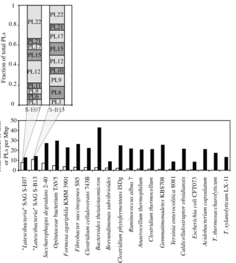
pathways and substrate level phosphorylation for coupled energy release and conservation. Both S-E07 and S-B13 encode a diverse array of carbohydrate active enzymes (CAZymes), with a conspicuous enrichment (Genes/Mbp), and diversity (number of different families) of poly-saccharide lyases (PLs) (Fig 2, Figure A inS1 File, Table B inS1 File). In contrast, the SAGs are relatively depauperate in genes encoding glycoside hydrolases (GHs) including enzymes in-volved in the degradation of cellulose (1 putative endoglucanase (GH5), 3 putativeβ -glucosi-dases (GH3, GH116, and GH9), and no putative cellobiohydrolase), and enzymes involved in the degradation of xylans (xylanases, andβ-xylosidases).
Fig 1. Updated taxonomic outline for candidate phylum“Latescibacteria”(A), and for the candidate order PBSIII_9 (B).Neighbor joining trees were constructed using Jukes-Cantor corrections in
MEGA6-Beta2 [100]. Bootstrap values (in percent) are based on 1000 replicates and are shown for branches with more than 50% bootstrap support. Numbers in parentheses represent the number of sequences in each WS3 candidate order.
doi:10.1371/journal.pone.0127499.g001
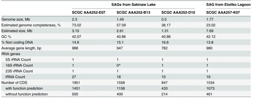
Table 1. General genomic features of“Latescibacteria”SAGs.
SAGs from Sakinaw Lake SAG from Etoliko Lagoon
SCGC AAA252-E07 SCGC AAA252-B13 SCGC AAA252-D10 SCGC AAA257-K07
Genome size, Mb 2.3 1.49 0.5 1.77
Estimated genome completeness, % 73.02 57.09 38.17 23.02
Estimated size, Mb 3.15 2.61 1.31 7.69
GC % 42.07 40.86 40.86 42.12
% Non coding DNA 14.6 15.1 16.6 13.8
Average gene length, bp 988 947 762 980
RNA genes
5S rRNA Count 1 1 1 1
16S rRNA Count 1 0* 1 1
23S rRNA Count 1 1 1 1
tRNA Count 27 18 10 19
Number of CDS 1951 1558 647 1534
with function prediction 1451 1158 433 1073
without function prediction 500 400 214 461
*The S-B13 16S rRNA couldn’t be retrieved via the whole genome shotgun approach, however the affiliation of S-B13 to CP-“Latescibateria”was confirmed through analyzing the amplified and Sanger-sequenced full-length 16S rRNA gene
Interestingly, many of the polymers that S-E07 and S-B13 are predicted to degrade are inte-gral components of cell walls of the green algal phyla Charophyta (most commonly encoun-tered in freshwater habitats), and Chlorophyta (widely distributed in freshwater, marine, and terrestrial habitats), as well as the brown algal Class Phaeophyceae. Green and brown algal cell walls are complex, with a diverse array of structural fibrillar polymers enmeshed in complex matrices with crystalline polymer components (Fig 3). Both S-E07 and S-B13 encode genes necessary for the conversion of these cell wall components, including pectin, alginate, ulvans, fucans, hydroxyproline-rich glycoproteins (HRGP), e.g. arabinogalactan proteins (AGP) and extensins, and xyloglucan (Table 2, Figure B inS1 File, Supplementary Text inS1 File). More-over, the SAGs also encode pathways mediating the conversion of soluble organic compounds commonly utilized for storage in algae (e.g. starch and trehalose). A more in depth description of these capabilities follows.
Algal cell wall degradation potential
1. Pectins. Pectins are components of the amorphous matrix and outer lattice of Charo-phyta cell wall (Fig 3A) [38]. Both S-E07 and S-B13 encode machinery for depolymerizing the pectic polysaccharide homogalacturonan (HG) (Table 2). They encode carboxyl esterases (CE8 Fig 2. Total number of PLs (white columns) and GHs (black columns) per Mbp of various pectinolytic and lignocellulolytic microorganisms’genomes.Note that, compared to other genomes,
“Latescibacteria”SAGS are enriched in PLs as opposed to GHs. The inset shows SAGs S-E07 and S-B13 different PL families as a fraction of total PLs.
and CE12) for the removal of accessory acetyl and methyl groups attached to the backbone, pectin lyase and pectate lyase (PL1, and PL10) to breakdown the backbone to oligosaccharides with 4-deoxy-α-D-galact-4-enuronosyl groups at their non-reducing ends, exopolygalacturo-nate lyase (PL9) to cleave digalacturoexopolygalacturo-nate unit, and oligogalacturonide lyase (PL22) to degrade the digalacturonate units to 5-dehydro-4-deoxy-D-glucuronate and galacturonic acid as the final end products of HG degradation [39,40]. In addition to HG, S-E07 and S-B13 encode all Fig 3. Schematic representation of algal cell walls.The cell wall composition differs between various algal groups [43]. Within the Charophyta (A), the wall is formed of an inner fibrillar layer made of cellulose microfibrils. The fibrillar layer is enmeshed in and surrounded by a middle amorphous matrix of pectin (homogalacturonan, HG, and rhamnogalacturonan I, RGI) that anchors the inner fibrillar cellulose layer to an outer lattice of homogalacturonan. Extracellular polymeric substances or mucilages are also present outside the outer lattice [38,43,101]. Similarly, cell walls of Chlorophyta (B) contain skeletal
polysaccharides enmeshed in a matrix. However, the skeletal polysaccharides in Chlorophyta cell walls form double fibrillar layers (inner layer and outer layer) with an amorphous matrix in between. The fibrillar layers vary in composition between cellulose,β-1,3-xylans orβ-1,4-mannans or complex
heteropolymers, and are rich in hydroproline-rich glycoprotein such as extensins and AGPs. The amorphous matrix polysaccharides are generally in the form of ulvans (e.g. in Ulva species). Brown algal cell walls (C) consist of a fibrillar framework of cellulose microfibrils present in layers parallel to the cell surface but with no clear orientation within each layer. Two such layers are depicted in the figure. All cellulose layers are enmeshed in acidic polysaccharides, e.g. alginates. The interfibrillar matrices are composed of alginates and fucans [41,43].
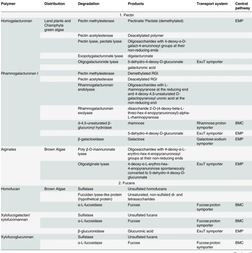
Table 2. Polymers potentially targeted by Latescibacteria, their distribution and occurrence in algae, structure, degradation enzymes encoded in the Latescibacterial SAGs, potential degradation products, their transport systems encoded in the SAGs, and ultimate central catabolic pathway.
Polymer Distribution Degradation Products Transport system Central
pathway
1. Pectin Homogalacturonan Land plants and
Charophyta green algae
Pectin methylesterase Pectinate/ Pectate (demethylated) EMP
Pectin acetylesterase Deacetylated polymer
Pectin lyase, pectate lyase Oligosaccharides with 4-deoxy-α -D-galact-4-enuronosyl groups at their non-reducing ends
Exopolygalacturonate lyase digalacturonate
Oligogalacturonide lyase 5-dehydro-4-deoxy-D-glucuronate ExuT symporter galacturonic acid
Rhamnogalacturonan I Pectin methylesterase Demethylated RGI Pectin acetylesterase Deacetylated RGI Rhamnogalacturonan
endolyase
Oligosaccharides with
L-rhamnopyranose at the reducing end and 4-deoxy-4,5-unsaturated D-galactopyranosyl uronic acid at the non-reducing end Rhamnogalacturonan exolyase disaccharide 2-O-(4-deoxy-beta-L- threo-hex-4-enopyranuronosyl)-alpha-L-rhamnopyranose
d-4,5-unsaturatedβ -glucuronyl hydrolase
rhamnose Rhamnose:proton
symporter
BMC
5-dehydro-4-deoxy-D-glucuronate ExuT symporter EMP
β-galactosidase Galactose Galactose:sodium
symporter
EMP
Alginates Brown Algae Polyβ-D-mannuronate lyase
Oligosaccharides with 4-deoxy-α -L-erythro-hex-4-enopyranuronosyl groups at their non-reducing ends Oligoalginate lyase 4-deoxy-α
-L-erythro-hex-4-enopyranuronose spontaneously converted to 5-dehydro-4-deoxy-D-glucuronate
ExuT symporter EMP
2. Fucans
Homofucan Brown Algae Sulfatase Unsulfated homofucans
Fucoidan lyase-like protein (hypothetical protein)
Unsaturated, non-sulfated di- and tetrasaccharides
α-L-fucosidase Fucose Fucose:proton
symporter
BMC
Xylofucogalactan/ xylofucomannan
Sulfatase Unsulfated fucans
α-L-fucosidase Fucose Fucose:proton
symporter
BMC
β-glucuronidase Glucuronic acid ExuT symporter EMP
Xylofucoglucuronan Sulfatase Unsulfated fucans
α-L-fucosidase Fucose Fucose:proton
symporter
BMC
Table 2. (Continued)
Polymer Distribution Degradation Products Transport system Central
pathway
3. Ulvans Chlorophyta Sulfatase Unsulfated ulvans
Heparin lyase Unsaturated, non-sulfated di- and tetrasaccharides
d-4,5-unsaturatedβ -glucuronyl hydrolase
5-dehydro-4-deoxy-D-glucuronate ExuT symporter EMP
Rhamnose Rhamnose:proton symporter BMC Xylose Xylose:proton symporter PPP
4. Xyloglucan Land plants, some green algae
endo-β-1,4-glucanase A mixture of oligosaccharides
α-1,2-fucosidase Fucose Fucose:proton
symporter
BMC
β-galactosidase Galactose Galactose:sodium
symporter
EMP
β-glucosidase Glucose Glucose:sodium
symporter
EMP
5. Hydroxyproline-rich glycoprotein (HRGP)
Extensin Land plants,
some green algae
β-L-arabinofuranosidase Arabinose ABC transporter PPP
Arabinogalactan protein (AGP)
endo-β-1,6-galactanase(?) galactan oligosaccharides
β-glucuronidase Glucuronic acid ExuT symporter EMP
α-Fucosidase Fucose Fucose:proton
symporter
BMC
α-rhamnosidase Rhamnose Rhamnose:proton
symporter
BMC
β-galactosidase Galactose Galactose:sodium
symporter
EMP
6. Others Extracellular proteins All organisms Non-specific
endopeptidases
Oligopeptides ABC transporter
Dipeptidases Dipeptides ABC transporter
Dipeptide peptidase, aminopeptidases, or Carboxypeptidases
Free amino acids Symporters for Pro, Ala, Asp, Glu, Gly, cationic aaABC transporter for Pro
EMP (Asp and Glu)
Starch Land plants, and
green algae storage compounds
α-amylase Oligosaccharides
α-glucosidase Glucose Glucose:sodium
symporter
EMP
Trehalose Brown algae
storage compound
Trehalase Glucose Glucose:sodium
symporter
EMP
Glucose-1-P Poly-D-galactosamine Some fungi such
asAspergillus, andNeurocrassa
Endo1,4-poly-D-galactosaminidase
Galactosamine PTS EMP
the necessary machinery to degrade rhamnogalacturonan I (RGI) (Table 2). These include car-boxyl esterases (CE8 and CE12), rhamnogalacturonan endolyase (PL11) that attack the back-bone to produce oligosaccharides with L-rhamnopyranose at the reducing end and 4-deoxy-4,5-unsaturated D-galactopyranosyl uronic acid at the non-reducing end, rhamnogalacturonan exolyase (PL11) that attacks those oligosaccharides to release the disaccharide 2-O-(4-deoxy-beta-L-threo-hex-4-enopyranuronosyl)-alpha-L-rhamnopyranose from the reducing end, and d-4,5-unsaturatedβ-glucuronyl hydrolase (GH88) that degrades those disaccharides to rham-nose and 5-dehydro-4-deoxy-D-glucuronate. The SAGs also encodeβ-galactosidase (GH42) for removal of galactosyl sugar substitutions [39,40].
2. Alginate. Alginates are present in the brown algal cell walls enmeshing fibrillar cellulose and also in the interfibrillar layers with fucans (Fig 3C) [41]. Both S-E07 and S-B13 encode PLs for the complete degradation of alginate (Table 2). These PLs include alginate lyases (PL6, PL15, PL17) that break down the alginate backbone producing oligosaccharides with
4-deoxy-α-L-erythro-hex-4-enopyranuronosyl groups at their non-reducing ends, as well as oligoalgi-nate lyase (PL15, and PL17) that exolytically cleave these oligosaccharides into monosaccha-rides and releases 4-deoxy-α-L-erythro-hex-4-enopyranuronose from the non-reducing end. The produced 4-deoxy-α-L-erythro-hex-4-enopyranuronose is spontaneously converted into 5-dehydro-4-deoxy-D-glucuronate as the final end product of alginate degradation [42].
3. Fucans. In addition to pectin and alginate, S-E07, and S-B13 also encode machinery for fucan degradation. Fucans are present, together with alginates, in brown algal cell walls interfi-brillar matrix (Fig 3C) [41]. Fucans exhibit wide variations in chemical structures, ranging from the highly sulfated homofucan polymers to the highly branched high-uronic-acid, low-sulfate-containing polymers (xylofucoglucan, xylofucogalactan, xylofucomannan, xylofucoglu-curonan) [41]. However, mechanistic details on the degradation of fucans are still in their in-fancy. Genomic analysis of“Latescibacteria”SAGs that S-E07, and S-B13 have the capacity to transform several fucans including homofucans, sulfated-xylofucoglucan, and sulfated-xylofu-coglucoronan. Indeed, a potential homofucan-degrading enzyme with sequence similarity to Mariniflexile fucanivoransfucoidan lyase could attack the backbone releasing unsaturated, non-sulfated fucan di- and tetrasaccharides. The SAGs also encode manyα-fucosidases (GH29, and GH95), that could attack those oligosaccharides and release fucosyl residues from the reducing end. Genomic evidence for the degradation of the highly branched high-uronic-acid, low-sulfate-containing polymers include manyα-fucosidases (GH29, and GH95), and oneα-glucuronidase (GH67).
4. Ulvans. Ulvans are present in the amorphous interfibrillar matrix of Chlorophyta cell walls (Fig 3B) [43–45]. Ulvan backbones are made of a few repeating disaccharides (Supple-mentary Text). However, the exact composition of ulvans is largely unknown. One important characteristic of ulvans is the presence of unusual sugars, e.g. iduronic acid, in its backbone [44]. Iduronic acid is also an important constituent of mammalian glycosaminoglycans (GAGs), e.g. heparan sulfate, dermatan sulfate, heparin [46].“Latescibacteria”SAGs harbor several PLs annotated as heparin and heparan lyase (PL12 and PL21). Structural similarity in sugar composition between ulvans and mammalian GAGs such as heparin suggest that those polysaccharide lyases (annotated as PL12 and PL21 with heparinase activity) might be poten-tial ulvan lyases responsible forulvan backbone cleavage to produce di- and tetrasaccharides with an unsaturatedβ-glucuronyl residue located at the non-reducing end [47]. SAGs also har-bor several copies of unsaturated glucuronyl hydrolases (GH88) that could potentially act on the oligosaccharides produced and release 5-dehydro-4-deoxy-D-glucuronate and other sugar residues, e.g. rhamnose, and xylose, as end products.
S-B13 encode machinery to degrade xyloglucan, a component of Charophyta and Chlorophyta cell walls usually present in association with cellulose microfibrils (Fig 3A and 3B) [48–50], in-cluding endo-β-1,4-glucanases (GH74), that cleave the xyloglucan backbone at locations of unsubstituted glycosyl moieties and give rise to a mixture of oligosaccharides,α-1,2-fucosidase (GH95), andβ-galactosidases (GH2, GH42) that attack those oligosaccharides to give rise to XXXG xyloglucans. The latter oligosaccharide can be attacked by oligoxyloglucanβ-glycosidase (GH3) generating isoprimeverose (Xyl-α(1,6)-Glu), and glucose. However, no homologs of oli-goxyloglucanβ-glycosidase were identified.
6. Hydroxyproline-rich, other O-linked, and N-linked glycoproteins. Hydroxyproline-rich glycoproteins (HRGP) are minor components in green algal cell walls (Fig 3) [51,52]. Both S-E07, and S-B13 SAGs encodeβ-L-arabinofuranosidase (GH127) that specifically targets arabinose residues attached to hydroxyproline in extensins [53] and release the sugar monomer arabinose. The SAGs also encode machinery for arabinogalactan protein (AGP) degradation including endo-β-1,6-galactanases (GH30) that hydrolyses theβ-1,6-galactan side chains and gives rise to galactan oligosaccharides,β-galactosidases (GH2, GH42),β-glucuronidase (GH79),α-fucosidase (GH29, GH95), andα-rhamnosidase (GH28, GH78, GH106) that attack the produced oligosaccharides and release substituting sugar monomers, e.g. galactose, glucu-ronic acid, fucose, and rhamnose [54]. In addition to HRGP degradation potential, the SAGs encode severalα-N-acetylgalactosaminidases (GH109) that specifically release N-acetylgalac-tosaminyl residues from O-linked glycoproteins [55], as well as severalα-mannosidases (GH38) that could potentially release mannosyl residues from N-linked glycoproteins [56]. Re-cently, sialic acid (neuraminic acid), a 9-carbon sugar acid was identified in green algal N-linked glycoproteins [57]. While a sialidase (GH33) homologue was not identified in the SAGs, they do encode for all the enzymes required for sialic acid degradation, including sialate O-acetylesterase, N-acetylneuraminate lyase, and N-acyl-D-glucosamine 2-epimerase that will collectively degrade sialic acid into pyruvate and N-acetyl-glucosamine (NAG).
7. Degradation of cell wall proteins. Both S-E07 and S-B13 SAGs encode multiple pepti-dases that can attack the peptide moiety of glycoproteins in algal cell walls (Table C inS1 File). The majority of these peptidases (~66% in S-E07, and 63.4% in S-B13) are thought to be nutri-tional, where they non-specifically break down proteins into oligopeptides (protease families C25, M06, M10, M20, M41, M48, M50, S01, S08, S09, S41, S54, and U62), dipeptides (protease family M19), and free amino acids (protease families M24, M28, S49, T03).
8. Sulfatase activity on sulfated polysaccharide. Both S-E07 and S-B13 encode multiple sulfatases (n = 14 in S-E07 and n = 3 in S-B13) belonging to the family of arylsulfatases (pfam 00884). Many of the polymers in marine algal cell walls are sulfated, e.g. ulvans, homofucans, sulfated-xylofucoglucan, and sulfated-xylofucoglucoronan [58]. Removal of the sulfate groups from such polysaccharides prior to their degradation facilitates access of GHs and PLs to side chains and backbones [59]. The SAGs also harbor the essential anaerobic sulfatase maturation enzyme-coding gene [60] for post-translational modification of a critical Cys or Ser in the ac-tive site to a C-α-formylglycine [61].
Degradation of algal storage compounds and additional polymers of
non-algal origin
intracellular storage compound in green algae and green plants [62], while trehalose is an intra-cellular storage compound in brown algae [41]. In addition, S-E07 and S-B13 encodeβ -fructo-furanosidase (GH32) specific for sucrose, and endo1,4-poly-D-galactosaminidase (GH114) specific for poly-D-galactosamine (Table 2).
Extracellular polymeric substance (EPS) as additional potential source
of energy for the
“
Latescibacteria
”
EPS forms extensive mucilaginous sheath outside the algal cell wall and function in adhesion, gliding motility, biofilm formation, and protection. Although the exact chemistry of EPS is not entirely known, EPS was shown to be composed mainly of polysaccharides (up to 75%), with minor protein content (2–10%). The polysaccharide fraction is rich in uronic acids, as well as monosaccharides, mainly glucose, galactose, mannose, xylose, arabinose, fucose, and rhamnose [63,64]. As mentioned above,“Latescibacteria”SAGs harbor genes involved in the uptake and catabolism of all such components.
“
Latescibacteria
”
SAGs harbor extensive transport systems for sugars,
and amino acids/oligopeptides uptake
Both S-E07 and S-B13 encode several non-specific porins for transport of substrates across the outer membrane, coupled to specialized transporters in the inner membrane, including multi-ple secondary (symport), ABC (ATP-binding cassette), and phosphotransferase system (PTS) transporters for the uptake of a wide array of monomers, e.g. those putatively produced from the degradation of all polymers described above (Fig 4,Table 2). Uronic acids and uronic acid derivatives are potentially imported using a single common transporter (a sugar phosphate permease transporter of the major facilitator superfamily similar to ExuT transporter of Ralsto-nia solanacearum[65]). Fucose, rhamnose, as well as xylose are potentially imported via dedi-cated proton symporters, while glucose and galactose are potentially imported via dedidedi-cated sodium symporters. Moreover, the SAGs encode components of dedicated ABC transporters for arabinose, ribose, and oligopeptides and dipeptides as well as components of the PTS spe-cific for N-acetylgalactosamine, fructose, and mannose import. The SAGs also encode a com-plete two-component signal transduction system for sensing di/tricarboxylates, e.g. malate, citrate, (DctBD), as well as a tripartite ATP-independent di/tricarboxylate transport system (TRAP) (DctPQM) [66].
Catabolism of imported sugars
Both S-E07 and S-B13 encode extensive pathways for the catabolism of a wide array of sugars, sugar acids, amino sugars, amino acids, as well as citrate and malate. Monomer degradation pathways in the SAGs are predicted to converge on one of three central metabolic routes, (i) feeding into the EMP pathway (for glucose, galactose, mannose, fructose, sugar acids, amino sugars, aspartate, and citrate and malate), (ii) feeding into PPP (for xylose, ribose, and arabi-nose), or (iii) the special fucose and rhamnose degradation pathways to propionate
and propanol.
subsequently converted to pyruvate and glyceraldehyde-3-phosphate (GAP), that feed into the EMP. In addition, the amino acid aspartate, as well as dicarboxylates (malate) and tricarboxy-lates (citrate) that could potentially serve as C and energy source are catabolized via conversion to oxaloacetate and subsequently to phosphoenolpyruvate (PEP). On the other hand, the C5 sugars xylose, ribose, and arabinose are metabolized via the non-oxidative branch of the pen-tose phosphate pathway by first conversion to xylulose-5-P. Collectively, the metabolism of these compunds via the EMP or the PPP results in the production of pyruvate. Pyruvate could potentially be converted to acetyl-CoA via the action of pyruvate:ferredoxin oxidoreductase. Indeed, as indicated previously, the SAGs encode the machinery necessary for substrate-level phosphorylation including acetyl CoA synthase, as well as propanediol transacetylase and ace-tate kinase, both of which convert acetyl-CoA to aceace-tate with concomitant ATP production (Fig 5).
Fucose and rhamnose metabolism requires a different catabolic pathway and partially oc-curs in an intracellular bacterial microcompartment (BMC) to protect against cellular damage Fig 4. Import systems in“Latescibacteria”predicted from the SAGs.Extracellular degradation of polymers, as detailed inTable 2, results in the production of monomers that could potentially be transported across the outer membrane (OM) of“Latescibacteria”cell wall through non-specific outer membrane porins (OMP). In the periplasm, those monomers are then transported across the inner membrane (IM) via dedicated transporters including (1) Secondary transporters: glucosamine (GluA), galactosamine (GalA), and 5-dehydro-4-deoxy-glucosamine (5-dehydro-4-deoxy-GluA) are potentially imported using a single common transporter ExuT. Fucose (Fuc), rhamnose (Rha), and xylose (Xyl) are imported via dedicated proton symporters, while glucose (Glu), and galactose (Gal) are imported via dedicated sodium symporters. (2) ATP-binding cassette (ABC) transporters: ribose (Rib) and arabinose (Ara) sugars, as well as oligopeptides and dipeptides have dedicated ABC transporters with specific periplasmic substrate binding protein (SBP), two membrane permeases (P), and an ATPase. And (3) Phosphotransferase system (PTS) transporters: mannose (Man), fructose (Fru), galactosamine (GalN), and N-acetyl galactosamine (N-Ac-GalN) are imported via dedicated PTS transporters with cytoplasmic enzyme-I component (E-I) and membrane associated enzyme II components (IIA, IIB, and IIC). Sugars are phosphorylated during this kind of transport. The SAGs also encode a dedicated signal transduction system, and a tripartite ATP-independnent transporter (TRAP) for sensing, and importing, respectively, dicarboxylates, e.g. malate, and tricarboxylates, e.g. citrate, across the inner membrane. The signal transduction system is composed of the sensor histidine kinase DctB, and the cytoplasmic response regulator DctD, while the TRAP transporter is composed of the periplasmic solute receptor (DctP), the membrane small permease component (DctQ), and the membrane large permease component (DctM). TonB-dependent import of vitamin B12 and iron complexes is also predicted from the SAGs. Several proteins with Plug domains could potentially act as the outer membrane receptor protein for vitamin B12 and iron complexes. Binding of the ligand to the receptor activates TonB-dependent import across the outer membrane via three proteins TonB, ExbB, and ExbD, that couple proton motive force to ligand transport across the outer membrane. In the periplasm, vitamin B12 or iron complexes are then transported across the inner membrane via a dedicated ABC transporter.
by containing the reactive intermediate propionaldehyde [67,68]. Both S-E07 and S-B13 en-code a dedicated pathway for the degradation of fucose and rhamnose to lactaldehyde and di-hydroxyacetone-phosphate. Several genes encoding for BMC structural shell proteins with BMC domains (pfam 00936, as well as pfam 03319) were identified in the SAGs consistent with a recent observation by Axen and colleagues exploring the taxonomic distribution of BMCs across bacterial phyla [69]. Inside the BMC, lactaldehyde is converted to 1,2-propane-diol (1, 2-PD). Although homologues for 1,2-PD dehydratase, the enzyme responsible for con-version of 1,2-PD to propionaldehyde, were not identified in S-E07 and S-B13, both SAGs harbor NAD-dependent aldehyde dehydrogenase, and NADH-dependent alcohol dehydroge-nase for conversion of propionaldehyde to propionyl-CoA, and propanol, respectively. Propio-nyl-CoA can then be converted to propionate with the concomitant production of 1 mole of ATP per propionate produced.
Fig 5. Metabolic reconstruction deduced from“Latescbacteria SAGs”.Metabolism is shown for the monomers produced during extracellular degradation of polymers (Table 2) followed by their transport across the outer and inner membranes as shown inFig 3. Three major routes are shown (depicted by red boxes) for the degradation of those monomers, Embden-Meyerhof-Paranas (EMP) pathway, Pentose phosphate pathway (PPP), and bacterial microcompartment (BMC) pathway. The BMC is depicted by an octahedral structure showing all reactions thought to occur inside of the BMC. All possible substrates potentially supporting growth are shown in blue, predicted final products are shown in red, and reactions with substrate level
phosphorylations are shown by red arrows. Abbreviations (other than those mentioned inFig 3legend): KDG, 2-dehydro-3-deoxy-D-gluconate; Pyr, pyruvate; Asp, aspartic acid; OAA, oxaloacetate;α-KG,α-ketoglutarate; Glu, glucose; Fru, fructose; Fru-1,6-PP, fructose-1,6-bisphosphate; DHAP, dihydroxyacetone phosphate; GAP, glyceraldehyde-3-phosphate; BPG, bisphosphoglycerate; G-3-P, 3-phosphoglycerate; G-2-P, 2-phosphoglycerate; PEP, phosphoenolpyruvate; Man, mannose; Gal, galactose; NAG, N-acetylglucosamine; NAGal, N-acetylgalactosamine; GluN, glucosamine; GalN, galactosamineRib, ribose; Ribu, ribulose; Xyl, xylose; Xylu, xylulose; Ara, arabinose; Rha, rhamnose; Fuc, fucose; L-Ald, lactaldehyde; 1,2-PD, 1,2-propanediol; P-ald, propionaldehyde; Prop-CoA, propionyl-CoA.
Additional genomic features
Both S-E07 and S-B13 encode machinery for pili and flagella production, enabling potential at-tachment to surfaces [70], as well as gas vesicles production for maintaining a position in the water column with the most favorable growth conditions [71] (Supplementary Text). In addi-tion, the SAGs encode multiple oxidative stress enzymes that counter harmful effects of chang-ing oxygen tension caused by vertical migration in the stratified water column while in pursuit of decaying algal cells or other food particles. These include rubrerythrin, rubredoxin, rubre-doxin oxidoreductase, superoxide reductase (desulfoferrerubre-doxin), ferritin-like protein,
NADPH-dependent alkyl hydroperoxide reductase, and glutathione peroxidase [72], as well as machinery for bacillithiol biosynthesis, a thiol implicated in peroxide sensing [72–75].
General features of Etoliko lagoon SAG E-K07
While the Etoliko lagoon SAG E-K07 shared similar metabolic potential with respect to algal cell wall polymer degradation to the Sakinaw Lake SAGs several unique features were apparent. In addition to harboring a large genome (estimated size 7.7 Mbp,Table 1) E-K07 encodes ma-chinery for the following: (1) Degradation of the amino acids Thr, D-Cys, Glu, and Met, (2) Neuraminidase (GH33) gene for cleavage of sialic acid residues from N-linked glycoproteins, and endo-β-1,4-glucuronan lyase (PL20) [76], that targetsβ-(1!4)-glucuronan, a minor poly-saccharide present in green algal cell walls [77], and (3) A papain (peptidase family C01), and a hyicolysin-like peptidase (family M30), possibly involved in matrix degradation. Also, E-K07 SAG encodes several stress response pathways, signal transduction, and defense mechanisms that were not identified in Sakinaw Lake SAGs. These include (1) oxidative stress enzymes cat-alase and ferroxidase, (2) CRISPR-associated genes including the 6 corecasgenes (cas1-cas6), as well as the CRISPR-associatedcsn1gene [78], and (3) type VI secretion system including ten of the thirteen coretssgenes [79].
Discussion
Our analysis of four“Latescibacteria”SAGs obtained from the anaerobic monimolimnion water column of Sakinaw Lake, and the anaerobic sediments of Etoliko lagoon revealed exten-sive saccharolytic and proteolytic capabilities, with preference for specific polysaccharides and glycoproteins such as pectins, alginates, fucans, ulvans, xyloglucans, starch, extensins, and ara-binogalactan protein originating from algal cell walls and EPS. While the degradation of some of these polymers (e.g. pectins and alginates) have been fairly well characterized at the genomic, enzymatic, and organismal levels [39,40,42], limited information is available regarding the pathways, genes, and microorganisms mediating the degradation of others (e.g. fucans, ulvans, extensins and arabinogalactan proteins) [44,46,47,53,54,80]. More importantly, our knowl-edge of the degradation of many of these compounds is based on the study of model aerobic or-ganisms with little knowledge of such pathways in anaerobes.
We argue that the observed patterns of polymer degradation, and monomer/oligomer trans-port and catabolism reflect niche specialization within“Latescibacteria”for survival and sub-strate acquisition in aquatic ecosystems. Specifically, we hypothesize that“Latescibacteria”
in Sakinaw Lake, with the water column being the main site for the degradation of fixed organic carbon [83]. The stratified nature and lack of upwelling within meromictic lakes results in greater accumulation of organic matter into the lake’s deeper anoxic layers [84]. The overall contribution of algal detritus to lacustrine sediments is often enhanced by the frequent occur-rence of algal blooms, an ecological phenomenon predicted to increase due to global warming trends, and the progressive increase in fertilizers usage [85]. This has been reported in the la-goon systems of Western Greece, where the occurrence of algal blooms and subsequent sedi-mentation of organic matter represent one of the driving forces for the observed progressive eutrophication and anoxia within this ecosystem [86,87].
It should also be noted that, in addition to polymers putatively degraded“Latescibacteria”, algal cells are known to produce considerable quantities of oils (up to 60% of their weight), es-pecially under unfavorable conditions (e.g. N and P starvation, temperature, salinity, or pH shifts, or heavy metal accumulation) [88,89]. Interestingly, the analyzed SAGs lack all enzymes of the fatty acid degradation pathway to acetyl CoA. Similarly, cellulose represents an impor-tant constituent of green and brown algal cell wall [43], but the analyzed“Latescibacteria” SAGs display an extremely sparse cellulose degradation capacity (Figure A inS1 File). We rea-son that readily degradable components within algal detritus, e.g. cellular lipids and fatty acids, free proteins, and cellulose, are promptly utilized by microorganisms in the algal phycosphere [90–92], as well as by aerobic and anaerobic copiotrophs in the surrounding water column dur-ing the sedimentation process. Thus“Latescibacteria”residing in the deeper anaerobic layers of Sakinaw lake and Etoliko lagoon sediments have evolved to specialize in the degradation of the more recalcitrant substrates that accumulate as algal detritus descends to deeper anoxic layers in stratified aquatic ecosystems. Indeed, studies in meromictic lakes have demonstrated that degradation of algal blooms occurs during sedimentation leading to biomass loss and chemical structure alteration of the algal blooms with depth [81,82].
The proposed ecological role for members of the“Latescibacteria”strongly suggests cellular attachment to sinking algal detritus.“Latescibacteria”SAGs encode genes for flagella and pili production, and formation of gas vesicles; traits that could enhance cellular capacity for track-ing and attachment to particulate organic matter. A recent survey of microbial communities in the oxygen starved Black Sea with considerable primary productivity within the upper oxic zone, shows higher relative abundance of“Latescibacteria”in particulate-associated samples derived from the deep anoxic zone when compared to water samples from the same location [24].
In addition to the major contribution to sinking organic matter in water bodies, algal bio-mass degradation under anaerobic conditions has recently received additional attention due to its potential use for biogas production [93–99]. Surprisingly, little is currently known regarding the microbial community involved in algal biomass degradation under anaerobic conditions [93]. Thus analysis of“Latescibacteria”SAGs directly contributes to our understanding of po-tential bacterial lineages involved in the anaerobic turnover of algal cell components.
habitats) should be considered when designing strategies for the isolation of members of the
“Latescibacteria”.
Supporting Information
S1 File. Supporting Information document containing supplementary text, Tables A-C, and Figures A-B, accompany this manuscript.Table A. Genbank accession numbers, candi-date order, and study site of all near-full-length 16S rRNA gene sequences affiliated with “Latescibacteria”that were used to construct phylogenetic trees shown inFig 1. Table B. Total number of glycosyl hydrolases (GHs), polysaccharide lyases (PLs), and carbohydrate esterases (CEs) in the two most complete“Latescibacteria”SAGs compared to other lignocellulolytic and alginolytic organisms. Table C. Number of peptidases belonging to various Merops pepti-dase families identified in“Latescibacteria”genomes and their possible physiological roles. Figure A. Total number of“Latescibacteria”genes belonging to the different families of glycosyl hydrolases (GHs) and polysaccharide lyases (PLs) shown on the X-axis for SAGs S-E07 and S-B13. Figure B. Schematic representation of polymers shown inTable 2.
(DOCX)
Author Contributions
Conceived and designed the experiments: NY CR TW ME. Performed the experiments: NY IF CR. Analyzed the data: NY IF CR SJH TW ME. Contributed reagents/materials/analysis tools: SJH. Wrote the paper: NY IF ME.
References
1. Quast C, Pruesse E, Yilmaz P, Gerken J, Schweer T, Yarza P, et al. The SILVA ribosomal RNA gene database project: improved data processing and web-based tools. Nucleic Acids Res. 2013; 41: D590–6. doi:10.1093/nar/gks1219PMID:23193283
2. McDonald D, Price MN, Goodrich J, Nawrocki EP, DeSantis TZ, Probst A, et al. An improved Green-genes taxonomy with explicit ranks for ecological and evolutionary analyses of bacteria and archaea. ISME J. 2012; 6:610–8. doi:10.1038/ismej.2011.139PMID:22134646
3. Winsley TJ, Snape I, McKinlay J, Stark J, van Dorst JM, Ji M, et al. The ecological controls on the prevalence of candidate division TM7 in polar regions. Front Microbiol. 2014; 5:345. doi:10.3389/ fmicb.2014.00345PMID:25076941
4. Farag IF, Davis JP, Youssef NH, Elshahed MS. Global patterns of abundance, diversity and commu-nity structure of the Aminicenantes (candidate phylum OP8). PloS one. 2014; 9:e92139. doi:10.1371/ journal.pone.0092139PMID:24637619
5. Portillo MC, Sririn V, Kanoksilapatham W, Gonzalez JM. Differential microbial communities in hot spring mats from Western Thailand. Extremophiles. 2009; 13:321–31. doi: 10.1007/s00792-008-0219-xPMID:19109691
6. Ferrari B, Winsley T, Ji M, Neilan B. Insights into the distribution and abundance of the ubiquitous can-didatusSaccharibacteriaphylum following tag pyrosequencing. Sci Rep. 2014; 4:3957. doi:10.1038/ srep03957PMID:24492458
7. Ohkuma M, Sato T, Noda S, Ui S, Kudo T, Hongoh Y. The candidate phylum 'Termite Group 1' of bac-teria: phylogenetic diversity, distribution, and endosymbiont members of various gut flagellated pro-tists. FEMS Microbiol Ecol. 2007; 60:467–76. PMID:17391329
8. Kamke J, Rinke C, Schwientek P, Mavromatis K, Ivanova N, Sczyrba A, et al. The candidate phylum Poribacteria by single-cell genomics: new insights into phylogeny, cell-compartmentation, eukaryote-like repeat proteins, and other genomic features. PloS one. 2014; 9:e87353. doi:10.1371/journal. pone.0087353PMID:24498082
9. Kantor RS, Wrighton KC, Handley KM, Sharon I, Hug LA, Castelle CJ, et al. Small genomes and sparse metabolisms of sediment-associated bacteria from four candidate phyla. mBio. 2013; 4: e00708–13. doi:10.1128/mBio.00708-13PMID:24149512
uncultivated phylum. Proc Natl Acad Sci USA. 2013; 110:E2390–9. doi:10.1073/pnas.1219809110 PMID:23754396
11. Wrighton KC, Castelle CJ, Wilkins MJ, Hug LA, Sharon I, Thomas BC, et al. Metabolic interdependen-cies between phylogenetically novel fermenters and respiratory organisms in an unconfined aquifer. ISME J. 2014; 8:1452–63. doi:10.1038/ismej.2013.249PMID:24621521
12. Takami H, Noguchi H, Takaki Y, Uchiyama I, Toyoda A, Nishi S, et al. A deeply branching thermophilic bacterium with an ancient acetyl-CoA pathway dominates a subsurface ecosystem. PloS one. 2012; 7:e30559. doi:10.1371/journal.pone.0030559PMID:22303444
13. Youssef NH, Rinke C, Stepanauskas R, Farag I, Woyke T, Elshahed MS. Insights into the metabo-lism, lifestyle and putative evolutionary history of the novel archaeal phylum'Diapherotrites'. ISME J. 2015; 9:447–60. doi:10.1038/ismej.2014.141PMID:25083931
14. Kamke J, Sczyrba A, Ivanova N, Schwientek P, Rinke C, Mavromatis K, et al. Single-cell genomics re-veals complex carbohydrate degradation patterns in poribacterial symbionts of marine sponges. ISME J. 2013; 7:2287–300. Epub 2013/07/12. doi:10.1038/ismej.2013.111PMID:23842652
15. Campbell JH, O’Donoghue P, Campbell AG, Schwientek P, Sczyrba A, Woyke T, et al. UGA is an ad-ditional glycine codon in uncultured SR1 bacteria from the human microbiota. Proc Natl Acad Sci. 2013; 110:5540–5. doi:10.1073/pnas.1303090110PMID:23509275
16. Wilson MC, Mori T, Ruckert C, Uria AR, Helf MJ, Takada K, et al. An environmental bacterial taxon with a large and distinct metabolic repertoire. Nature. 2014; 506:58–62. doi:10.1038/nature12959 PMID:24476823
17. Dojka MA, Hugenholtz P, Haack SK, Pace NR. Microbial diversity in a hydrocarbon- and chlorinated-solvent-contaminated aquifer undergoing intrinsic bioremediation. Appl Environ Microbiol. 1998; 64:3869–77. PMID:9758812
18. Pereira AD, Leal CD, Dias MF, Etchebehere C, Chernicharo CA, de Araujo JC. Effect of phenol on the nitrogen removal performance and microbial community structure and composition of an anammox re-actor. Bioresour Technol. 2014; 166:103–11. doi:10.1016/j.biortech.2014.05.043PMID:24907569
19. Schabereiter-Gurtner C, Saiz-Jimenez C, Pinar G, Lubitz W, Rolleke S. Phylogenetic diversity of bac-teria associated with Paleolithic paintings and surrounding rock walls in two Spanish caves (Llonin and La Garma). FEMS Microbiol Ecol. 2004; 47:235–47. doi:10.1016/S0168-6496(03)00280-0 PMID:19712338
20. Ikenaga M, Guevara R, Dean AL, Pisani C, Boyer JN. Changes in community structure of sediment bacteria along the Florida coastal everglades marsh-mangrove-seagrass salinity gradient. Microb Ecol. 2010; 59:284–95. doi:10.1007/s00248-009-9572-2PMID:19705193
21. Reed AJ, Lutz RA, Vetriani C. Vertical distribution and diversity of bacteria and archaea in sulfide and methane-rich cold seep sediments located at the base of the Florida Escarpment. Extremophiles. 2006; 10:199–211. PMID:16465452
22. Hernandez-Raquet G, Budzinski H, Caumette P, Dabert P, Le Menach K, Muyzer G, et al. Molecular diversity studies of bacterial communities of oil polluted microbial mats from the Etang de Berre (France). FEMS Microbiol Ecol. 2006; 58:550–62. PMID:17117996
23. Briggs BR, Pohlman JW, Torres M, Riedel M, Brodie EL, Colwell FS. Macroscopic biofilms in fracture-dominated sediment that anaerobically oxidize methane. Appl Environ Microbiol. 2011; 77:6780–7. doi:10.1128/AEM.00288-11PMID:21821755
24. Fuchsman CA, Kirkpatrick JB, Brazelton WJ, Murray JW, Staley JT. Metabolic strategies of free-living and aggregate-associated bacterial communities inferred from biologic and chemical profiles in the Black Sea suboxic zone. FEMS Microbiol Ecol. 2011; 78:586–603. doi:10.1111/j.1574-6941.2011. 01189.xPMID:22066565
25. Kormas KA, Meziti A, Dahlmann A, De Lange GJ, Lykousis V. Characterization of methanogenic and prokaryotic assemblages based on mcrA and 16S rRNA gene diversity in sediments of the Kazan mud volcano (Mediterranean Sea). Geobiology. 2008; 6:450–60. doi:10.1111/j.1472-4669.2008. 00172.xPMID:19076636
26. Lee OO, Yang J, Bougouffa S, Wang Y, Batang Z, Tian R, et al. Spatial and species variations in bac-terial communities associated with corals from the Red Sea as revealed by pyrosequencing. Appl En-viron Microbiol. 2012; 78:7173–84. PMID:22865078
27. Carbonetto B, Rascovan N, Alvarez R, Mentaberry A, Vazquez MP. Structure, composition and meta-genomic profile of soil microbiomes associated to agricultural land use and tillage systems in Argen-tine Pampas. PloS one. 2014; 9:e99949. doi:10.1371/journal.pone.0099949PMID:24923965
29. Rinke C, Schwientek P, Sczyrba A, Ivanova NN, Anderson IJ, Cheng JF, et al. Insights into the phy-logeny and coding potential of microbial dark matter. Nature. 2013; 499:431–7. doi:10.1038/ nature12352PMID:23851394
30. Kanehisa M, Goto S, Sato Y, Kawashima M, Furumichi M, Tanabe M. Data, information, knowledge and principle: back to metabolism in KEGG. Nucleic Acids Res. 2014; 42:D199–205. doi:10.1093/ nar/gkt1076PMID:24214961
31. Caspi R, Altman T, Billington R, Dreher K, Foerster H, Fulcher CA, et al. The MetaCyc database of metabolic pathways and enzymes and the BioCyc collection of Pathway/Genome Databases. Nucleic Acids Res. 2014; 42:D459–D71. doi:10.1093/nar/gkt1103PMID:24225315
32. Bland C, Ramsey TL, Sabree F, Lowe M, Brown K, Kyrpides NC, et al. CRISPR recognition tool (CRT): a tool for automatic detection of clustered regularly interspaced palindromic repeats. BMC Bio-informatics. 2007; 8:209. PMID:17577412
33. Anonymous. PILER Genomic repeat analysis software. 2009.
34. Rawlings ND, Waller M, Barrett AJ, Bateman A. MEROPS: the database of proteolytic enzymes, their substrates and inhibitors. Nucleic Acids Res. 2014; 42:D503–9. doi:10.1093/nar/gkt953PMID: 24157837
35. Saier MH Jr, Reddy VS, Tamang DG, Vastermark A. The transporter classification database. Nucleic Acids Res. 2014; 42:D251–8. doi:10.1093/nar/gkt1097PMID:24225317
36. Yin Y, Mao X, Yang J, Chen X, Mao F, Xu Y. dbCAN: a web resource for automated carbohydrate-ac-tive enzyme annotation. Nucleic Acids Res. 2012; 40:W445–51. doi:10.1093/nar/gks479PMID: 22645317
37. Lombard V, Golaconda Ramulu H, Drula E, Coutinho PM, Henrissat B. The carbohydrate-active en-zymes database (CAZy) in 2013. Nucleic Acids Res. 2014; 42:D490–5. doi:10.1093/nar/gkt1178 PMID:24270786
38. Domozych DS, Sorensen I, Popper ZA, Ochs J, Andreas A, Fangel JU, et al. Pectin metabolism and assembly in the cell wall of the charophyte green algaPenium margaritaceum. Plant Physiol. 2014; 165:105–18. doi:10.1104/pp.114.236257PMID:24652345
39. Abbott DW, Boraston AB. Structural biology of pectin degradation by Enterobacteriaceae. Microbiol Mol Biol Rev. 2008; 72:301–16. doi:10.1128/MMBR.00038-07PMID:18535148
40. Benoit I, Coutinho P, Schols H, Gerlach J, Henrissat B, de Vries R. Degradation of different pectins by fungi: correlations and contrasts between the pectinolytic enzyme sets identified in genomes and the growth on pectins of different origin. BMC Genomics. 2012; 13:321. PMID:22812459
41. Michel G, Tonon T, Scornet D, Cock JM, Kloareg B. The cell wall polysaccharide metabolism of the brown algaEctocarpus siliculosus. Insights into the evolution of extracellular matrix polysaccharides in Eukaryotes. New Phytol. 2010; 188:82–97. doi:10.1111/j.1469-8137.2010.03374.xPMID: 20618907
42. Kabisch A, Otto A, Konig S, Becher D, Albrecht D, Schuler M, et al. Functional characterization of polysaccharide utilization loci in the marine Bacteroidetes 'Gramella forsetii' KT0803. ISME J. 2014; 8:1492–502. doi:10.1038/ismej.2014.4PMID:24522261
43. Domozych DS. Algal cell walls. eLS. Chichester: John Wiley & Sons, Ltd; 2011.
44. Chiellini F, Morelli A. Ulvan: A Versatile Platform of Biomaterials from Renewable Resources: INTECH Open Access Publisher; 2011.
45. Jiao G, Yu G, Zhang J, Ewart HS. Chemical structures and bioactivities of sulfated polysaccharides from marine algae. Mar Drugs. 2011; 9:196–223. doi:10.3390/md9020196PMID:21566795
46. Ferro DR, Provasoli A, Ragazzi M, Casu B, Torri G, Bossennec V, et al. Conformer populations of L-iduronic acid residues in glycosaminoglycan sequences. Carbohydr Res. 1990; 195:157–67. PMID: 2331699
47. Nyvall Collen P, Sassi JF, Rogniaux H, Marfaing H, Helbert W. Ulvan lyases isolated from the Flavo-bacteriaPersicivirga ulvanivoransare the first members of a new polysaccharide lyase family. J Biol Chem. 2011; 286:42063–71. doi:10.1074/jbc.M111.271825PMID:22009751
48. Domozych DS, Sorensen I, Willats WG. The distribution of cell wall polymers during antheridium de-velopment and spermatogenesis in the Charophycean green alga,Chara corallina. Ann Bot. 2009; 104:1045–56. doi:10.1093/aob/mcp193PMID:19696037
49. Ikegaya H, Hayashi T, Kaku T, Iwata K, Sonobe S, Shimmen T. Presence of xyloglucan-like polysac-charide inSpirogyraand possible involvement in cell–cell attachment. Phycol Res. 2008; 56:216–22.
51. Estevez JM, Fernandez PV, Kasulin L, Dupree P, Ciancia M. Chemical and in situ characterization of macromolecular components of the cell walls from the green seaweedCodium fragile. Glycobiology. 2009; 19:212–28. doi:10.1093/glycob/cwn101PMID:18832454
52. Domozych DS, Ciancia M, Fangel JU, Mikkelsen MD, Ulvskov P, Willats WG. The cell walls of green algae: a journey through evolution and diversity. Front Plant Sci. 2012; 3:82. doi:10.3389/fpls.2012. 00082PMID:22639667
53. Fujita K, Sakamoto S, Ono Y, Wakao M, Suda Y, Kitahara K, et al. Molecular cloning and characteri-zation of a beta-L-arabinobiosidase inBifidobacterium longumthat belongs to a novel glycoside hy-drolase family. J Biol Chem. 2011; 286:5143–50. doi:10.1074/jbc.M110.190512PMID:21149454
54. Knoch E, Dilokpimol A, Geshi N. Arabinogalactan proteins: focus on carbohydrate active enzymes. Front Plant Sci. 2014; 5:198. doi:10.3389/fpls.2014.00198PMID:24966860
55. Bakunina I, Nedashkovskaya O, Balabanova L, Zvyagintseva T, Rasskasov V, Mikhailov V. Compar-ative analysis of glycoside hydrolases activities from phylogenetically diverse marine bacteria of the genusArenibacter. Mar Drugs. 2013; 11:1977–98. doi:10.3390/md11061977PMID:23752354
56. Cobucci-Ponzano B, Conte F, Strazzulli A, Capasso C, Fiume I, Pocsfalvi G, et al. The molecular characterization of a novel GH38 alpha-mannosidase from the crenarchaeonSulfolobus solfataricus revealed its ability in de-mannosylating glycoproteins. Biochimie. 2010; 92:1895–907. doi:10.1016/j. biochi.2010.07.016PMID:20696204
57. Mamedov T, Yusibov V. Green algaeChlamydomonas reinhardtiipossess endogenous sialylated N-glycans. FEBS Open Bio. 2011; 1:15–22. doi:10.1016/j.fob.2011.10.003PMID:23650571
58. Popper ZA, Michel G, Herve C, Domozych DS, Willats WG, Tuohy MG, et al. Evolution and diversity of plant cell walls: from algae to flowering plants. Ann Rev Plant Biol. 2011; 62:567–90. doi:10.1146/ annurev-arplant-042110-103809PMID:21351878
59. Gerken HG, Donohoe B, Knoshaug EP. Enzymatic cell wall degradation ofChlorella vulgarisand other microalgae for biofuels production. Planta. 2013; 237:239–53. doi:10.1007/s00425-012-1765-0 PMID:23011569
60. Benjdia A, Leprince J, Guillot A, Vaudry H, Rabot S, Berteau O. Anaerobic sulfatase-maturating en-zymes: radical SAM enzymes able to catalyze in vitro sulfatase post-translational modification. J Am Chem Soc. 2007; 129:3462–3. PMID:17335281
61. Dierks T, Miech C, Hummerjohann J, Schmidt B, Kertesz MA, von Figura K. Posttranslational forma-tion of formylglycine in prokaryotic sulfatases by modificaforma-tion of either cysteine or serine. J Biol Chem. 1998; 273:25560–4. PMID:9748219
62. Deschamps P, Haferkamp I, d’Hulst C, Neuhaus HE, Ball SG. The relocation of starch metabolism to chloroplasts: when, why and how. Trends Plant Sci. 2008; 13:574–82. doi:10.1016/j.tplants.2008.08. 009PMID:18824400
63. Bellinger BJ, Gretz MR, Domozych DS, Kiemle SN, Hagerthey SE. Composition of extracellular poly-meric substances from periphyton assemblages in the Florida everglades. J Phycol. 2010; 46:484–
96.
64. Mishra A, Kavita K, Jha B. Characterization of extracellular polymeric substances produced by micro-algaeDunaliella salina. Carbohydr Pol. 2011; 83:852–7.
65. Gonzalez ET, Allen C. Characterization of aRalstonia solanacearumoperon required for polygalactur-onate degradation and uptake of galacturonic acid. Mol Plant-Micr Int. 2003; 16:536–44.
66. Valentini M, Storelli N, Lapouge K. Identification of C(4)-dicarboxylate transport systems in Pseudo-monas aeruginosaPAO1. J Bacteriol. 2011; 193:4307–16. doi:10.1128/JB.05074-11PMID: 21725012
67. Havemann GD, Bobik TA. Protein content of polyhedral organelles involved in coenzyme B12-depen-dent degradation of 1,2-propanediol inSalmonella entericaserovarTyphimuriumLT2. J Bacteriol. 2003; 185:5086–95. PMID:12923081
68. Petit E, LaTouf WG, Coppi MV, Warnick TA, Currie D, Romashko I, et al. Involvement of a bacterial microcompartment in the metabolism of fucose and rhamnose byClostridium phytofermentans. PloS One. 2013; 8:e54337. doi:10.1371/journal.pone.0054337PMID:23382892
69. Axen SD, Erbilgin O, Kerfeld CA. A taxonomy of bacterial microcompartment loci constructed by a novel scoring method. PLoS Comput Biol. 2014; 10:e1003898. doi:10.1371/journal.pcbi.1003898 PMID:25340524
70. Dunne WM. Bacterial adhesion: Seen any good biofilms lately? Clin Microbiol Rev. 2002; 15:155–66. PMID:11932228
72. Briukhanov AL, Netrusov AI. Aerotolerance of strictly anaerobic microorganisms and factors of de-fense against oxidative stress: a review. Prikladnaia Biokhimiia i Mikrobiologiia. 2007; 43:635–52. PMID:18173105
73. Liu R, Ochman H. Stepwise formation of the bacterial flagellar system. Proc Natl Acad Sci USA. 2007; 104:7116–21. PMID:17438286
74. Pfeifer F. Distribution, formation and regulation of gas vesicles. Nat Rev Microbiol. 2012; 10:705–15. doi:10.1038/nrmicro2834PMID:22941504
75. Takhar HK, Kemp K, Kim M, Howell PL, Burrows LL. The platform protein is essential for type IV pilus biogenesis. Journal Biol Chem. 2013; 288:9721–8. doi:10.1074/jbc.M113.453506PMID:23413032
76. Konno N, Ishida T, Igarashi K, Fushinobu S, Habu N, Samejima M, et al. Crystal structure of polysac-charide lyase family 20 endo-β-1,4-glucuronan lyase from the filamentous fungusTrichoderma reesei. FEBS Lett. 2009; 583:1323–6. doi:10.1016/j.febslet.2009.03.034PMID:19306878
77. Redouan E, Cedric D, Emmanuel P, Mohamed EG, Bernard C, Philippe M, et al. Improved isolation of glucuronan from algae and the production of glucuronic acid oligosaccharides using a glucuronan lyase. Carbohydr Res. 2009; 344:1670–5. doi:10.1016/j.carres.2009.05.031PMID:19616199
78. Horvath P, Barrangou R. CRISPR/Cas, the immune system of bacteria and archaea. Science. 2010; 327:167–70. doi:10.1126/science.1179555PMID:20056882
79. Coulthurst SJ. The Type VI secretion system–a widespread and versatile cell targeting system. Res Microbiol. 2013; 164:640–54. doi:10.1016/j.resmic.2013.03.017PMID:23542428
80. Camacho C, Coulouris G, Avagyan V, Ma N, Papadopoulos J, Bealer K, et al. BLAST+: architecture and applications. BMC Bioinformatics. 2009; 10:421. doi:10.1186/1471-2105-10-421PMID: 20003500
81. Meyers PA. Preservation of elemental and isotopic source identification of sedimentary organic mat-ter. Chem Geol. 1994; 114:289–302.
82. Meyers PA, Ishiwatari R. Lacustrine organic geochemistry—an overview of indicators of organic mat-ter sources and diagenesis in lake sediments. Org Geochem. 1993; 20:867–900.
83. Perry KA. The chemical limnology of two meromictic lakes with emphasis on pyrite formation. Van-couver: University of British Columbia; 1990.
84. Bresciani M, Bolpagni R, Laini A, Matta E, Bartoli M, Giardino C. Multitemporal analysis of algal blooms with MERIS images in a deep meromictic lake. Eur J Remote Sens. 2013; 46:445–58.
85. Paerl HW, Scott JT. Throwing fuel on the fire: synergistic effects of excessive nitrogen inputs and global warming on harmful algal blooms. Environ Sci Technol. 2010; 44:7756–8. doi:10.1021/ es102665ePMID:20804137
86. Kormas KA, Nicolaidou A, Reizopoulou S. Temporal variations of nutrients, chlorophyll A and particu-late matter in three coastal lagoons of Amvrakikos Gulf (Ionian Sea, Greece). Mar Ecol. 2001; 22:201–13.
87. Diapoulis A, Haritonidis S. Marine algae of West Greek Coasts. Acta Adriatica. 1987; 28:85–101.
88. Chisti Y. Biodiesel from microalgae. Biotechnol Adv. 2007; 25:294–306. PMID:17350212
89. Sharma KK, Schuhmann H, Schenk PM. High lipid induction in microalgae for biodiesel production. Energies. 2012; 5:1532–53.
90. Buchan A, LeCleir GR, Gulvik CA, Gonzalez JM. Master recyclers: features and functions of bacteria associated with phytoplankton blooms. Nat Rev Microbiol. 2014; 12:686–98. doi:10.1038/
nrmicro3326PMID:25134618
91. Sapp M, Schwaderer AS, Wiltshire KH, Hoppe HG, Gerdts G, Wichels A. Species-specific bacterial communities in the phycosphere of microalgae? Microb Ecol. 2007; 53:683–99. PMID:17264999
92. Mann AJ, Hahnke RL, Huang S, Werner J, Xing P, Barbeyron T, et al. The genome of the alga-associ-ated marine flavobacteriumFormosa agariphilaKMM 3901T reveals a broad potential for degradation of algal polysaccharides. Appl Environ Microbiol. 2013; 79:6813–22. doi:10.1128/AEM.01937-13 PMID:23995932
93. Ward AJ, Lewis DM, Green FB. Anaerobic digestion of algae biomass: A review. Alg Res. 2014; 5:204–14.
94. Chisti Y. Biodiesel from microalgae beats bioethanol. Trends Biotechnol. 2008; 26:126–31. doi:10. 1016/j.tibtech.2007.12.002PMID:18221809
96. Sutherland DL, Turnbull MH, Broady PA, Craggs RJ. Effects of two different nutrient loads on microal-gal production, nutrient removal and photosynthetic efficiency in pilot-scale wastewater high rate almicroal-gal ponds. Water Res. 2014; 66c:53–62.
97. Prajapati SK, Kaushik P, Malik A, Vijay VK. Phycoremediation and biogas potential of native algal iso-lates from soil and wastewater. Bioresource Technol. 2013; 135:232–8. doi:10.1016/j.biortech.2012. 08.069PMID:22985826
98. Vergara-Fernández A, Vargas G, Alarcón N, Velasco A. Evaluation of marine algae as a source of bio-gas in a two-stage anaerobic reactor system. Biomass Bioenergy. 2008; 32:338–44.
99. Mahadevaswamy M, Venkataraman LV. Bioconversion of poultry droppings for biogas and algal pro-duction. Agric Wastes. 1986; 18:93–101.
100. Tamura K, Stecher G, Peterson D, Filipski A, Kumar S. MEGA6: Molecular Evolutionary Genetics Analysis version 6.0. Mol Biol Evol. 2013; 30:2725–9. doi:10.1093/molbev/mst197PMID:24132122
![Fig 3. Schematic representation of algal cell walls. The cell wall composition differs between various algal groups [43]](https://thumb-eu.123doks.com/thumbv2/123dok_br/18306223.348245/6.918.61.656.120.772/schematic-representation-algal-walls-composition-differs-various-groups.webp)