DNA-Dependent Protein Kinase As
Molecular Target for Radiosensitization of
Neuroblastoma Cells
M. Emmy M. Dolman1*, Ida van der Ploeg1, Jan Koster1, Laurel Tabe Bate-Eya1, Rogier Versteeg1, Huib N. Caron2, Jan J. Molenaar1
1Department of Oncogenomics, Academic Medical Center, University of Amsterdam, Amsterdam, the Netherlands,2Department of Pediatric Oncology, Emma Kinderziekenhuis, Academic Medical Center, University of Amsterdam, Amsterdam, the Netherlands
Abstract
Tumor cells might resist therapy with ionizing radiation (IR) by non-homologous end-joining (NHEJ) of IR-induced double-strand breaks. One of the key players in NHEJ is DNA-depen-dent protein kinase (DNA-PK). The catalytic subunit of DNA-PK, i.e. DNA-PKcs, can be inhibited with the small-molecule inhibitor NU7026. In the current study, thein vitropotential of NU7026 to radiosensitize neuroblastoma cells was investigated. DNA-PKcs is encoded by thePRKDC (protein kinase,DNA-activated,catalytic polypeptide)gene. We showed thatPRKDClevels were enhanced in neuroblastoma patients and correlated with a more advanced tumor stage and poor prognosis, making DNA-PKcs an interesting target for radiosensitization of neuroblastoma tumors. Optimal dose finding for combination treatment with NU7026 and IR was performed using NGP cells. One hour pre-treatment with 10μM NU7026 synergistically sensitized NGP cells to 0.63 Gy IR. Radiosensitizing effects of NU7026 increased in time, with maximum effects observed from 96 h after IR-exposure on. Combined treatment of NGP cells with 10μM NU7026 and 0.63 Gy IR resulted in apoptosis, while no apoptotic response was observed for either of the therapies alone. Inhibition of IR-induced DNA-PK activation by NU7026 confirmed the capability of NGP cells to, at least partially, resist IR by NHEJ. NU7026 also synergistically radiosensitized other neuroblas-toma cell lines, while no synergistic effect was observed for low DNA-PKcs-expressing non-cancerous fibroblasts. Results obtained for NU7026 were confirmed byPRKDCknockdown in NGP cells. Taken together, the current study shows that DNA-PKcs is a promising target for neuroblastoma radiosensitization.
Introduction
The DNA damage response plays a dual role in cancer since it prevents genomic instabilities that can cause cancer, while on the other hand it might protect tumors from therapy-induced DNA damage [1–3]. Under normal circumstances, cells have a variety of repair pathways for the repair of DNA single- and double-strand breaks (SSBs and DSBs) to maintain genomic
OPEN ACCESS
Citation:Dolman MEM, van der Ploeg I, Koster J, Bate-Eya LT, Versteeg R, Caron HN, et al. (2015) DNA-Dependent Protein Kinase As Molecular Target for Radiosensitization of Neuroblastoma Cells. PLoS ONE 10(12): e0145744. doi:10.1371/journal. pone.0145744
Editor:Sue Cotterill, St. Georges University of London, UNITED KINGDOM
Received:July 22, 2015
Accepted:December 8, 2015
Published:December 30, 2015
Copyright:© 2015 Dolman et al. This is an open access article distributed under the terms of the
Creative Commons Attribution License, which permits unrestricted use, distribution, and reproduction in any medium, provided the original author and source are credited.
Data Availability Statement:mRNA profiling data for the cohort of 88 neuroblastoma tumors are available at the Gene Expression Omnibus under accession GSE16476. Additional profiling datasets are available within the open bioinformatics platform R2 at (http://r2.amc.nl) using the following accession numbers: GSE12460, GSE7307, GSE3526, GSE8514, and GSE28019. Other relevant data are within the paper and its Supporting Information files.
stability [4]. DNA DSBs are in general very destructive and are primarily restored by non-homologous end-joining (NHEJ) or non-homologous recombination (HR). The choice between NHEJ and HR depends on the nature of the DNA damage and the cell cycle stage of the cells [5,6]. NHEJ is the major DSB repair pathway and is active in all phases of the cell cycle, while HR is only active in the S/G2 phase of the cell cycle. Broken DNA ends are directly ligated in NHEJ, without the presence of a homologous sequence [6–8]. DNA-dependent protein kinase (DNA-PK), consisting of the DNA end-binding heterodimer Ku70/80 and the catalytic subunit DNA-PKcs, plays a key role in NHEJ. It recognizes DSBs, facilitates DNA ligation and recruits and activates proteins that are responsible for the processing and final ligation of the broken DNA ends [9–12].
Many therapeutic strategies applied in cancer treatment, including ionizing radiotherapy, aim to kill cancer cells by inducing DNA damage [12]. Restoration of damaged DNA by the DNA damage response then might result in decreased compound efficacy or resistance [13,
14]. Resistance of cancer cells to radiotherapy has been observed for different types of cancer, including neuroblastoma [15–17]. High-risk neuroblastoma patients are often treated with external beam radiotherapy for the primary tumor and in some protocols with131I-MIBG (metaiodobenzylguanidine) prior to chemotherapy. A promising strategy to radiosensitize neu-roblastoma cancer cells and overcome therapy resistance is to combine radiotherapy with a DNA repair inhibitor that selectively interferes in the repair cascade [12,13,18,19]. This strat-egy might even be more efficacious when the activity of the targeted DNA repair pathway is elevated in cancer and cancer cells are depending on the higher levels of these proteins. Ioniz-ing radiation (IR) destroys tumor cells by inducIoniz-ing lethal DSBs, among other DNA damagIoniz-ing effects [12,18]. Since these IR-induced DSBs can be repaired by DNA-PK-mediated NHEJ [20], co-administration of a DNA-PK inhibitor might be advantageous.
Several small-molecule inhibitors of DNA-PKcs are developed, which have successfully been evaluatedin vitrofor their potency to sensitize cells to chemo- and radiotherapy [21–28].
Among these inhibitors is the selective ATP-competitive DNA-PKcs inhibitor NU7026, which has been shown to radiosensitize ovarian, pancreatic and gastric tumor cellsin vitro[29–31].
The current paper shows that DNA-PKcs is a promising target for neuroblastoma radiosen-sitization, as low radiation doses might be used in combination with NU7026 and this combi-nation does not affect low DNA-PKcs expressing cells.
Materials and Methods
Chemicals
NU7026 and gemcitabine were purchased from TOCRIS Bioscience (Bristol, UK) and Sigma Aldrich (Zwijndrecht, the Netherlands), respectively. Stock solutions of 10 mM were prepared in DMSO (NU7026) and demineralised water (gemcitabine) and aliquots were stored at -20°C till further use.
Affymetrix microarray analysis
Affymetrix microarray analyses were performed to determinePRKDCmRNA expression levels
in neuroblastoma cell lines and a neuroblastic tumor panel consisting of 88 neuroblastoma samples derived from primary tumors of untreated patients [32]. Patient material was obtained during surgery and immediately frozen in liquid nitrogen. Total RNA isolation, Affymetrix mRNA profiling and data analysis were performed as described previously [33]. The Affyme-trix microarray profiling results for the cohort of 88 neuroblastoma tumors have been depos-ited at the Gene Expression Omnibus under accession GSE16476. As a second neuroblastic tumor panel, the public available dataset of Delattre was used [34]. Affymetrix expression data
Dutch Children Cancer Free Foundation, Villa Joep and the Tom Voûte Fund.
from normal tissues in adults were derived from the Expression Project for Oncology database from the International Genomics Consortium.
Cell culture
Human fibroblast lines F1366, F1641, F0577 and F2112 were isolated from patient materials under informed consent at the department of oncogenomics from the university of Amsterdam (the Netherlands), as previously described [35,36]. Human neuroblastoma cell lines (NGP, SHSY5Y, SHEP2, SJNB12, LAN5 and SKNBE(2)) [37] and fibroblast lines were grown in Dul-becco’s modified Eagle’s medium (DMEM) containing 4.5 g/L D-glucose, glutamate and sup-plemented with 10% (v/v) foetal calf serum, 2 mM L-glutamine, 10 U/mL penicillin, 10μg/mL
streptomycin and MEM non-essential amino acids (1x). Cells were maintained at 37°C under 5% CO2in humidified air. Penicillin and streptomycin were obtained from Sigma Aldrich, all
other cell culture related materials were obtained from PAA Laboratories GmbH (Pasching, Austria).
Lentiviral short hairpin RNA production and transduction optimization
Lentiviral particles were produced in HEK293T cells by transduction of lentiviral vector con-taining the short hairpin RNA (shRNA) with lentiviral packaging plasmids pMD2G, pRRE, and pRSV/REV using FuGene HD. Supernatant of the HEK293T cells was harvested at 48 and 72 h after transduction, and purified by filtration and ultracentrifugation. Cells were seeded at 10–20% confluence. After 24 h cells were transduced with lentiviralPRKDCshRNA (Sigma,
TRCN0000197152) in various volumes (5, 10, 20, 40 and 80μL). SHC002 shRNA
(non-target-ing shRNA: AACAAGATGAAGAGCACCAA) was used as a negative control. Twenty-four hours after transduction, medium was refreshed and puromycin (1μg/mL) was added to
deter-mine the efficacy of transduction. Protein was harvested 48 h after transduction and used for Western Blot analysis of DNA-PKcs to determine the volume required for the optimal trans-duction efficiency and knockdown.
PRKDC
knockdown
NGP cells were seeded in 6-cm culture dishes at various seeding densities and transduced after 24 h withPRKDCshRNA or a non-targeting shRNA control (SHC002). After another 24 h,
medium was refreshed by medium supplemented with puromycin. Knockdown ofPRKDC
mRNA and DNA-PKcs protein levels was confirmed by Q-PCR (144 h after transduction) and Western Blot analysis (168 h after transduction), respectively. For target validation studies, cells were exposed to 0.63 Gy IR at 48 h after transduction. The occurrence of apoptosis was studied at indicated time points after IR-exposure by Western Blot and FACS analysis.
Q-PCR
Total RNA of NGP cells transduced withPRKDCshRNA or SHC002 was extracted using
Tri-zol reagent (Invitrogen, Breda, the Netherlands) according to the manufacturers protocol. RNA concentration was determined using the NanoDrop ND-1000. cDNA was prepared using 1μg of total RNA, 125 pmol oligo(dT)12 primer, 0.36 mM deoxyribonucleotide triphosphates,
1.4 mM MgCl2, 10x Reverse Transcriptase buffer and 200 U/μL superscript III in a total volume
of 35μL (Invitrogen). One microliter cDNA was used for Q-PCR. A fluorescence based kinetic
Following primers were used:β-actin forward: CCCAGCACAATGAAGATCAA reverse: ACATCTGCTGGAAGGTGGAC,PRKDCforward: TGCAGCTGATTCACTGGTTC reverse:
TCGAATACACCGACCACAAA.
Irradiation
To induce DSBs, an external Caesium 137 (137Cs) gamma irradiator was used. Cells were exposed to various amounts of ionizing radiation (IR) as described.
Optimal dose finding for combination treatment
Evaluation of the most optimal drug-IR combinations has been performed in NGP cells because of their highPRKDCmRNA expression. NGP cells were seeded at a 20% confluence in
96-well plates and cultured overnight. For the combination of NU7026 and IR, cells were pre-incubated with NU7026 in a dose range of 0–50μM for 1 h under normal culture conditions
and subsequently exposed to IR in a dose range of 0–6.25 Gy. NU7026 was left in the culture medium and cell viability was established at 48, 72 and 96 h after IR-exposure. For the combi-nation of gemcitabine and IR, NGP cells were pre-incubated with gemcitabine in a dose range of 0–10μM for 3 or 24 h and subsequently exposed to 0.63 Gy IR. Cell viability was established
at 96 h after IR-exposure. Most optimal drug-IR combinations were used for further analyses.
MTT cell proliferation assay
Cell viability was established at indicated time points using the 3-(4,5-dimethylthiazol-2-yl)-2,5-diphenyltetrazolium bromide (MTT) colorimetric assay [38]. Statistical analysis was performed using one-way analysis of variance (ANOVA), with p<0.05 as the minimum level of significance.
Soft agar colony formation assay
Anchorage-independent soft agar colony formation assays on NGP cells were performed in 24-well plates. First, base layers of 0.5% (w/v) agar in culture medium were prepared (250μL/well).
After solidification, single cell suspensions in 0.4% agar in culture medium (20000 cells/mL) were prepared and cells were added on top of the base layers (5000 cells in 250μL/well). After overnight
incubation at normal culture conditions, cells were 1 h pre-incubated with 0, 2, 10 or 20μM
NU7026 by adding 500μL 0.4% agar in culture medium supplemented with DMSO or NU7026.
Next, cells were exposed to 0 or 0.63 Gy IR. The following 3 weeks, 500μL/well fresh 0.4% agar in
culture medium supplemented with DMSO or NU7026 was added to the cells once a week. Colo-nies were visualized by 4 h incubation with 250μL 1 mg/mL MTT in PBS.
FACS analysis
Attached and floating cells were fixated with 100% ethanol at -20°C. Cells were stained with 0.05 mg/mL propidium iodide and 0.05 mg/ml RNAse A in PBS. After 1 h incubation in the dark at room temperature (RT), DNA content of the nuclei was analyzed using a fluorescence-activated cell sorter. A total of 20,000 nuclei were counted per sample. The cell cycle distribu-tion and the apoptotic sub-G1 fracdistribu-tion were determined using the AccuriTMC6 Flow Cytome-ter with CFlow Plus software (BD Biosciences, Erembodegem, Belgium).
Western Blot analysis
Cells were lysed at indicated time points using Laemmli buffer (i.e. H2O/glycerol/20% sodium
determined using the Bio-RadDCProtein Assay. Equal protein amounts were diluted with 5x
reducing sampling buffer (i.e. Laemmli buffer/β-mercaptoethanol 3:1 (v/v) with bromophenol blue sodium salt). Diluted samples were boiled (5 min; 95°C) and centrifuged (13000 rpm; 2 min). Proteins were separated by SDS-polyacrylamide gel electrophoresis on 10% (DNA-PKcs and cleaved PARP) or 12% (p-DNA-(DNA-PKcs andβ-actin) Mini-Protean1Tris-glycince extended precast gels (Bio-Rad). Seperated proteins were transferred onto Immobilon1-FL polyvinylidene fluoride (PVDF) membranes by overnight wet blotting at 30 V. Transfer buffer consisted of 20% (v/v) methanol, 3.025 g/L Tris and 14.4 g/L glycine in demineralized water.
Forβ-actin andα-tubulin, membranes were blocked with Odyssey blocking buffer (OBB) (LI-COR, Westburg BV, Leusden, the Netherlands) 1:1 diluted in PBS (1 h; RT). After block-ing, membranes were incubated with mouse anti-humanβ-actin (clone AC-15) monoclonal antibody (1:20000; Abcam) or mouse anti-humanα-tubulin (clone DM1A) monoclonal anti-body (1:10000) in OBB with 0.1% (v/v) Tween-20 (OBB-T) (overnight; 4°C). Next, membranes were incubated with IRDye 800CW goat anti-mouse secondary antibody (1:5000; Li-COR) in OBB-T (1 h; RT) and visualized on the LI-COR Odyssey.
For cleaved PARP, membranes were blocked with 5% (w/v) nonfat dry milk in Tris-buffered saline (TBS) (1 h; RT) and subsequently incubated with mouse anti-human PARP monoclonal antibody (1:1000; BD Biosciences) in TBS containing 0.1% (v/v) Tween-20 (TBS-T) (overnight; 4°C). After incubation with IRDye 800CW goat anti-mouse secondary antibody (1:5000; Li-COR) in TBS-T (1 h; RT), membranes were visualized on the LI-COR Odyssey.
For DNA-PKcs and p-DNA-PK, membranes were blocked with 5% (w/v) nonfat dry milk in PBS containing 0.1% (v/v) Tween-20 (PBS-T, 1 h; RT) and subsequently incubated with rab-bit anti-human DNA-PKcs (clone Y393) monoclonal antibody (1:1000; Abcam, Cambridge, UK) or rabbit anti-human p-DNA-PK (S2056) polyclonal antibody (1:400; Abcam) in 5% (w/v) bovine serum albumin (BSA) in PBS-T (1 h; RT). Membranes were then incubated with Amersham horseradish peroxidase (HRP)-conjugated ECLTMdonkey anti-rabbit secondary antibody (1:5000; GE Healthcare, Hoevelaken, the Netherlands) (1 h; RT). Proteins were visu-alized by a chemiluminescence-based detection reagent (SuperSignal1West Femto; Thermo Fischer Scientific, Etten-Leur, the Netherlands) and band density was determined on the Ima-geQuant LAS 4000 mini (GE Healthcare, Diegem, Belgium).
Results
Enhanced
PRKDC
expression in neuroblastoma patients correlates with
poor prognosis
For clinical purposes it would be desirable that anticancer agents exert their effects specifically on cancer cells, so unwanted side effects in healthy organs will be limited. We therefore evalu-atedPRKDCmRNA expression levels in human neuroblastoma patients and compared these
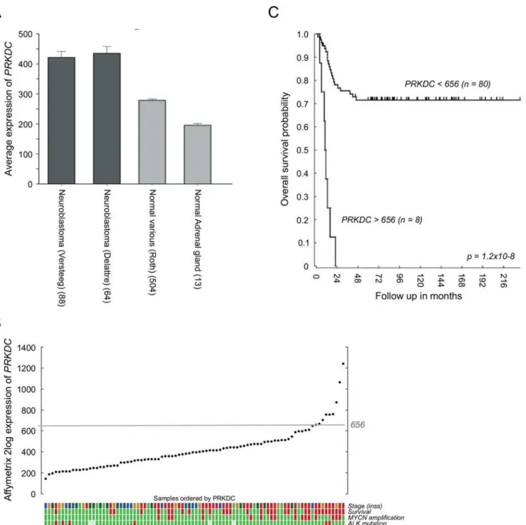
expression levels with expression levels in non-diseased (normal) tissues. As shown inFig 1A,
PRKDCmRNA expression levels in two independent neuroblastoma tumor datasets were
sig-nificantly higher than the expression levels in a compiled library of normal tissues and nonma-lignant adrenal glands (the main primary tumor site) [39]. ElevatedPRKDCmRNA levels in
neuroblastoma were associated with poor clinical characteristics (Fig 1B) and a significant reduction in overall survival (Fig 1C).
NU7026 sensitizes NGP cells to low IR doses
Fig 1. EnhancedPRKDCexpression in neuroblastoma patients correlates with a poor prognosis.(A) Comparison between the average Affymetrix PRKDCmRNA expression levels in two independent neuroblastoma datasets (dark grey) versus expression levels in a compiled library of normal tissues and in nonmalignant adrenal glands (light grey). (B) AffymetrixPRKDCmRNA expression in neuroblastoma of 88 individual patients, ordered by the2log fold
transformed level ofPRKDC. Color coded tracks below the YY plot give information about neuroblastoma stage (INSS; green = stage 1, dark green = stage 2, light brown = stage 3, blue = stage 4s and red = stage 4), patient survival (green = yes and red = no) and the presence ofMYCNamplification andALK mutation (green = yes and red = no). (C) Kaplan-Meier curve of the survival of neuroblastoma patients with an AffymetrixPRKDCmRNA expression above 656 (n = 8) versus neuroblastoma patients with an AffymetrixPRKDCmRNA expression below 656 (n = 80). The cutoff of 656 was determined using the Kaplan scanner tool in the R2 web application (http://r2.amc.nl). Samples were sorted according to the expression ofPRKDCand divided into two groups on the basis of a cutoff expression value. All cutoff expression levels and the resulting groups were analyzed for survival, with the provision that each group included at least eight samples. For each cutoff level and grouping, the log-rank significance of projected survival was calculated. The graph depicts the best pvalue corrected for multiple testing (Bonferroni correction).
anticancer activity is obtained after combination treatment. Optimal doses for combination treatment with NU7026 and IR were studied in the neuroblastoma cell line NGP because of the highPRKDCmRNA expression in this cell line, as determined by Affymetrix microarray
analy-sis (S1 Fig).Fig 2shows the cell viability of NGP cells pre-treated with 0, 2, 10 or 20μM
NU7026 before being exposed to increasing doses of IR up to 6.25 Gy. Effects on cell viability after combination treatment with NU7026 and IR increased in time, with maximum effects observed from 96 h after IR-exposure on (Fig 2A). As anticipated, cytotoxic effects of mono-therapy with NU7026 or IR increased with increasing dose (S1 Table).
Combination treatment of NGP cells with 10μM NU7026 and 0.63 Gy IR resulted in a
decrease in cell viability of 89% (at 96 as well as 120 h after IR-exposure), while maximum inhibitory effects achieved with 10μM NU7026 and 0.63 Gy IR monotherapy were no more
than 38 and 46%, respectively (Fig 2B). To quantify the interaction between 10μM NU7026
and 0.63 Gy IR, combination indices (CIs) were determined according to Chou and Talalay [40]. From these CIs (i.e. 0.25 and 0.26 at 96 and 120 h after IR-exposure, respectively) it could be concluded that synergistic radiosensitization of NGP cells was obtained. CIs for all combina-tions are presented inS1 Table.
Radiosensitizing effects of NU7026 were confirmed by analysis of the long-term effects on colony formation. In line with the effects on cell viability, NU7026 monotherapy dose-depen-dently inhibited the colony forming capacity of NGP cells (Fig 2CandS2A Fig). While irradia-tion with 0.63 Gy alone did not result in any effect, pre-treatment of NGP cells with 10 or 20μM NU7026 before irradiation resulted in less colonies compared with NU7026
monother-apy. Further combination studies were performed using 10μM NU7026 and 0.63 Gy IR.
Combination treatment with NU7026 and IR induces apoptosis of NGP
cells
As the cell viability and colony forming responses shown inFig 2represent combined effects on cell proliferation and cell death, additional analyses were performed to study specific effects on apoptosis. Combination treatment of NGP cells with 10μM NU7026 and 0.63 Gy IR
resulted in a remarkable increase in sub-G1 fraction (i.e. + 18% versus untreated cells), while hardly any effect was obtained after monotherapy with NU7026 or IR (i.e. + 0% and 1% versus untreated cells, respectively) (Fig 3A and 3B). Most strikingly, the synergistic effect between NU7026 and IR is much more pronounced for the apoptotic response than for the cell viability response shown inFig 2. Observed effects on sub-G1 fraction increased in time (Fig 3B), con-firming the time-dependent effects on cell viability. Apoptosis of co-treated NGP cells was additionally shown by PARP cleavage at 96 h after IR-exposure (Fig 3C).
DNA-PKcs was rapidly activated upon cell exposure to 0.63 Gy IR, as shown by the increase in DNA-PKcs autophosphorylation at serine 2056 observed at 1 h after irradiation. IR-induced DNA-PKcs activation was inhibited by concomitant treatment with 10μM NU7026, added to
the cells at 1 h prior to IR-exposure (Fig 3D). These results confirm that NGP cells can partially evade cytotoxicity of IR by activating NHEJ.
NU7026 does not radiosensitize low DNA-PKcs-expressing
non-cancerous fibroblasts
Radiosensitizing effects of NU7026 were investigated in five additional neuroblastoma cell lines (i.e. SHSY5Y, SHEP2, SJNB12, LAN5 and SKNBE(2)) with varyingPRKDCmRNA
cell lines were subsequently compared with the radiosensitizing effects of NU7026 for a panel of non-cancerous fast-proliferating fibroblasts (i.e. F1366, F1641, F0577 and F2112) (Fig 4A). In non-cancerous fibroblasts an additive inhibitory effect on cell viability of NU7026 in combi-nation with IR was observed, with CIs between 0.96 and 1.02. This differential effect might be explained by up-regulated DNA-PKcs protein levels in neuroblastoma cells, as only limited lev-els of DNA-PKcs could be detected in the fibroblast cell lines (Fig 4BandS2B Fig).
assays were performed to study inhibitory effects on cell viability. Data represent the mean (n = 4) +/- SD. (B) Cell viability of NGP cells after treatment with 10μM NU7026 and/or 0.63 Gy IR at 96 and 120 h after IR-exposure. Data represent the mean (n = 4) +/- SD. Statistical differences between untreated and treated NGP cells are indicated as*(p<0.05). (C) Effects of NU7026 plus IR combination therapy versus monotherapy on the colony forming capacity of NGP cells. Cells in 0.4% agar in culture medium were seeded on top of a hardened 0.5% agar base layer. After overnight incubation at normal culture conditions, cells were 1 h pre-incubated with 0, 2, 10 or 20μM NU7026 in 0.4% agar in culture medium before exposure to 0 or 0.63 Gy IR. The following 3 weeks, fresh DMSO or NU7026 in 0.4% agar in culture medium was added to the cells once a week. Colonies were subsequently visualized by 4 h incubation with MTT and counted. The number of colonies formed by untreated NGP cells has been set at 100%. Data represent the mean (n = 3) +/- SD.
doi:10.1371/journal.pone.0145744.g002
Fig 3. Combination treatment with NU7026 and IR induces apoptosis in NGP cells.(A) Effects of NU7026 plus IR combination therapy versus monotherapy on the cell cycle status of NGP cells. For combination treatment, cells were pre-treated with 10μM NU7026 for 1 h before exposure to 0.63 Gy IR. Samples were analyzed by FACS analysis at 96 h after treatment. (B) FACS analysis of the effects of combination therapy versus monotherapy on the sub-G1 fraction of cells at 48, 72 and 96 h after treatment. (C) Western Blot detection of total DNA-PKcs and activated DNA-PKcs (via phosphorylation at serine 2056) levels and PARP cleavage after combination therapy versus monotherapy with 10μM NU7026 and/or 0.63 Gy IR, at 1 h and 96 h after IR-exposure, respectively.β-actin protein levels were used as loading control.
Gemcitabine is a chemotherapeutic agent which is currently evaluated extensively in clinical trials for its additional radiosensitizing potential [41]. It is suggested that gemcitabine exerts its radiosensitizing effect via inhibition of ribonucleotide reductase by its active metabolite gemcita-bine diphosphate, thereby reducing the synthesis of deoxynucleoside triphosphates [41]. Current study compared the radiosensitizing effects of NU7026 in neuroblastoma cell lines with the radiosensitizing effects of gemcitabine. As for NU7026, we first investigated the optimal gemcita-bine dose for combination treatment with 0.63 Gy IR in NGP cells. Based on literature [41–43], NGP cells were pre-treated with 0, 1, 5, 10 or 100 nM or 1 or 10μM gemcitabine for 3 or 24 h
before irradiation. Effects on cell viability were studied at 96 h after IR-exposure of the cells. Twenty-four hours pre-treatment with gemcitabine did not improve the inhibitory effect of low-dose IR treatment on the cell viability of NGP cells, while 3 h pre-treatment did (Fig 5A). Mono-therapy of NGP cells with100 nM gemcitabine furthermore resulted into almost complete cell death (i.e. 97–99%). For the comparison with NU7026, gemcitabine was therefore administered to the cells in doses below 100 nM (i.e. 1, 5 and 10 nM) at 3 h before IR-exposure.
Radiosensitizing effects of gemcitabine were studied for the same neuroblastoma cell line panel as used for NU7026. While NU7026 synergistically radiosensitized all tested neuroblas-toma cell lines to 0.63 Gy IR (i.e. mean CI = 0.60), only an additive effect on cell viability was observed for the combination between 1 nM gemcitabine and 0.63 Gy IR (i.e. 0.97CI1.08; mean CI = 1.02). Except for neuroblastoma cell line LAN5, higher doses of gemcitabine seemed to even result in less favorable effects (i.e. mean CI for 5 nM gemcitabine = 1.11; mean CI for 10 nM gemcitabine = 1.14) (Fig 5B). So in this experimental setup the DNA-PKcs inhibitor NU7026 exerted a more significant radiosensitizing effect in neuroblastoma cell lines than gemcitabine.
Radiosensitization is DNA-PKcs dependent
Small-molecule inhibitors, also when they are called specific, often possess off–target effects. To further investigate if the radiosensitizing effects observed for NU7026 are caused by
Fig 4. NU7026 does not radiosensitize low DNA-PKcs-expressing non-cancerous cells.(A) Radiosensitizing effect of NU7026 in neuroblastoma cell lines NGP, SHSY5Y, SHEP2, SJNB12, LAN5 and SKNBE(2) compared with radiosensitizing effects in non-cancerous fast-proliferating fibroblast cell lines F1366, F1641, F0577 and F2112. Co-treated cells were exposed to 0.63 Gy IR after 1 h pre-treatment with 10μM NU7026. At 96 h after IR-exposure, MTT cell proliferation assays were performed to assess cell viability (n = 4 for each cell line). Radiosensitizing effects were estimated by calculating combination indices (CI) according to Chou and Talalay [40], given on the Y-axis. CI>1.1 is antagonistic (dark grey), 1.1CI0.9 is additive (white) and CI<0.9 is
synergistic (light grey). Horizontal lines between the symbols represent the mean combined effect obtained for all neuroblastoma- or fibroblast cell lines. (B) Western Blot analysis of DNA-PKcs protein levels in neuroblastoma cell lines NGP, SHSY5Y, SHEP2, SJNB12, LAN5 and SKNBE(2) and fibroblast cell lines F1366, F1641, F0577 and F2112.β-actin protein levels were used as loading control.
Fig 5. NU7026 more potently radiosensitizes neuroblastoma cells than gemcitabine.(A) Cell viability of NGP cells after co-treatment with gemcitabine and IR. Cells were pre-treated with 0, 1, 5, 10 or 100 nM or 1 or 10μM gemcitabine for 3 or 24 h before exposure to 0.63 Gy IR. At 96 h after IR-exposure, MTT cell
proliferation assays were performed to study effects on cell viability. Data represent the mean (n = 4) +/- SD. (B) Radiosensitizing effects of 1, 5 and 10 nM gemcitabine for neuroblastoma cell lines NGP, SHSY5Y, SHEP2, SJNB12, LAN5 and SKNBE(2). Cells were pre-treated with gemcitabine for 3 h before exposure to 0.63 Gy IR. At 96 h after IR-exposure, MTT cell proliferation assays were performed to assess cell viability (n = 4 for each cell line). Radiosensitizing effects were estimated by calculating combination indices (CI) according to Chou and Talalay [40], given on the Y-axis. CI>1.1 is antagonistic (dark grey), 1.1CI0.9 is additive (white) and CI<0.9 is synergistic (light grey). Horizontal lines between the symbols represent the mean combined effects.
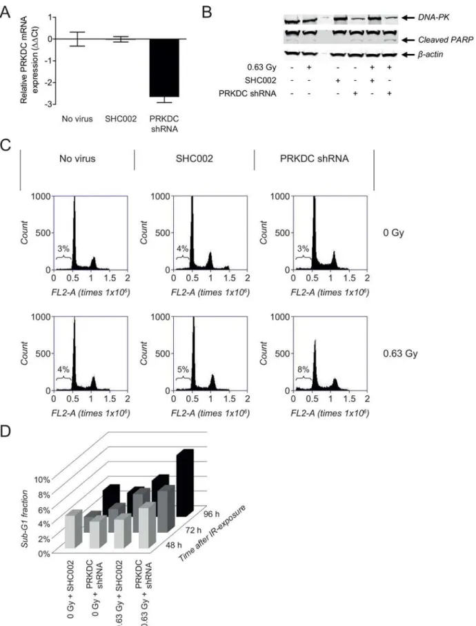
DNA-PKcs inhibition, validation studies were performed using NGP cells transduced with a
PRKDCshRNA or a nonspecific control shRNA (SHC002).PRKDCgene silencing was
observed at 144 h after transduction of the NGP cells withPRKDCshRNA, while SHC002 did
not affect the gene expression level ofPRKDC(Fig 6A). Effects on gene level were confirmed
on protein level by Western Blot analysis of DNA-PKcs (Fig 6B).
No apoptotic responses were observed after transduction of NGP cells with SHC002 or
PRKDCshRNA, as shown by the lack of significant changes in sub-G1 fraction (Fig 6C). While
only a 1% increase in sub-G1 fraction was observed after irradiation of SHC002-transduced NGP cells, exposure ofPRKDCshRNA-transduced NGP cells to 0.63 Gy IR resulted in an
increase in sub-G1 fraction of 4% at 96 h after irradiation (Fig 6C). As was seen for the combi-nation of NU7026 and IR, the apoptotic response was more pronounced at 96 h than at 48 or 72 h after treatment (Fig 6D). Effects on sub-G1 fraction were confirmed by Western Blot anal-ysis of PARP cleavage, with the highest cleaved PARP levels being observed after irradiation of NGP cells withPRKDCknockdown (Fig 6B).
Although the knockdown studies showed less potent effects, these results confirmed that the observed radiosensitizing effects of NU7026 are at least partially caused due to inhibition of its target DNA-PKcs.
Discussion
This is the first manuscript reporting thein vitroradiosensitization of neuroblastoma cells by
pre-treatment with a DNA-PKcs inhibitor. Radiosensitization of tumor cells by preventing NHEJ of DSBs has successfully been studied for different tumor types (e.g. breast cancer, ovar-ian cancer, colon cancer, cervical cancer and pancreatic cancer [29,31,44–46]), after it was shown that DSB repair by NHEJ is the predominant mechanism by which tumor cells might escape from radiation-induced cell death [8]. Currently, DNA-PKcs and DNA ligase IV are the only druggable targets in the NHEJ pathway [27]. As DNA-PKcs is one of the most upstream key players in NHEJ [27], it is not surprising that DNA-PKcs inhibitors are more frequently tested for their potency to radiosensitize tumor cells and overcome radioresistance than DNA ligase IV inhibitors. In the current manuscript, we studied the radiosensitizing potential of DNA-PKcs inhibitor NU7026 for neuroblastoma treatment.
Synergistic responses between IR and NU7026 were initially calculated based on cell viabil-ity assays which could consist of a growth inhibitory effect combined with a cell killing response. We showed that apoptosis-induced cell killing showed a much stronger synergistic response compared to the cell viability assays. This implies that neuroblastoma cells show a much stronger induction of cell death if radiation therapy is combined with a DNA-PKcs inhibitor and therefore a switch could be made from a cytostatic to a cytotoxic effect on neuro-blastoma cells. Of note, irradiated cells might also be killed through mechanisms other than apoptosis, i.e. mitotic catastrophe, autophagy and necrosis [47]. These mechanisms of cell death have not been investigated in the current study.
Although promising, the increased risk of toxic effects on healthy tissues should be taken into account when adding a DSB repair inhibitor to radiation therapy. Strategies to restrict side effects on healthy tissues are limiting doses and/or using a radiosensitizer acting on a differen-tially expressed target in neuroblastoma tumors, resulting in a tumor-specific sensitizing effect. In determining the most optimal dose for combination treatment with NU7026 and IR, a bal-ance was made between the effects obtained with NU7026 or IR alone or in combination. For example, 20μM NU7026 more potently sensitized NGP cells to 0.63 Gy IR than 10μM of the
activity was only 4% (i.e. 93 versus 89%). Similarly, equal combined effects could be obtained when combining lower NU7026 doses with higher IR doses, but in many of these cases weaker cytotoxic effects obtained with NU7026 were compensated by stronger cytotoxic effects of IR. Comparing our results with previously published results for other cancer types showed that NU7026 doses most potently sensitizing NGP cells to IR were comparable with NU7026 doses radiosensitizing ovarian, pancreatic and gastric tumor cells [29–31]. However, in the other studies similar NU7026 doses were combined with much higher IR doses (i.e. 2–5 Gy) [29–31]. We showed that exposure of NGP cells pre-treated with 10μM NU7026 to 6.25 instead of 0.63
Gy IR only increased the combined inhibitory effect on cell viability from 89 to 95%, equal to the effect observed without NU7026 pre-treatment.
As NU7026 inhibits DNA-PKcs, elevated DNA-PKcs levels in cancer cells might contribute to a more tumor cell-specific radiosensitizing effect. DNA-PKcs is expressed in normal tissues such as colon, pancreas and kidneys [48]. However, various tumor types have been associated with elevatedPRKDCmRNA- and/or DNA-PKcs levels [28,49]. We demonstrated that also
neuroblastoma express higher levels ofPRKDCmRNA compared with normal tissues.
Radio-sensitizing effects of NU7026 were evaluated for neuroblastoma cell lines expressing different
PRKDCmRNA levels, including cell lines with the lowest (i.e. SHSY5Y) and highest (i.e. NGP) PRKDCexpression (S1 Fig). No direct correlation could be found betweenPRKDCmRNA
lev-els and extent of radiosensitization. Synergistic radiosensitizing effects of NU7026 observed for all tested neuroblastoma cell lines indicate that even the lowestPRKDCmRNA level observed
in neuroblastoma cell lines was high enough to induce radiosensitization. NU7026 did not radiosensitize low DNA-PKcs-expressing fast-proliferating fibroblasts that share some of their characteristics with cancer cells. Future studies need to focus on thePRKDCmRNA cutoff
val-ues above which NU7026 radiosensitizes neuroblastoma cells and on the correlation between
PRKDCmRNA levels and DNA-PKcs protein levels in neuroblastoma.
NU7026 is one of the first DNA-PKcs inhibitors derived from the nonspecific DNA-PKcs inhibitor LY294002 to obtain a more potent inhibitor with improved specificity towards DNA-PKcs [8,26]. Due to similarities between the kinase domains of DNA-PKcs and PI3K, LY294002 potently inhibits both kinases [8]. NU7026 inhibits DNA-PKcs much more potently than PI3K with IC50values of 0.23μM and 13μM, respectively [26,27,31]. Currently,
LY294002 has been used as a backbone for the development of several other DNA-PKcs inhibi-tors including NU7441 [8,26]. NU7441 is more potent and specific than NU7026 with IC50
values against DNA-PKcs and PI3K of 14 nM and 5μM, respectively, [49,50] and possesses
more favorable pharmacokinetic characteristics leading to an improved residence time in the tumor [26]. Together with its radiosensitizing effects observed in pre-clinical studies for other cancer types [45,46,51], these properties make NU7441 a valuable compound for adjuvant testing in neuroblastoma cells. At this moment, NU7026 and NU7441 not yet reached clinical phase.
Results obtained with the small-molecule inhibitor NU7026 were confirmed byPRKDC
knockdown, proving that NU7026 radiosensitizes neuroblastoma cells due to DNA-PKcs inhi-bition. But the radiosensitizing effect ofPRKDCknockdown was less potent than the
values were normalized to the household geneβ-actin. NormalizedΔCt values of untreated non-transduced NGP cells (no virus) were set at zero, giving ΔΔCt. Data represent the mean (n = 4) +/- SD. (B) FACS analysis of the effects of treatment with 0.63 Gy IR on the sub-G1 fraction of normal NGP cells and NGP cells transduced with SHC002 orPRKDCshRNA. Results are obtained at 96 h after IR-exposure of the cells. (C) Sub-G1 fraction of NGP cells transduced with SHC002 orPRKDCshRNA after non-treatment and after treatment with 0.63 Gy IR, at 48, 72 and 96 h after irradiation. (D) Western Blot analysis of protein levels of DNA-PKcs and cleaved PARP in non-irradiated and irradiated normal NGP cells and NGP cells transduced with SHC002 or PRKDCshRNA. Results were obtained at 120 h after irradiation, which was equal to 168 h after transduction.β-actin protein levels were used as loading control.
radiosensitizing effects of NU7026. This might relate to incomplete DNA-PKcs silencing or the fact that NU7026 inhibits additional targets [31,52,53].
Taken together, this paper demonstrates that DNA-PKcs inhibition synergistically sensi-tized neuroblastoma cells to low radiation doses. Combination therapy with 10μM NU7026
and 0.63 Gy IR resulted in one of the most favorable effects, leading to the effective killing of neuroblastoma cells. In contrast, NU7026 did not radiosensitize low DNA-PKcs expressing non-cancerous fibroblasts. Together with the observation that neuroblastoma patients have increased levels of the gene encoding for DNA-PKcs (i.e.PRKDC), these results make
DNA-PKcs a promising target for neuroblastoma radiosensitization.
Supporting Information
S1 Fig.PRKDCmRNA expression levels in neuroblastoma cell lines.PRKDCmRNA
expres-sion levels were analyzed by Affymetrix microarrays. Black symbols represent the expresexpres-sion levels in the neuroblastoma cell lines included in the current study.
(TIF)
S2 Fig. NU7026 radiosensitizes high DNA-PKcs-expressing NGP cells.(A) Effects of NU7026 plus IR combination therapy versus monotherapy on the colony forming capacity of NGP cells. Cells in 0.4% agar in culture medium were seeded on top of a hardened 0.5% agar base layer. After overnight incubation at normal culture conditions, cells were 1 h pre-incu-bated with 0, 2, 10 or 20μM NU7026 in 0.4% agar in culture medium before exposure to 0 or
0.63 Gy IR (n = 3 per condition). The following 3 weeks, fresh DMSO or NU7026 in 0.4% agar in culture medium was added to the cells once a week. Colonies were subsequently visualized by 4 h incubation with MTT. (B) Western Blot analysis of DNA-PKcs protein levels in neuro-blastoma cell lines NGP and SKNBE(2) and fibroblast cell lines F2112 and F1366.α-Tubulin protein levels were used as loading control. Separate analysis of the fibroblast cell lines showed that the non-cancerous fast-proliferating fibroblast cell lines F2112 and F1366 express low lev-els of DNA-PKcs (right pictures).
(TIF)
S1 Table. Sensitivity of NGP cells to NU7026 plus IR combination therapy versus mono-therapy.Percentage inhibition of the cell viability after monotherapy or combination therapy of NGP cells with indicated doses of NU7026 and/or IR. Combination indices (CIs) are given between brackets and calculated according to Chou and Talalay [40]. CI>1.1 is antagonistic,
1.1CI0.9 is additive and CI<0.9 is synergistic.
(DOCX)
Author Contributions
Conceived and designed the experiments: MEMD JJM. Performed the experiments: MEMD IP LTBE. Analyzed the data: MEMD IP JK. Wrote the paper: MEMD RV HNC JJM.
References
1. O'Connor MJ, Martin NM, Smith GC. Targeted cancer therapies based on the inhibition of DNA strand break repair. Oncogene. 2007; 26(56): 7816–24. PMID:18066095
2. Plummer R. Perspective on the pipeline of drugs being developed with modulation of DNA damage as a target. Clin Cancer Res. 2010; 16(18): 4527–31. doi:10.1158/1078-0432.CCR-10-0984PMID:
20823148
4. Madhusudan S, Hickson ID. DNA repair inhibition: a selective tumour targeting strategy. Trends Mol Med. 2005; 11(11): 503–11. PMID:16214418
5. Sonoda E, Hochegger H, Saberi A, Taniguchi Y, Takeda S. Differential usage of non-homologous end-joining and homologous recombination in double strand break repair. DNA Repair (Amst). 2006; 5(9–
10): 1021–9.
6. Valerie K, Povirk LF. Regulation and mechanisms of mammalian double-strand break repair. Onco-gene. 2003; 22(37): 5792–812. PMID:12947387
7. Shibata A, Conrad S, Birraux J, Geuting V, Barton O, Ismail A, et al. Factors determining DNA double-strand break repair pathway choice in G2 phase. EMBO J. 2011; 30(6): 1079–92. doi:10.1038/emboj.
2011.27PMID:21317870
8. Mukherjee B, Choy H, Nirodi C, Burma S. Targeting nonhomologous end-joining through epidermal growth factor receptor inhibition: rationale and strategies for radiosensitization. Semin Radiat Oncol. 2010; 20(4): 250–7. doi:10.1016/j.semradonc.2010.05.002PMID:20832017
9. Chen BP, Uematsu N, Kobayashi J, Lerenthal Y, Krempler A, Yajima H, et al. Ataxia telangiectasia mutated (ATM) is essential for DNA-PKcs phosphorylations at the Thr-2609 cluster upon DNA double strand break. J Biol Chem. 2007; 282(9): 6582–7. PMID:17189255
10. Burma S, Chen DJ. Role of DNA-PK in the cellular response to DNA double-strand breaks. DNA Repair (Amst). 2004; 3(8–9): 909–18.
11. Zha S, Jiang W, Fujiwara Y, Patel H, Goff PH, Brush JW, et al. Ataxia telangiectasia-mutated protein and DNA-dependent protein kinase have complementary V(D)J recombination functions. Proc Natl Acad Sci U S A. 2011; 108(5): 2028–33. doi:10.1073/pnas.1019293108PMID:21245310
12. Salles B, Calsou P, Frit P, Muller C. The DNA repair complex DNA-PK, a pharmacological target in can-cer chemotherapy and radiotherapy. Pathol Biol (Paris). 2006; 54(4): 185–93.
13. Helleday T, Petermann E, Lundin C, Hodgson B, Sharma RA. DNA repair pathways as targets for can-cer therapy. Nat Rev Cancan-cer. 2008; 8(3): 193–204. doi:10.1038/nrc2342PMID:18256616
14. Luqmani YA. Mechanisms of drug resistance in cancer chemotherapy. Med Princ Pract. 2005; 14 Suppl 1: 35–48. PMID:16103712
15. Frosina G. DNA repair and resistance of gliomas to chemotherapy and radiotherapy. Mol Cancer Res. 2009; 7(7): 989–99. doi:10.1158/1541-7786.MCR-09-0030PMID:19609002
16. Park JR, Eggert A, Caron H. Neuroblastoma: biology, prognosis, and treatment. Pediatr Clin North Am. 2008; 55(1): 97–120. doi:10.1016/j.pcl.2007.10.014PMID:18242317
17. Morrison R, Schleicher SM, Sun Y, Niermann KJ, Kim S, Spratt DE, et al. Targeting the mechanisms of resistance to chemotherapy and radiotherapy with the cancer stem cell hypothesis. J Oncol. 2011; 2011: 941876. doi:10.1155/2011/941876PMID:20981352
18. Lord CJ, Garrett MD, Ashworth A. Targeting the double-strand DNA break repair pathway as a thera-peutic strategy. Clin Cancer Res. 2006; 12(15): 4463–8. PMID:16899589
19. Verissimo CS, Molenaar JJ, Fitzsimons CP, Vreugdenhil E. Neuroblastoma therapy: what is in the pipe-line? Endocr Relat Cancer. 2011; 18(6): R213–31. doi:10.1530/ERC-11-0251PMID:21971288 20. Mahaney BL, Meek K, Lees-Miller SP. Repair of ionizing radiation-induced DNA double-strand breaks
by non-homologous end-joining. Biochem J. 2009; 417(3): 639–50. doi:10.1042/BJ20080413PMID:
19133841
21. Amrein L, Loignon M, Goulet AC, Dunn M, Jean-Claude B, Aloyz R, et al. Chlorambucil cytotoxicity in malignant B lymphocytes is synergistically increased by 2-(morpholin-4-yl)-benzo[h]chomen-4-one (NU7026)-mediated inhibition of DNA double-strand break repair via inhibition of DNA-dependent pro-tein kinase. J Pharmacol Exp Ther. 2007; 321(3): 848–55. PMID:17351105
22. Veuger SJ, Curtin NJ, Richardson CJ, Smith GC, Durkacz BW. Radiosensitization and DNA repair inhi-bition by the combined use of novel inhibitors of DNA-dependent protein kinase and poly(ADP-ribose) polymerase-1. Cancer Res. 2003; 63(18): 6008–15. PMID:14522929
23. Tavecchio M, Munck JM, Cano C, Newell DR, Curtin NJ. Further characterisation of the cellular activity of the DNA-PK inhibitor, NU7441, reveals potential cross-talk with homologous recombination. Cancer Chemother Pharmacol. 2011. Epub 2011/06/02. doi:10.1007/s00280-011-1662-4
24. Davidson D, Coulombe Y, Martinez-Marignac VL, Amrein L, Grenier J, Hodkinson K, et al. Irinotecan and DNA-PKcs inhibitors synergize in killing of colon cancer cells. Invest New Drugs. 2011. Epub 2011/ 01/12. doi:10.1007/s10637-010-9626-9
26. Davidson D, Amrein L, Panasci L, Aloyz R. Small Molecules, Inhibitors of DNA-PK, Targeting DNA Repair, and Beyond. Front Pharmacol. 2013; 4: 5. doi:10.3389/fphar.2013.00005PMID:23386830
27. Jekimovs C, Bolderson E, Suraweera A, Adams M, O'Byrne KJ, Richard DJ. Chemotherapeutic com-pounds targeting the DNA double-strand break repair pathways: the good, the bad, and the promising. Front Oncol. 2014; 4:86. doi:10.3389/fonc.2014.00086PMID:24795863
28. Hosoya N, Miyagawa K. Targeting DNA damage response in cancer therapy. Cancer Sci. 2014; 105 (4): 370–88. doi:10.1111/cas.12366PMID:24484288
29. Li YH, Wang X, Pan Y, Lee DH, Chowdhury D, Kimmelman AC. Inhibition of non-homologous end join-ing repair impairs pancreatic cancer growth and enhances radiation response. PLoS One. 2012; 7(6): e39588. doi:10.1371/journal.pone.0039588PMID:22724027
30. Niazi MT, Mok G, Heravi M, Lee L, Vuong T, Aloyz R, et al. Effects of dna-dependent protein kinase inhibition by NU7026 on dna repair and cell survival in irradiated gastric cancer cell line N87. Curr Oncol. 2014; 21(2): 91–6. doi:10.3747/co.21.1509PMID:24764698
31. Nutley BP, Smith NF, Hayes A, Kelland LR, Brunton L, Golding BT, et al. Preclinical pharmacokinetics and metabolism of a novel prototype DNA-PK inhibitor NU7026. Br J Cancer. 2005; 93(9): 1011–8.
PMID:16249792
32. Molenaar JJ, Koster J, Ebus ME, van Sluis P, Westerhout EM, de Preter K, et al. Copy number defects of G1-cell cycle genes in neuroblastoma are frequent and correlate with high expression of E2F target genes and a poor prognosis. Genes Chromosomes Cancer. 2012; 51(1): 10–9. doi:10.1002/gcc.
20926PMID:22034077
33. Molenaar JJ, Domingo-Fernandez R, Ebus ME, Lindner S, Koster J, Drabek K, et al. LIN28B induces neuroblastoma and enhances MYCN levels via let-7 suppression. Nat Genet. 2012; 44(11): 1199–206.
doi:10.1038/ng.2436PMID:23042116
34. Fix A, Lucchesi C, Ribeiro A, Lequin D, Pierron G, Schleiermacher G, et al. Characterization of ampli-cons in neuroblastoma: high-resolution mapping using DNA microarrays, relationship with outcome, and identification of overexpressed genes. Genes Chromosomes Cancer. 2008; 47(10): 819–34. doi:
10.1002/gcc.20583PMID:18553563
35. Laureys G, Speleman F, Versteeg R, van der Drift P, Chan A, Leroy J, et al. Constitutional translocation t(1;17)(p36.31-p36.13;q11.2-q12.1) in a neuroblastoma patient. Establishment of somatic cell hybrids and identification of PND/A12M2 on chromosome 1 and NF1/SCYA7 on chromosome 17 as breakpoint flanking single copy markers. Oncogene. 1995; 10(6): 1087–93. PMID:7700633
36. Molenaar JJ, Ebus ME, Geerts D, Koster J, Lamers F, Valentijn LJ, et al. Inactivation of CDK2 is syn-thetically lethal to MYCN over-expressing cancer cells. Proc Natl Acad Sci U S A. 2009; 106(31): 12968–73. doi:10.1073/pnas.0901418106PMID:19525400
37. Thiele CJ. Neuroblastoma cell lines. In: Masters J, editor. Human cell culture. Lancaster, UK: Kluwer Academic Publishers; 1998. pp. 21–53.
38. Twentyman PR, Luscombe M. A study of some variables in a tetrazolium dye (MTT) based assay for cell growth and chemosensitivity. Br J Cancer. 1987; 56(3): 279–85. PMID:3663476
39. Nuchtern JG. Perinatal neuroblastoma. Semin Pediatr Surg. 2006; 15(1): 10–6. PMID:16458841 40. Chou TC, Talaly P. A simple generalized equation for the analysis of multiple inhibitions of
Michaelis-Menten kinetic systems. J Biol Chem. 1977; 252(18): 6438–42. PMID:893418
41. Morgan MA, Parsels LA, Maybaum J, Lawrence TS. Improving gemcitabine-mediated radiosensitiza-tion using molecularly targeted therapy: a review. Clin Cancer Res. 2008; 14(21): 6744–50. doi:10.
1158/1078-0432.CCR-08-1032PMID:18980967
42. Pauwels B, Korst AE, Andriessen V, Baay MF, Pattyn GG, Lambrechts HA, et al. Unraveling the mech-anism of radiosensitization by gemcitabine: the role of TP53. Radiat Res. 2005; 164(5): 642–50. PMID:
16238441
43. Pacini S, Milano F, Pinzani P, Pazzagli M, Gulisano M, Ruggiero M, et al. Effects of gemcitabine in nor-mal and transformed human lung cell cultures: cytotoxicity and increase in radiation sensitivity. Tumori. 1999; 85(6): 503–7. PMID:10774574
44. Fuhrman CB, Kilgore J, LaCoursiere YD, Lee CM, Milash BA, Soisson AP, et al. Radiosensitization of cervical cancer cells via double-strand DNA break repair inhibition. Gynecol Oncol. 2008; 110(1): 93–8.
doi:10.1016/j.ygyno.2007.08.073PMID:18589211
45. Ciszewski WM, Tavecchio M, Dastych J, Curtin NJ. DNA-PK inhibition by NU7441 sensitizes breast cancer cells to ionizing radiation and doxorubicin. Breast Cancer Res Treat. 2014; 143(1): 47–55. doi:
10.1007/s10549-013-2785-6PMID:24292814
46. Zhao Y, Thomas HD, Batey MA, Cowell IG, Richardson CJ, Griffin RJ, et al. Preclinical evaluation of a potent novel DNA-dependent protein kinase inhibitor NU7441. Cancer Res. 2006; 66(10): 5354–62.
47. Golden EB, Pellicciotta I, Demaria S, Barcellos-Hoff MH, Formenti SC. The convergence of radiation and immunogenic cell death signaling pathways. Front Oncol. 2012; 2: 88. doi:10.3389/fonc.2012. 00088PMID:22891162
48. Moll U, Lau R, Sypes MA, Gupta MM, Anderson CW. DNA-PK, the DNA-activated protein kinase, is dif-ferentially expressed in normal and malignant human tissues. Oncogene. 1999; 18(20): 3114–26.
PMID:10340383
49. Hsu FM, Zhang S, Chen BPC. Role of DNA-dependent protein kinase catalytic subunit in cancer devel-opment and treatment. Translational Cancer Research. 2012; 1(1): 22–34. PMID:22943041
50. Shaheen FS, Znojek P, Fisher A, Webster M, Plummer R, Gaughan L, et al. Targeting the DNA double strand break repair machinery in prostate cancer. PLoS One. 2011; 6(5): e20311. doi:10.1371/journal. pone.0020311PMID:21629734
51. Yu L, Shang ZF, Hsu FM, Zhang Z, Tumati V, Lin YF, et al. NSCLC cells demonstrate differential mode of cell death in response to the combined treatment of radiation and a DNA-PKcs inhibitor. Oncotarget. 2015; 6(6): 3848–60. PMID:25714019
52. Gupta AK, Cerniglia GJ, Mick R, Ahmed MS, Bakanauskas VJ, Muschel RJ, et al. Radiation sensitiza-tion of human cancer cells in vivo by inhibiting the activity of PI3K using LY294002. Int J Radiat Oncol Biol Phys. 2003; 56(3): 846–53. PMID:12788194
53. Liu Y, Cui B, Qiao Y, Zhang Y, Tian Y, Jiang J, et al. Phosphoinositide-3-kinase inhibition enhances radiosensitization of cervical cancer in vivo. Int J Gynecol Cancer. 2011; 21(1): 100–5. doi:10.1097/