ISSN 0100-879X
BIOMEDICAL SCIENCES
www.bjournal.com.br
www.bjournal.com.br
Volume 45 (8) 681-791 August 2012
Braz J Med Biol Res, August 2012, Volume 45(8) 693-700
doi:
10.1590/S0100-879X2012007500060
Tissue transglutaminase (TG2) activity regulates osteoblast
differentiation and mineralization in the SAOS-2 cell line
Xiaoxue Yin, Zhongqiang Chen, Zhongjun Liu and Chunli Song
Institutional Sponsors
The Brazilian Journal of Medical and Biological Research is partially financed by
Faculdade de Medicina de Ribeirão Preto Campus
Ribeirão Preto
Explore High - Performance MS Orbitrap Technology In Proteomics & Metabolomics
Tissue transglutaminase (TG2) activity
regulates osteoblast differentiation and
mineralization in the SAOS-2 cell line
Xiaoxue Yin, Zhongqiang Chen, Zhongjun Liu and Chunli Song
Department of Orthopaedics, Peking University Third Hospital, Beijing, China
Abstract
Tissue transglutaminase (type II, TG2) has long been postulated to directly promote skeletal matrix calcification and play an important role in ossification. However, limited information is available on the expression, function and modulating mechanism of TG2 during osteoblast differentiation and mineralization. To address these issues, we cultured the well-established human osteosarcoma cell line SAOS-2 with osteo-inductive conditioned medium and set up three time points (culture days 4, 7, and 14) to represent different stages of SAOS-2 differentiation. Osteoblast markers, mineralization, as well as TG2 expression and activity, were then assayed in each stage. Furthermore, we inhibited TG activity with cystamine and then checked SAOS-2 differentiation and mineralization in each stage. The results showed that during the progression of osteoblast differentiation SAOS-2 cells presented significantly high levels of osteocalcin (OC) mRNA, bone morphogenetic protein-2 (BMP-2) and col -lagen I, significantly high alkaline phosphatase (ALP) activity, and the increased formation of calcified matrix. With the same tendency, TG2 expression and activity were up-regulated. Furthermore, inhibition of TG activity resulted in a significant decrease of OC, collagen I, and BMP-2 mRNA and of ALP activity and mineralization. This study demonstrated that TG2 is involved in osteoblast differentiation and may play a role in the initiation and regulation of the mineralization processes. Moreover, the modulating effects of TG2 on osteoblasts may be related to BMP-2.
Key words: Transglutaminase; Osteoblast; Differentiation; Mineralization; Bone morphogenetic protein-2 (BMP-2)
Introduction
Correspondence: Zhongqiang Chen, Department of Orthopaedics, Peking University Third Hospital, #49 Hua-Yuan North Road, Haidian District, Beijing 100191, China. Fax: +8610-8226-5557. E-mail: luckyemail2008@sina.com or zhongqiangchen@tom.com
Received November 25, 2011. Accepted April 9, 2012. Available online April 27, 2012. Published August 3, 2012.
Transglutaminase enzymes (TGs; EC 2.3.2.13) are a group of enzymes whose main function is to stabilize and assemble their substrate proteins into large polymers by creating covalent γ-(glutamyl)-ε-lysyl bonds (i.e., isopep-tide crosslinks) between glutamine and lysine residues in a calcium ion-dependent reaction (1). To date, the transglutaminase family consists of 9 different TG genes (2). Tissue transglutaminase (TG2, type II) is one of the best-characterized, skeletal tissue-related TGs (3,4) and may be involved in the initiation of mineralization and play an important role in ossification (5,6).
The biological function of TG2 has yet to be determined. However, there is now increasing evidence suggesting that TG2 can act at the cell surface facilitating cell adhesion, cell spreading and the modification of the extracellular matrix (ECM) (7,8). Moreover, TG2 is a highly selective enzyme, with only a few native proteins identified as its substrates (9). In mineralized tissues, collagen, fibronectin,
osteopontin, and bone sialoprotein are all TG2 substrates, which assemble into polymeric forms to participate in matrix stabilization, chondrocyte and osteoblast differentiation and matrix mineralization in the presence of TG2 (3).
TG2 is expressed in cartilage, bone, and teeth, the most extensively studied being cartilage (3,5,10,11). It has been demonstrated that TG2 expression correlates with chon-drocyte differentiation and matrix mineralization. Moreover, chondrocyte cultures from TG2-knockout mice show lack of induction of matrix calcification (12),while addition of exogenous TG2 increases chondrocyte hypertrophy and mineralization (10,13).
TG2 enzyme activity is also found in extracts of intramem-branous bone, and has been identified in osteoblast-like bone cells in vitro, with results suggesting an involvement in
694 Xiaoxue Yin et al.
FXIIIA, and Wosniak et al. (15) showed that mechanical strain increased osteopath mineralization by the action of TG2. In addition, the TG-catalyzed Nε (γ-glutamyl) lysine crosslinks are abundant in the bone matrix and also seem to correlate with ectopic mineralization in pathological pro-cesses such as atherosclerosis (17) and osteoarthritis (18). Our previous study also showed that TG2 expression and enzyme activity are up-regulated in the ossification cells of the ligamentum flavum (OLF) and TG2 may be involved in the pathologic process of OLF (19).Collectively, these findings indicate that TG2 activity is critical to osteoblast differentiation and matrix maturation.
Although TG2 has been noted in osteoblasts, limited information is available on the expression, function and modulating mechanism of TG2 during osteoblast differen-tiation and mineralization. We intended to address these issues in vitro using the well-established, matrix-producing
and mineralizing human osteosarcoma cell line SAOS-2 (20).In this study, we set up three times (days 4, 7, and 14) to represent different stages of SAOS-2 differentiation and mineralization, and assayed TG2 expression and ac-tivity in each stage. Furthermore, we inhibited TG acac-tivity with cystamine and then checked SAOS-2 differentiation and subsequent mineralization in each stage. The results provided insight into the contribution of TG2 to the cas-cade of events leading to bone differentiation and matrix maturation.
Material and Methods
Cell culture
SAOS-2 cells were a generous gift from Dr. David R. Eyre (University of Washington Medical Center, Seattle, WA, USA). Cells were routinely grown in Dulbecco’s modi-fied Eagle’s medium (DMEM; Gibco, USA), supplemented with 10% fetal bovine serum (Gibco), and incubated in a humidified atmosphere of 95% air and 5% CO2 at 37°C. The culture medium was changed every second day. Dif-ferentiation and mineralization were induced by the osteo-inductive conditioned medium, which was composed of 10% FBS DMEM supplemented with 50 µg/mL L-ascorbic acid (AA; Sigma, USA) and 10 mM β-glycerophosphate (β-GP,
Sigma). Cells treated with 10% FBS DMEM normal medium were used as controls for all experiments.
Osteoblast differentiation and mineralization assays
SAOS-2 cells were cultured in osteo-inductive con-ditioned medium for 4, 7, and 14 days and were then harvested individually. Subsequent assays of osteoblast differentiation and mineralization were carried out as described below.
mRNA expression of osteoblast markers
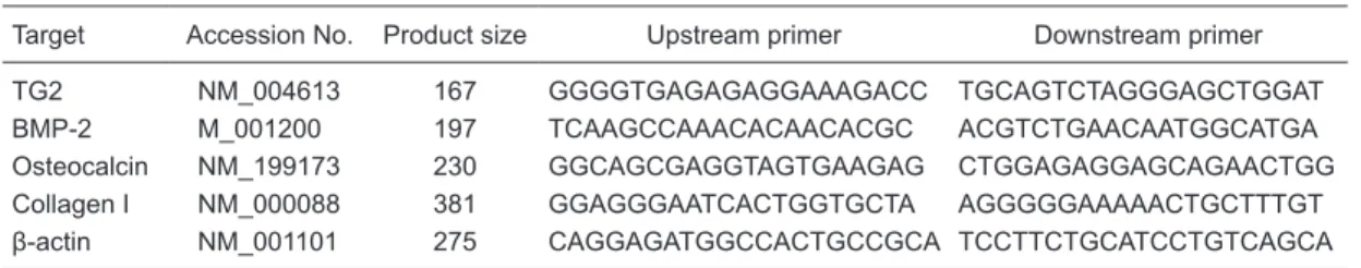
Total RNA of cells was extracted with Trizol reagent. cDNA synthesis was carried out with 5 µg total RNA, 1 µL random primer, 2 µL dNTPs and 200 U M-MLV reverse tran-scriptase (Promega, USA) at 37°C for 1 h. The osteocalcin (OC), bone morphogenetic protein-2 (BMP-2), collagen I, and TG2 genes in mRNA were detected by semiquantitative RT-PCR, with the amplification of β-actin as an internal con-trol. The specific primers (Invitrogen, USA) were designed by Primer Premier and described in Table 1.
Amplification was performed using the Platinum PCR SuperMix (Invitrogen). PCR was carried out for 33 cycles, each at 95°C for 30 s, at 58°C for 30 s, at 72°C for 1 min, with a final extension at 74°C for 7 min. The PCR products were analyzed by 3% agarose gel electrophoresis and visu-alized with ethidium bromide staining, and the densitometry values of the bands were quantified and analyzed using the Image J software.
Alkaline phosphatase activity assay
Alkaline phosphatase (ALP) activity was assayed us-ing the LabAssay™ ALP kit (Wako, Japan). SAOS-2 cells were seeded onto 6-well plates at a density of 2 х 105 cells per well and treated for the designated time. Cells were then obtained and completely lysed by sonication for 10 min with a sonifer cell disruptor (Cosmo Bio, Japan). The sonicates were centrifuged for 10 min at 20,142 g, and the supernatants were used as samples for the ALP activity assay. The protein concentrations were determined with the bicinchoninic (BCA) protein assay reagent. The relative activity of the sample is reported as the ratio of activity and the corresponding protein concentration (U/mg).
Table 1. Primers for RT-PCR used to detect osteoblast markers and TG2 in SAOS-2.
Target Accession No. Product size Upstream primer Downstream primer
TG2 NM_004613 167 GGGGTGAGAGAGGAAAGACC TGCAGTCTAGGGAGCTGGAT
BMP-2 M_001200 197 TCAAGCCAAACACAACACGC ACGTCTGAACAATGGCATGA
Osteocalcin NM_199173 230 GGCAGCGAGGTAGTGAAGAG CTGGAGAGGAGCAGAACTGG
Collagen I NM_000088 381 GGAGGGAATCACTGGTGCTA AGGGGGAAAAACTGCTTTGT
β-actin NM_001101 275 CAGGAGATGGCCACTGCCGCA TCCTTCTGCATCCTGTCAGCA
Mineralization assay
Calcified nodules on the cells were determined by Alizarin red staining. SAOS-2 cells were seeded into 3.5-cm dishes at a density of 2 х 105 cells per dish and treated for the designed time. After incubation, the cells were rinsed with PBS(without Mg2+ and Ca2+) andfixed in ice-cold 95%
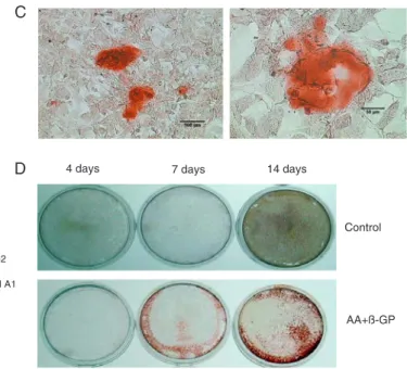
ethanol for 30 min at -20°C.Subsequently, the cells and the matrix were stainedwith 40 mM Alizarin red-S, pH 4.2, for1 h at room temperature. The stained nodules that appeared bright red in color represented physiological mineralization but not dystrophic mineralization, and they were identified by light microscopy. To compare the stained region of min -eralization, the whole dish was photographed.
TG2 enzyme assay
Protein expression of TG2 by Western blot. SAOS-2 cells
were cultured on 100-mm culture dishes for the designed time. Total cellular protein was isolated from cultured cells using radio-immunoprecipitation assay extraction buffer. Protein concentration was determined with the BCA protein assay reagent.
A total of 25 μg protein was loaded per well, separated on 10% SDS-PAGE gels and subsequently transferred to polyvinylidene fluoride membranes. Membranes were blocked overnight at 4°C with 3% bovine serum albumin/ Tris-buffered saline and Tween 20 (BSA/TBST) and then incubated with primary antibodies against TG2 (rabbit poly -clonal antibody, sc-20621, Santa Cruz Biotechnology Inc., USA), followed by the secondary antibody (IRDye 800cw conjugated Goat (polyclonal) anti-rabbit IgG, 926-32211, LI-COR Biosciences, USA). Bands were visualized using an infrared fluorescent scan imaging system (Odyssey, USA). GAPDH was used as the internal control. The den-sitometry values of the fluorescent bands were quantified and analyzed using the Image J software.
TG2 enzyme activity assay
SAOS-2 cells were cultured on 100-mm culture dishes for the designated time. TGase activity was measured by a previously described method (19). Specifically, we coated 96-well ImmunoModule plates with 200 μL 20 mg/ mL N,N-dimethylcasein for 1 h at 23°C. The N,N-dime -thylcasein was removed and nonspecific protein binding was blocked by adding 3% BSA in 100 mM Tris, pH 8.5, 150 mM NaCl, 0.05% Tween-20 (TBST) to each well for an additional 1 h at 23°C. Subsequently, aliquots of 25 μg total cellular protein that had been lysed and sonicated (in 5 mM Tris-HCl, 0.25 M sucrose, 0.2 mM MgSO4, 2 mM dithiothreitol, 0.4 mM phenylmethyl sulfonyl fluoride, 0.4% Triton X-100, pH 7.5) were added to the plate in triplicate. Fifty microliters of solution A (100 mM Tris, pH 8.5, and 20 mM CaCl2) was added to all samples, followed by the addition of 50 µL of solution B (100 mM Tris, pH 8.5, 40 mM dithiothreitol, and freshly added 2 mM 5-(biotinamido) pentylamine). The plates were incubated for 1 h at 37°C.
The wells were washed once with TBST containing 1 mM ethylenediaminetetraacetic acid and then three times with TBST. One hundred microliters of a 1:5000 dilution of streptavidin-peroxidase in 3% BSA/TBST was added to each well for 1 h at 23°C. The wells were washed twice with TBST, and 100 µL TMB working solution was added to each well. Absorbance was measured at 450 nm for 15 min after adding 0.5 M H2SO4 to stop the reaction. Puri-fied guinea pig liver TGase (Sigma) was used to prepare a standard curve. TG enzyme activity was designated as the amount of 5-(biotinamido)pentylamine incorporated into casein.
SAOS-2 differentiation and mineralization assay when TG activity was inhibited by cystamine
SAOS-2 cells were seeded on 100-mm dishes (for RT-PCR) or 48-well plates (for ALP assay) or 3.5-cm dishes (for Alizarin red staining) with osteo-inductive conditioned medium and were allowed to attach overnight. The cells were then treated with 0.5 mM cystamine (Sigma-Aldrich, USA) for the indicated days, i.e., 0-14 or 4-14. Cells cultured with conditioned medium but without cystamine were used as controls and cells cultured with normal medium as the blank control. Collagen I, OC, and BMP-2 mRNA were assayed by semiquantitative RT-PCR, ALP activity with the LabAssay™ ALP kit and mineralization by Alizarin red staining.
Statistical analysis
Data were analyzed for statistical significance by one-way ANOVA using the Dunnett test and the SPSS software. P < 0.05 was considered to be statistically significant.
Results
Expression of TG2 by SAOS-2 cells
696 Xiaoxue Yin et al.
Semiquantitative RT-PCR and Western blot were per-formed to examine TG2 mRNA and its protein expression at the three differentiation stages. TG2 mRNA expression appeared to be up-regulated with the progression of os-teoblast differentiation. Compared to the controls and to the early stage at day 4, TG2 mRNA significantly increased at days 7 and 14 (Figure 2A). Furthermore, the results of Western blot showed that TG2 mRNA was transcribed into protein and the protein levels were significantly high at days 7 and 14, with the same tendency observed for TG2 mRNA level (Figure 2B).
TG2 activity in SAOS-2 was dramatically increased when cultured with osteo-inductive conditioned medium, even at the early stage of day 4. Compared to control cultures, TG2 activity was enhanced by about 3- to 4-fold when treated with conditioned medium, which was
inde-pendent of culture time (Figure 2C).
Inhibition of TG activity blocks osteoblast differentiation and mineralization
To gain insight into the function of TG2 in the osteoblast program, we inhibited TG activity during cell differentiation and mineralization with the well-known TG inhibitor cysta -mine (CYS) (21).CYS treatment (0.5 mM) does not disturb cell growth (16).However, our data showed that the treat-ment resulted in a significant decrease of OC, collagen I and BMP-2 mRNA, an obvious reduction of ALP activity and disturbance of mineralization, regardless of the inhibition of CYS between days 4 and 14, or between days 0 and 14. But the inhibition for the full 14-day period was more effective in blocking mineral deposition as visualized directly by the absence of Alizarin red staining (Figure 3A-C).
Discussion
SAOS-2 is an established human osteosarcoma cell line, which possesses a typical osteoblastic phenotype, including elevated ALP, parathyroid hormone-stimulatable adenylate cyclase, synthesis and secretion of type I collagen, OC, and osteopontin, and production of mineralized matrix (20).SAOS-2 represents a useful experimental model for studying osteoblastic properties and osteoblast-produced molecules and is supposed to provide significantly more
698 Xiaoxue Yin et al.
mRNA and protein levels remain constant throughout the differentiation program in MC3T3-E1 cells, we found that in this human osteoblast cell line, the expression of TG2 protein and mRNA, which was consistent with the expres-sion of osteoblast markers, was dramatically elevated with the progression of osteoblast differentiation, especially at the fully differentiated and mineralized stage. Furthermore, TG2 activity responded (increased) to the osteo-inductive conditioned medium. Even in the early differentiation phase (day 4), TG2 activity was increased by about 3-fold in cells cultured with conditioned medium, as compared to cells cultured with normal medium. Furthermore, we demon -strated that when TG (including TG2) activity was inhibited by CYS, the expression of osteoblast markers, ALP activity and mineralization of SAOS-2 was reduced synchronously
and significantly. Compared to inhibition between day 4 and day 14, the full 14-day inhibition was more effective, resulting in an almost complete block of mineralization. This suggested that TG2 activity at the early phase (0-4 days) is very important for mineralization. Collectively, our results directly linked the progression of osteoblast differentiation and mineralization to the up-regulated TG2, demonstrating that TG2 is involved in osteoblast differentiation and may play a role in the initiation and regulation of the mineraliza-tion processes.
Among 9 family members of TGs, TG2 and FXIIIA have long been linked to the formation of skeletal elements, and growing evidence indicates that these two enzymes have similar and/or overlapping, but not necessary identical, functions in connective tissue cells (23). However, Al-Jallad Figure 3. Inhibition of TG2 activity by cystamine (CYS) blocks the differentiation and mineralization of SAOS-2 cells. Cells were
et al. (16) reported that FXIIIA could play a major role in MC3T3-E1 cell differentiation and collagen I matrix forma -tion, and TG2 crosslinking activity did not contribute to osteoblast differentiation (16,24). However, in the current study, although we did not assay the expression of FXIIIA, we did find that TG2 levels continued to increase steadily during osteoblast differentiation. The difference might be attributed to the diversity of two different cell lines. Alterna-tively, human osteoblasts may have a different mechanism than mouse osteoblasts to activate TGase activity. Additional effort is now being directed towards understanding the difference of TG2 effect by using different cell lines from different species and also primary cells.
It has been demonstrated that TG2 has some main physiological functions independent of its crosslink activity, such as signal transduction, cell adhesion and interaction with transforming growth factor-beta (TGF-β) (1,25). Our results showed that during the progression of SAOS-2 dif-ferentiation, BMP-2 mRNA increased significantly and TG2 expression and activity were up-regulated at the same time. However, when TG2 was inhibited, BMP-2 expression de-creased dramatically. As we all know, BMP-2 belongs to the
TGF-β superfamily and is a potent bone cell-differentiating factor as well as bone-formation stimulator. Our results showed that there is some relationship between BMP-2 and TG2. The stimulatory effect of TG2 regarding the osteoblast differentiation and mineralization may be possibly mediated in part by the up-regulation of BMP-2.
The present study established a specific linkage be -tween TG2 and the differentiation and mineralization of human osteoblasts. TG2 levels and activity steadily increase and correlate with the expression of osteoblast differentia-tion markers during osteoblast maturadifferentia-tion. When TG2 is inhibited, the program of SAOS-2 differentiation and min-eralization is obstructed, implying that TG2 is essential for osteoblast differentiation. Moreover, the modulating effects of TG2 on osteoblasts could be related to BMP-2. Our finding is highly relevant to the understanding of the role of TG2 in the differentiation and mineralization of osteoblasts.
Acknowledgments
Research supported by the National Natural Science Foundation of China (#81101334).
1. Lorand L, Graham RM. Transglutaminases: crosslinking enzymes with pleiotropic functions. Nat Rev Mol Cell Biol 2003; 4: 140-156.
2. Grenard P, Bates MK, Aeschlimann D. Evolution of trans-glutaminase genes: identification of a transtrans-glutaminase gene cluster on human chromosome 15q15. Structure of the gene encoding transglutaminase X and a novel gene family member, transglutaminase Z. J Biol Chem 2001; 276: 33066-33078.
3. Kaartinen MT, El-Maadawy S, Rasanen NH, McKee MD. Tissue transglutaminase and its substrates in bone. J Bone Miner Res 2002; 17: 2161-2173.
4. Nurminskaya M, Kaartinen MT. Transglutaminases in miner-alized tissues. Front Biosci 2006; 11: 1591-1606.
5. Rosenthal AK, Masuda I, Gohr CM, Derfus BA, Le M. The transglutaminase, Factor XIIIA, is present in articular chon-drocytes. Osteoarthritis Cartilage 2001; 9: 578-581. 6. Thomazy VA, Davies PJ. Expression of tissue
transglutami-nase in the developing chicken limb is associated both with apoptosis and endochondral ossification. Cell Death Differ 1999; 6: 146-154.
7. Jones RA, Nicholas B, Mian S, Davies PJ, Griffin M. Re-duced expression of tissue transglutaminase in a human endothelial cell line leads to changes in cell spreading, cell adhesion and reduced polymerisation of fibronectin. J Cell Sci 1997; 110 (Part 19): 2461-2472.
8. Barsigian C, Stern AM, Martinez J. Tissue (type II) trans-glutaminase covalently incorporates itself, fibrinogen, or fibronectin into high molecular weight complexes on the ex-tracellular surface of isolated hepatocytes. Use of 2-[(2-oxo-propyl)thio] imidazolium derivatives as cellular transglutami-nase inactivators. J Biol Chem 1991; 266: 22501-22509.
References
9. Aeschlimann D, Paulsson M. Transglutaminases: protein cross-linking enzymes in tissues and body fluids. Thromb Haemost 1994; 71: 402-415.
10. Johnson KA, Terkeltaub RA. External GTP-bound transglu-taminase 2 is a molecular switch for chondrocyte hypertro-phic differentiation and calcification. J Biol Chem 2005; 280: 15004-15012.
11. Orlandi A, Oliva F, Taurisano G, Candi E, Di Lascio A, Melino G, et al. Transglutaminase-2 differently regulates cartilage destruction and osteophyte formation in a surgical model of osteoarthritis. Amino Acids 2009; 36: 755-763.
12. Johnson KA, van Etten D, Nanda N, Graham RM, Terkeltaub RA. Distinct transglutaminase 2-independent and transglu-taminase 2-dependent pathways mediate articular chondro-cyte hypertrophy. J Biol Chem 2003; 278: 18824-18832. 13. Nurminskaya M, Magee C, Faverman L, Linsenmayer TF.
Chondrocyte-derived transglutaminase promotes matura-tion of preosteoblasts in periosteal bone. Dev Biol 2003; 263: 139-152.
14. Heath DJ, Downes S, Verderio E, Griffin M. Characterization of tissue transglutaminase in human osteoblast-like cells. J Bone Miner Res 2001; 16: 1477-1485.
15. Wozniak M, Fausto A, Carron CP, Meyer DM, Hruska KA. Mechanically strained cells of the osteoblast lineage organize their extracellular matrix through unique sites of alphavbeta3-integrin expression. J Bone Miner Res 2000; 15: 1731-1745.
700 Xiaoxue Yin et al.
epsilon(gamma-glutamyl)lysine crosslinks in atherosclerotic aortas. Atherosclerosis 1994; 111: 247-253.
18. Johnson K, Hashimoto S, Lotz M, Pritzker K, Terkeltaub R. Interleukin-1 induces pro-mineralizing activity of cartilage tissue transglutaminase and factor XIIIa. Am J Pathol 2001; 159: 149-163.
19. Yin X, Chen Z, Guo Z, Liu X, Yu H. Tissue transglutaminase expression and activity in human ligamentum flavum cells derived from thoracic ossification of ligamentum flavum. Spine 2010; 35: E1018-E1024.
20. Rodan SB, Imai Y, Thiede MA, Wesolowski G, Thompson D, Bar-Shavit Z, et al. Characterization of a human osteosar-coma cell line (Saos-2) with osteoblastic properties. Cancer Res 1987; 47: 4961-4966.
21. Jeitner TM, Delikatny EJ, Ahlqvist J, Capper H, Cooper AJ. Mechanism for the inhibition of transglutaminase 2 by cys-tamine. Biochem Pharmacol 2005; 69: 961-970.
22. Xiaoxue Y, Zhongqiang C, Zhaoqing G, Gengting D, Qingjun
M, Shenwu W. Immortalization of human osteoblasts by transferring human telomerase reverse transcriptase gene. Biochem Biophys Res Commun 2004; 315: 643-651. 23. Cordell PA, Kile BT, Standeven KF, Josefsson EC, Pease
RJ, Grant PJ. Association of coagulation factor XIII-A with Golgi proteins within monocyte-macrophages: implications for subcellular trafficking and secretion. Blood 2010; 115: 2674-2681.
24. Al-Jallad HF, Myneni VD, Piercy-Kotb SA, Chabot N, Mulani A, Keillor JW, et al. Plasma membrane factor XIIIA transglu-taminase activity regulates osteoblast matrix secretion and deposition by affecting microtubule dynamics. PLoS One 2011; 6: e15893.