385
Pakistan Veterinary Journal
ISSN: 0253-8318 (PRINT), 2074-7764 (ONLINE)
Accessible at: www.pvj.com.pk
Recent Advances in the Management of Foreign Body Syndrome in Cattle and Buffaloes: A
Review
Ashraf M. Abu-Seida1* and Oday S. Al-Abbadi2
1Department of Surgery, Anesthesiology and Radiology, Faculty of Veterinary Medicine, Cairo University. Giza, P.O. Box: 12211, Egypt; 2Ministry of Agriculture, Iraq
*Corresponding author: [email protected]
ARTICLE HISTORY (15-470) A B S T R A C T
Received: Revised: Accepted: Published online:
November 14, 2015 May 06, 2016 June 20, 2016 July 04, 2016
Foreign body syndrome (FBS) is a fairly common disease of cattle and buffaloes, especially in the developing countries. This disease is caused by ingestion of indigestible metallic and non-metallic blunt or sharp foreign objects. It is associated with high economic losses and therefore an urgent science-based policy is required to control and manage this syndrome. Indiscriminate feeding habits, feed scarcity, industrialization and mechanization of agriculture are predisposing factors for FBS in bovine and bubaline. The condition is difficult to diagnose solely on the basis of clinical signs and physical examination. However, laboratory diagnosis and imaging techniques like radiography and ultrasonography can be of high diagnostic value in detecting the condition. Anemia, increased packed cell volume, neutrophilia with a left shift, increased total protein, globulin, total bilirubin, Alanine Aminotransferase, Alkaline Phosphatase, Phosphorus and decreased albumin/globulin ratio and Calcium are the common abnormal laboratory findings. Recently, ultrasonography has replaced radiography for diagnosis of FBS in bovine and bubaline due to its availability and accuracy in evaluation of features of the reticulum, detection of penetrating metallic objects, diagnosis and assessment of various sequelae of FBS including; local and diffuse traumatic reticuloperitonitis, reticular, splenic, hepatic, abdominal and thoracic abscesses, diaphragmatic hernia, traumatic pericarditis and pleuropneumonia. Although, FBS is ideally treated with rumenotomy, it can be prevented to a large extent by proper management practices, increasing the awareness among the livestock keepers, oral administration of rumen magnets at the age of one year and reapplication of a new magnet every 4 years in animals at high risk.
©2016 PVJ. All rights reserved Key words:
Buffaloes Cattle
Foreign body syndrome Hardware disease
Traumatic reticuloperitonitis Ultrasonography
To Cite This Article: Abu-Seida AM and Al-Abbadi OS, 2016. Recent advances in the management of foreign body syndrome in cattle and buffaloes: A Review. Pak Vet J, 36(4): 385-393.
INTRODUCTION
Ingestion of indigestible foreign materials by cattle and buffaloes is a common problem worldwide, known as foreign body syndrome (FBS) (Aref and Abdel-Hakiem, 2013). This syndrome is more common in bovine than in small ruminants because they do not use their lips for prehension and are more likely to eat chopped feed (Misk and Semieka, 2001; Ashfaq et al., 2015). Moreover, indiscriminate feeding habits, feed scarcity, industrialization and mechanization of agriculture are predisposing factors for FBS (Semieka, 2010).
The non-metallic foreign body syndrome is a silent killer disease resulting from ingestion of polywastes, rubber, plastics, leather materials, ropes, clothes and
cement bags (Reddy and Sasikala, 2012). The presence of foreign bodies in the rumen and reticulum hampers the absorption of volatile fatty acids, consequently leading to reduction in the rate of animal fattening (Igbokwe et al., 2003).
hernia. Other consequences include pericarditis, reticular fistulation, reticular, diaphragmatic, mediastinal, hepatic, splenic, lateral and ventral abdominal wall abscesses, vagal indigestion, rupture of left gastro-epiploic artery, traumatic pneumonia and pleurisy (Roth and King, 1991; Floeck and Baumgartner, 2001; Abu-Seida and Al-Abbadi, 2014). These complications depend mainly upon the nature, length and direction of penetration of the swallowed foreign body (Abouelnasr et al., 2012).
This condition produces devastating economic losses due to severe reduction in milk and meat production, treatment costs, potential fatalities and fetal losses in affected pregnant animals (Nugusu et al., 2013). It may prove lethal because the bacteria and protozoa can contaminate the body cavity resulting in peritonitis and the heart and diaphragm may be punctured by the ingested object, causing their failure (Abu-Seida and Al-Abbadi, 2015).
Due to its high economic importance in dairy animals, FBS is still a matter of concern worldwide, therefore several recent advances in the diagnosis and prevention of the disease have been recorded. This review highlights the recent advances in FBS in cattle and buffaloes.
Incidence: Sharp foreign body syndrome is one of the most common diseases of the digestive tract of cattle and buffaloes. It was recorded in 59.14 and 56.88% of the examined cattle and buffaloes in Pakistan (Khan et al., 1999; Anwar et al., 2013), 23.1% of the examined cattle in Ethiopia (Nugusu et al., 2013), 25, 23.38 and 22.9% of the examined buffaloes in Egypt (Aref and Abdel-Hakeim, 2013), India (Ramprabhu et al., 2003) and Iraq (Abu-Seida and Al-Abbadi, 2015), respectively. This high incidence in developing countries is attributed to unsatisfactory animal management and practice of livestock rearing based on hand feeding (Misk et al., 1984). In addition, buffaloes are more prone to FBS than cattle at the same area. Recently calved and senile buffaloes have higher incidence of FBS than dry and lactating buffaloes (Ramprabhu et al., 2003). In addition, dairy cattle are more commonly affected than beef cattle because they are more likely to be fed a chopped feed, such as silage or hay (Kahn, 2005). Moreover, a high incidence of FBS is recorded during drought and harvest season (Sharma and Kumar, 2006).
The mean age of the affected buffaloes is usually 4.0±0.8 years. Moreover, 99.1% of the affected animals are commonly females (Abu-Seida and Al-Abbadi, 2015). In cattle, FBS was recorded in 36.23% of animals ≤3 years old, 33.33% of adult animals (3-7 years old) and 30.43% of old animals (7-11 years of age). In addition, cows are more commonly affected (65.8%) than bulls (37.03%; Anwar et al., 2013).
Indigestible foreign bodies are found either in rumen (58.5%), reticulum (19.3%) or rumen and reticulum (22.2%) of the affected cattle (Anwar et al., 2013). Reportedly, 49.38% of the diseased buffaloes had foreign bodies in the reticulum and only 7.5% had foreign bodies in the rumen. Moreover, the total number of foreign bodies in the reticulum of buffaloes was higher than that in the rumen. Conversely, the total weight of the ruminal
foreign bodies was greater than that in the reticulum (Khan et al., 1999).
Etiology: The mode of animal prehension, indiscriminate feeding habits, bad nutritional management, heavy industrialization and human habitations are major predisposing factors for the occurrence of such condition in bovine (Khan et al., 1999). In addition, pregnancy, tenesmus and vigorous reticular contraction increase the potential for development of SFBS in bovine. Therefore, numerous animals have ruminal and reticular foreign bodies without development of clinical signs and sometimes these foreign bodies pass into feces.
Mostly, the ingested foreign objects are lodged in the reticulum without harm due to their fixation by the honeycomb like reticular cells. The typical foreign body is a metallic object longer than 2.5 cm.
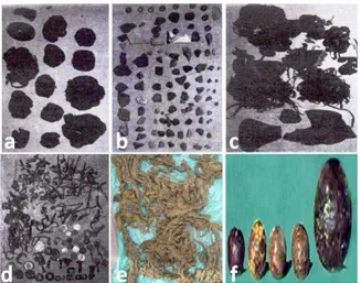
The foreign objects recovered from buffaloes are classified into sharp metallic foreign objects (knives, tin openers, wires, nails, needles and pieces of iron; 75.7%), mixed foreign objects (20.7%), blunt non-metallic objects (sand, stones, gravels, electrical wires, nylon ropes, pieces of clothes, socks, rubber pieces, polyethylene bags and hair balls) (2.1%), blunt metallic foreign objects such as coins (0.9%) and sharp non-metallic foreign objects like bones, feathers and pieces of glass (0.6%; Abu-Seida and Al-Abbadi, 2014; 2015). Sharp foreign bodies, both sharp and blunt foreign bodies or blunt foreign bodies have been recorded in 76.6, 20.7 and 2.7 of the diseased buffaloes, respectively (Abu-Seida and Al-Abbadi, 2015). In cattle, the detected foreign bodies were clothes, plastic, nails, ropes, hair, leather, wires and nails (Tesfaye and Chanie, 2012) (Fig. 1).
Diagnosis: Without an accurate case history and when the victim is admitted after several days of ingestion of a metal object, the diagnosis is more difficult (Ramin et al., 2011). Differential diagnosis of SFBS is considered a challenge since the diseased animals show signs similar to those of several other diseases. Uterine or vaginal trauma, metritis, perforating abomasal ulcers, grain overload, ketosis, abomasal displacement, hepatic abscesses, pyelonephritis, intestinal adhesions to the abdominal wall and volvulus, should be considered during the differential diagnosis of SFBS. Thus the diagnosis should be based on case history, clinical symptoms, clinical examination, laboratory diagnosis, electrocardiography, radiography, ultrasonography, necropsy and histopathological diagnosis.
Animals with large amount of blunt foreign bodies show anorexia, depression, intermittent respiratory distress, recurrent rumen tympany, rumen stasis, dehydration, reduced milk yield, distended left para lumbar fossa and sometimes vomition (Reddy and Sasikala, 2012; Abu-Seida and Al-Abbadi, 2014).
Clinical signs of traumatic pericarditis (TP) include tachycardia, muffled heart sounds, absence of lung sound in the ventral thorax, asynchronous abnormal heart sounds, distension of the jugular veins and pulsation, submandibular, brisket and ventral abdominal edema (Figs. 2d and e).
Clinical examination: Tympanic sounds are heard on percussion with simultaneous auscultation of paralumbar fossa. By stethoscope, muffled heart beats, reduced gut sounds, and rapid breathing may be heard. Moreover, rectal palpation is a reliable method of diagnosing the rumen impaction in bovine and to exclude uterine or vaginal trauma and metritis (Reddy et al., 2014). Electronic metal detector can identify reticular metals but does not distinguish between perforating, perforating and non-magnetic foreign bodies (Reddy and Sasikala, 2012).
Briefly, pain tests include pinching of withers, walking on downhill and side stick method. Affected animals will not reflex ventrally when their withers are punched. They may also exhibit pain when a large bar
placed under the animal’s sternum is forced upwards.
Laboratory diagnosis: Laboratory tests may be helpful in diagnosis of FBS. Hemogram of the diseased animals shows anemia, increased packed cell volume and neutrophilia with a left shift (Reddy et al., 2014). Serum biochemical parameters of diseased animals show increased total protein, globulin, total bilirubin, ALT, ALP, P and decreased albumin/globulin ratio and Ca (Ghanem, 2010). Hyperproteinemia is noticed in buffaloes with acute and chronic local TRP, and reticular abscess. Hyper-beta-globulinemia is noticed in animals with chronic local TRP, reticular abscess and purulent pericarditis. Hyper-gamma-globulinemia is evident in animals with acute and chronic local TRP, reticular abscess and purulent pericarditis. Hypoproteinemia associated with severe hypoalbuminemia and very low A/G ratios characterizes animals with acute diffuse TRP, purulent and fibrinous pericarditis (Saleh et al., 2008)
Animals with indigestible plastic materials in the rumen show mild hypocalcemia, hypophosphatemia, hypoglycemia and hypoproteinemia and increase in BUN, and increased values of Methylene Blue Reduction Test (MBRT), total volatile fatty acids and sedimentation activity test (Reddy et al., 2014). Severely affected animals may have coagulation abnormalities, as evidenced by prolonged prothrombin time, thrombin time, and activated partial thromboplastin time. Rumen liquor examination reveals a pH of 7.0-8.0 and nil or low protozoal motility and counts (Reddy and Sasikala, 2012).
On hematological examination of animals with TP, erythrocytopenia, pronounced leukocytosis, with shift to left accompanied with neutrophilia, eosinopenia, monocytosis, lymphocytopenia, basopenia and anemia are usual. Hyperfibrinogenemia, hemoconcentration and increased serum AST, ALT, LDH, CPK and bilirubin are
also reported. Moreover, cardiac troponin proteins have a value in determining the degree of heart damage (Gunes, 2008).
Pericardiocentesis at the left 4th or 5th intercostal space may be applied under echocardiography to collect a sample of pericardial fluid. However, pneumothorax, fatal arrhythmia, pleuritis and cardiac puncture are potential complications. Protein concentration >3.5 g/dL and WBC
count >2500/μL with straw yellow to slightly blood
tinged, foamy, and foul smelling pericardial fluid are characteristics of TP (Elhanafy and French, 2012). Electrocardiography: Electrocardiography (ECG) is one of the most important parameters for an animal suffering from a cardiovascular problem (Reddy et al., 2015). Buffaloes should be kept in a standing position in a stock without any sedation (Mousavi et al., 2007). After 30 min rest, 3 channel ECG system and base apex lead was used for obtaining ECG in about 30 sec. For ECG in buffaloes the positive electrode of lead I is attached to the skin of the left 5th intercostal space just caudal to the olecranon process, while the negative electrode is placed on the jugular furrow in the caudal third of the right neck. The ground electrode is placed remote from the heart. To ensure good adherence of electrodes to the skin, shaving and cleaning the skin with alcohol prior to the application of alligator clips and gel should be done (Hasanpour et al., 2008). ECG showed that 70% of normal buffaloes had arrhythmia. Wandering pacemaker (57%), sinus arrhythmia (29%), second degree atrioventicular block (9%) and electrical alternance (5%) were the most common arrhythmias in buffaloes (Mashhadi et al., 2014). Electrocardiography can aid in the diagnosis of TP. Decreased amplitude, electric alternans, slurring, atrial fibrillation are the most common ECG changes in animals with TP (Foss, 1985; Gunes, 2008). Moreover buffaloes with TRP had cardiac arrhythmias (55.6%) including; sinus tachycardia, premature atrial beat, atrioventricular block I and atrioventricular block II (Mousavi et al. 2007). ECG is not useful for diagnosis of the early stages of cardiac diseases except for arrhythmias. Meanwhile, the costly echocardiogram is more classical tool for the investigation of the preclinical forms of cardiomyopathies in buffaloes (Reddy et al., 2015).
Radiographic examination: Standing lateral radiographs of the cranioventral abdomen can detect metallic materials in the reticulum but should be taken only after oral administration of a magnet. It is contraindicated to place the affected animal in dorsal recumbency to obtain radiographs, because such position induces stress on adhesions and may lead to diffuse peritonitis.
Fig. 1: Compact plastic materials (a), pieces of stone, bones and marbles (b), pieces of rubber and ropes (c), metallic objects (d), pieces of cloths (e) and hair balls (f) recovered from buffaloes. Sources: a, b, c and d: Khan et al. (1999), e: Nugusu et al. (2013) and f: Abu-Seida and Al-Abbadi (2014).
Fig. 2: a) A cow with TRP showing arched back and reluctance to move. b) A buffalo with TRP showing a localized abscess at the left cranioventral abdomen. c) A buffalo with TRP showing a fistula and perforating foreign object at the right cranioventral abdomen. d) A cow with TP showing abduction of fore limbs, jugular venous distention, brisket and submandibular edema and e) A cow with TP showing the classic pear-shaped abdomen. Sources a: Ghanem (2010), b and c: Abdelaal and Floeck (2015) and d and e: Imran et al. (2011).
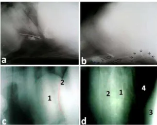
Radiographic examination could be a helpful tool in the diagnosis of diaphragmatic hernia in buffaloes. Intra thoracic circumscribed swelling of soft tissue density representing the reticulum and overlying the caudal border of heart, discontinuation of the diaphragm (Fig. 3c) and sometimes presence of metallic foreign object are the main radiographic findings in diaphragmatic hernia (Aref and Abdel-Hakiem, 2013).
Radiography reveals good thoracic details in early stages of TP, while poor differentiation of thoracic structures is evident at progressive stages. Moreover, gas and fluid accumulation and metallic foreign objects are usually detected in the pericardium and cranial reticulum or caudal thorax (Fig. 3d), respectively. However, it is difficult to distinguish between pleural and pericardial effusion on the basis of radiography (Misk and Semieka, 2001; Masseau et al., 2008).
Ultrasonographic examination: Ultrasound has proven a useful diagnostic tool for assessment of different surgical
affections in bovine (Abu-Seida, 2012; 2016; Abdelaal et al., 2016). The reticulum and surrounding structures are examined by using a 3.5-MHz linear or convex transducer in the area from 6th to 8th intercostal spaces, the transducer is applied to the ventral aspect of the thorax on the left and right of the sternum as well as to the left and right lateral thorax up to the level of elbow (Braun, 2003). The examination is conducted on standing, non-sedated animals for visualization of physiological (Abouelnasr et al., 2014) and pathological conditions of the reticulum in cattle and buffaloes with TRP (Floeck, 2006).
Various sequelae of TRP in both cattle and buffaloes are clarified by ultrasound (Abdelaal et al., 2009). The most common ultrasonographic finding in animals with TRP is the corrugation of the reticular wall (Fig. 4a) due to the accumulation of fibrinous deposits interspersed with fluid pockets on the reticular serosa. The thickness of reticular wall, distance between reticulum and abdominal wall and relaxation period are significantly increased in animals with SFBS. Conversely, the reticular motility and amplitude of contraction are significantly decreased. The contraction period shows no significant change.
The ultrasonographic findings vary according to the complications of SFBS (Mostafa et al., 2015). Ultrasono-graphically, the reticular abscesses have an echogenic capsule of varying thickness and surrounded with a homogeneous hypoechogenic to moderately echogenic center (Fig. 4b). The foreign bodies are seen by ultrasono-graphy as hyper echogenic structures penetrating the reticular wall with comet tail artifact (Fig. 4a and b). In certain cases, it is possible to drain abscesses through an ultrasound-guided transcutaneous incision (Braun et al., 1998).
Animals with local peritonitis show echogenic strands interspersed with anechoic fluid between the reticulum and/or liver, rumen, spleen (Fig. 4c and d), and abomasum. Animals with diffused peritonitis show diffuse echogenic strands interspersed with anechoic fluid involving the whole abdomen (Fig. 4e). The rumen is displaced 2 to 15 cm from the abdominal wall by hypoechogenic fluid with echogenic fibrin in case of suppurative peritonitis(Braun et al., 1998).
Abdominal abscesses of different sizes (2 to 20 cm) appear as anechoic to echogenic center surrounded by echogenic wall. These abscesses usually locate between the reticulum and rumen, abomasum, spleen and/or liver. Meanwhile, thoracic abscesses are usually imaged at 3rd. and 4th. intercostal spaces as circumscribed masses (2 to 20 cm) with echogenic capsule and hypoechoic contents and sometimes they are partitioned by echogenic septa (Mohamed and Oikawa, 2007). Only pulmonary abscesses near the pleura can be imaged by ultrasound (Elhanafy and French, 2012). Other ultrasonographic findings of pulmonary abscess are broadened and hyperechogenic pleura and medium echogenic lungs resembling the liver. Aspiration of the abdominal and thoracic abscesses is minimally invasive and cost-effective technique to obtain an aspirate for confirmation (Tharwat, 2011).
Fig. 3: a) Lateral radiograph of the reticulum showing metallic foreign objects cranial to diaphragm. b) Lateral radiograph of the reticulum showing indistinct diaphragmatic line, intra thoracic metallic densities (hollow arrows) and gas density (black arrows) with embedded metallic object. c) Lateral radiograph of diaphragmatic hernia in a buffalo showing intra thoracic reticulum (1) overlying the caudal border of heart with discontinuation of the diaphragm- drawing red line (2). (d) Lateral radiograph of TP in a buffalo showing radio-opaque metallic foreign object (1) at the caudal border of the heart (2). (3): reticulum and (4): lung. Sources a and b: Athar et al. (2010) and c and d: Aref and Abdel-Hakiem (2013).
Fig. 4: a) Ultrasonogram of the reticulum (R) of a hardware diseased buffalo showing uneven surface of the reticulum and echogenic foreign body (FB) with a comet tail artifact (CT). F: fibrin; E: exudates. b) Ultrasonogram of the reticulum (R) of a hardware diseased buffalo showing hypoechoic abdominal abscess (AB) and two echogenic foreign objects (FB) with comet tail artifacts (CT). Ultrasonograms of local peritonitis showing corrugated thick reticulum and echogenic fibrin strands (arrows) interspersed with anechoic peritoneal exudates (PE) between the reticulum and liver (c), reticulum and spleen (d) and reticulum and abdominal wall (e). (f) Ultrasonogram of traumatic pleuropneumnia showing hypoechoic exudates in the pleural cavity (PLE), absence of reverberation artifact, presence of echogenic foreign bodies, comet tail artifacts (CT) and hepatized lung. GB: gall bladder, AW: abdominal wall. Sources:. a and b: Abdelaal and Floeck (2015); c, d and e: Imran et al. (2011).
Echocardiography is a simple, well-established and rapidly method for assessment of the heart size and function (Hassan and Torad, 2015). It was previously applied in both normal (Torad et al., 2016) and diseased buffaloes (Tharwat and Buczinski, 2011; Hussein and Staufenbiel, 2014)
Echocardiography is the method of choice for confirmation of pericarditis, differentiation between effusive and fibrinous pericarditis, detection of impact of
pericardial effusions on the cardiac chambers or function, differentiation between pericardial and pleural effusion and choosing the optimal site of pericardiocentesis (Buczinski, 2009; Athar et al., 2012).
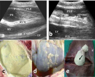
Animals with suppurative TP usually have a large amount of hypoechogenic to echogenic pericardial fluid (Fig. 5a). Meanwhile fibrinous TP shows echogenic deposits and strands of fibrin on the epicardium and sometimes in the fluid between the epicardium and pericardium (Fig. 5b). Other ultrasonographic findings include; thick pericardial layer, compressed cardiac ventricles and lungs, medial and dorsal displaced lungs, bright hyperechoic pinpoint echoes representing free gas, vegetations of the tricuspid, mitral and pulmonary valves (Ghanem, 2010), a large hypoechoic area with irregular margins in the cranioventral thorax (Elhanafy and French, 2012) and sometimes obscured heart by the effusion (Schweizer et al., 2003). Abdominal ultrasonography in animals with TP reveals reticular changes typical of TRP, hepatomegaly and moderate to severe ascites. Caudal vena cava usually appears dilated and round to oval instead of triangular in cross section. Ultrasonography of the presternal edema reveals excessive accumulation of anechoic fluid separated with echogenic septa.
Reticular diaphragmatic hernias in bovine have been diagnosed by radiography and ultrasonography (Saini et al., 2007). However, ultrasonography is still the method of choice for detecting and confirming the diagnosis (Athar et al., 2010). Herniated reticulum can be imaged at left 3rd to 5th intercostal spaces beneath the heart or beneath the lung (Abdelaal et al., 2014). Other ultrasonographic findings include; half-moon shaped reticulum, low amplitude contraction and hypoechoic exudates between the reticulum and surrounding thoracic organs (Ramprabhu et al., 2003). When the motility patterns of the abdominal and herniated reticulum match, the animal is declared positive for reticular diaphragmatic hernia (Mohindroo et al, 2007). Sometimes, thick hyperechoic fibrous bands originate from the herniated mass and extend towards the sternum or the pericardium.
Traumatic suppurative splenitis occurs in 2–14% of cattle with SFBS. Encapsulated masses (3–4 cm diameter) with an echogenic center inside the splenic parenchyma are seen in cattle with traumatic suppurative splenitis (Skarda, 1996).
Ultrasound can assess the stratification of ruminal contents and contraction by moving the probe dorsoventrally either parallel to the last rib or in the left paralumbar fossa from lumbar transverse processes to ventral midline (Imran et al., 2011; Braun et al., 2013). Therefore, ultrasound can be helpful for diagnosis of foreign bodies inside the rumen.
Fig. 5: a) Echocardiogram of a cow with suppurative TP showing anechoic pericadial effusion (PE) with echogenic sediment, pleural effusion (PLE), thickened pericardium (P) and increased right ventricle (RV). RVW; right ventricular wall, IVS: inter ventricular septum, LV: left ventricle. b) Echocardiogram of a cow with fibrinous TP showing pleural effusion (PLE) and thickened pericardium (P) with fibrin shreds (FS). LVW: left ventricular wall, LV: left ventricle, IVS: inter ventricular septum. c) Postmortem photograph of the heart of a cow with suppurative TP. d) Postmortem photograph of the liver of cow with TRP showing hepatomegaly with fibrin shreds on its parietal surface. e) Postmortem photograph of the liver showing traumatic hepatic abscess (arrow). Sources a, b, c and d: Imran et al. (2011) and e: Ismail and Abdullah (2014).
Fig. 6: a) Hepatic abscess showing liquifactive necrosis (blue arrow) and surrounded by a fibrous capsule (yellow arrow) (H & E, 165X). b) Foreign body granuloma showing granulomatous inflammatory exudates infiltrated by giant cell (black arrows) and mononuclear inflammatory cells (yellow and blue arrows) (H & E 450X). c) Heart of a cow with TP showing thickened pericardium (1) and accumulation of fibrinous inflammatory exudates (2) between the pericardium and myocardium (3) (H & E 200X). d) Myocardium of a cow with TP showing atrophy and hyalinosis (yellow arrows) (H & E 400X). Sources: a and b: Ismail and Abdullah (2014); c and d: Ghanem (2010).
Advantages of the laparoscopy include practicality, rapid postoperative recovery and low risk of complications (Rees et al., 2015). On the other hand the main disadvantages are the cost of the instruments and inability to perform laparoscopy in animals with cardiopulmonary diseases and abdominal adhesions (Seeger et al., 2006).
There are three techniques for laparoscopy in bovine including; right and left paralumbar fossa in the standing animals and cranioventral midline laparoscopy with the animal positioned in dorsal recumbency (Anderson et al.,
1993; Steiner and Zulauf, 1999). Right flank laparoscopy using a flexible fiberoptic laparoscope, 14 mm diameter and 1120 mm working length, is an easy, rapid and reliable diagnostic aid for TRP (Bakos and Vörös, 2011).
Laparoscopies were performed using a combination of Xylazine and local infiltration anesthesia in standing buffaloes (Ambrose et al., 1993). Before laparoscopic examination, the animals' tail is tied to its hock or neck on the opposite side of the surgery. Both paralumbar fossa are prepared for aseptic surgery then the abdominal wall is infiltrated with 8–10 mL of local anesthetic solution at portal sites. The laparoscopic cannula is inserted in the middle of the fossa, dorsally to the crus of the internal oblique muscle. When cannula is placed in the body wall, the trocar is replaced with a 10-mm rigid, which has an installed video camera and light source, and is connected to the laptop via a USB cable. Tubing connected to the insufflators is attached to the cannula, insufflation is stopped when organs are visualized, and the laparoscope can freely move in the abdomen. In buffaloes with peritonitis, inflammatory signs are visible in the form of congested blood vessel varying degrees of fibrin production and adhesion between abdominal organs and body wall depending upon the stage of peritonitis. Increased peritoneal fluid that changed in color and turbidity and strand of free fibrin in peritoneal fluid are also visible. In few cases heavy adhesion between organ and abdominal wall decreases further exploration of abdominal cavity after laparoscopic examination, the endoscope is removed and CO2 gas/air is released through the open cannula. Then the cannula is removed and the portal sites are closed. Systemic antibiotics and nonsteroidal anti-inflammatory drugs are required for only 24 h after the surgery (Safarchi et al., 2014).
Laparoscopy successfully diagnosed peritonitis of the cranial parts of the abdomen, caused by foreign bodies or laparotomy (Franz et al., 2000), left abomasal displacement (Janowitz, 1998; Tolasi et al., 2015) and occasionally the reticular abscesses in cattle (Wilson and Ferguson, 1984). Moreover, laparoscopy has been used for surgical correction of left displaced abomasum (van Leeuwen et al., 2009; Doga et al., 2010; Sickinger et al., 2013).
In contrary, laparoscopy is not suitable for examination of the ventral abdomen nor for diagnosing non-inflammatory intestinal affections (Franz et al., 2000). Further studies are warranted to evaluate the reliability of laparoscope in diagnosis of SFBS in large animals. Adhesions produced by SFBS may be the main obstacle during laparoscopic examination in bovine. Necropsy and histopathological examination: Shrunken rumen, strangulated foreign bodies, congested ruminal mucosa and ulceration are the common necropsy findings in animals with FBS. Acute cases of TP show distention of pericardial sac with foul-smelling grayish fluid containing flakes of fibrin and heavy fibrin deposits on the
pericardium giving the appearance of "scrambled eggs”
foreign body is not possible due to extensive adhesions (Singh et al., 2008).
Postmortem findings of SFBS may include; fibrinous adhesions between reticulum and/or diaphragm, spleen, abomasum, rumen, liver (Fig. 5d) and left abdominal wall, large amount of foul-smelling pus containing fibrin clots, adhesions between the intestinal loops and omentum (Chanie and Tesfaye, 2012). Also splenic, pulmonary, reticular, abdominal and hepatic abscesses (Fig. 5e), pleural effusions and foreign bodiescan be seen(Sobti et al., 1989).
Pathologically, hepatic, pulmonary and splenic abscesses have liquifactive necrosis infiltrated with inflammatory cells mainly, macrophages, neutrophils and lymphocytes and surrounded with fibrous capsule (Fig. 6a). Foreign body granulomas with necrotic center infiltrated by plasma cells, epitheloid cells, macrophages and foreign body giant cells are reported (Fig, 6b). Pathognomonic lesions of TP are thick pericardium, fibrinous network trapping inflammatory cells, atrophy and hyalinosis of myocardium (Figs. 6c and d).
Treatment: Mostly, conservative treatment is indicated in acute cases of SFBS.However, in chronic cases and late pregnant animals, conservative treatment is unsuccessful. Conservative treatments of SFBS include; external massage of the xiphoid process, oral administration of purgative, elevation of the fore limbs, fasting, supportive therapy, intra peritoneal antibiotic injection, and administration of a cage magnet (Horney and Wallace, 1984).However, it is unlikely that the magnet will move into the reticulum due to ruminal stasis. Rumen inoculation with 4–8 L of ruminal fluid from a healthy animal is beneficial in animals with prolonged ruminal stasis. If the conservative treatments fail to improve the animal after 3 days, rumenotomy is indicated.
Rumenotomy is a rapid and successful procedure for diagnosis and treatment of FBS. The direct approach to the reticulum through a mid-line incision just posterior to the xiphoid is not preferable due to the possibility of diffuse peritonitis (Dehghani and Ghadrdani, 1995). However, a standing laparotomy is desirable due to the difficult manipulation of such large organ. The site of laparorumenotomy incision, size of the animal, and length
of the surgeon’s arm should be considered to perform
successful rumenotomy (Horney and Wallace, 1984). Moreover, gentle manipulation is recommended to minimize the spreading of infection in animals with traumatic reticulitis. Usually, there is little or no benefit from breaking down chronic adhesion because it tends to reform very rapidly. Also breakdown of recent adhesion is not advised because it may mask and surround an abscess (Weaver et al., 2005).
Several techniques for laparorumenotomy including; skin suture fixation, stay suture technique, skin clamp
technique, rumenotomy ring, Weingarth’s technique and
the Gabel rumen retractor (rumen board) have been applied. All techniques are conducted through an approach in the left paralumbar fossa for successful access, exteriorization and securing of the rumen and minimizing contamination. The main difference between these techniques is the method by which the rumen is secured to the abdominal wall or skin (Niehaus, 2008).
The stay suture technique is inferior to other techniques due to high incidence of infection while Weingarth’s technique is superior to other techniques (Dehghani and Ghadrdani, 1995).
In the skin suture fixation technique, the rumen is sutured to the skin using a continuous Connell suture pattern to invert the skin edges under the rumen to minimize contamination. Inthe stay suture technique, four stay stitches at the cranial, caudal, dorsal and ventral parts of the incision are performed to fix the rumen to the skin. Fixation of the rumen to the skin dorsally and ventrally and fixation of the ruminal incision to the skin incision cranially and caudally are applied by 6-8 towel clamps in the skin clamp technique. An aluminum ring with a rubber ring attached to its inner circumference is used during rumenotomy ring technique to fix the rumen to this rubber ring. In Weingarth's technique, a Weingarth's frame is fixed to the dorsal commissure of the incision by its thumb screw following laparotomy. Then the ruminal incision is fixed to the frame by multiple hooks. The Gabel rumen retractor depends mainly upon a device having a central hole that the rumen is pulled through. The rumen is fixed to the board by a series of bolts (Dehghani and Ghadrdani, 1995). Post-operative complications such as suture abscess, wound dehiscence, subcutaneous emphysema and local peritonitis at the surgical site are recorded (Nugusu et al., 2013).
Reticular abscesses may be drained through an ultrasound-guided transcutaneous paracentesis or via rumenotomy into the rumen. Treatment of TP is often unrewarding and usually is addressed toward salvage or short term survival to calving. Diuretics are effective in eliminating the severity of peripheral edema, reducing venous return and preload in animals with pericarditis (Buczinski et al., 2010). Rarely, medical therapy with systemic antibiotics and drainage of pericardial sac permanently cures affected animals.
years has become a popular preventive measure for hardware disease (Weaver et al., 2005). After oral administration, most magnets drop firstly into the rumen then move to the reticulum by ruminoreticular contractions. Metallic foreign bodies are attracted and fixed to the magnets, and consequently do not penetrate the reticulum as easily as when they are free. The extensive prophylactic use of these has reduced the incidence of TRP by 90-98% in cattle and 89-91% in buffaloes. A time dependent increase in the proportion of buffaloes developing TRP is noticed after 4 years of magnet administration, not due to loss of magnetic power but due to fixation of numerous foreign objects on the magnet. Therefore, reapplication of a second new magnet is recommended after four years of the first one particularly in animals at high risk (Al-Abbadi et al., 2014).
Conclusions: Foreign body syndrome of cattle and buffaloes is still a challenge in veterinary field all over the world particularly in developing countries. Although the condition has no specific clinical signs, it has several hematological, biochemical and radiographic characteristics. Moreover, ultrasonography provides recent advances in diagnosis and prognosis of this syndrome.
Author’s contribution: BothAMA and OSA conceived
and designed the review, interpreted the data, revised the manuscript and approved the final version.
REFERENCES
Abdelaal AM and Floeck M, 2015. Clinical and sonographical findings in buffaloes (Bubalus bubalis) with traumatic reticuloperitonitis. Vet Arch, 85: 1-9.
Abdelaal AM, Floeck M, El Maghawry S and Baumgartner W, 2009. Clinical and ultrasonographic differences between cattle and buffaloes with various sequelae of traumatic reticuloperitonitis. Vet Med- Czech, 54: 399-406.
Abdelaal A, Gouda S, Ismail A and Gomaa M, 2014. Reticular diaphragmatic hernia in Egyptian buffaloes: clinical, haemato-biochemical and ultrasonographic findings. Pak Vet J, 34: 541-545. Abdelaal AM, Al-Abbadi OS and Abu-Seida AM, 2016. Transcutaneous
and transrectal ultrasonography in buffalo calves with urine retention. Asian J Anim Vet Adv, 11: 79-88.
Abouelnasr KS, Mosbah E, Karrouf GI and Zaghloul AE, 2012. Comparative ultrasonographic findings of traumatic reticulitis, perireticular abscess and diaphragmatic hernia in buffalo (Bubalus
Bubalis). J Amer Sci, 12:590-595.
Abouelnasr K, Mosbah E, Karrouf G and Zaghloul A, 2014. Ultrasonography of normal reticulum in 30 healthy buffalo (Bubalus
bubalis). J App Anim Res, 42: 153-159.
Abu-Seida AM, 2012. Ultrasonographic diagnosis of some scrotal swellings in bulls. Pak Vet J, 32: 378-381.
Abu-Seida AM, 2016.Current status and prospect of ultrasonographic application in buffaloes. Asian J Anim Vet Adv, 11: 144- 157. Abu-Seida AM and Al-Abbadi OS, 2014. Recurrent rumen tympany
caused by trichobezoars in buffaloes (Bubalus bubalis): A series report. Thai J Vet Med, 44: 147-151.
Abu-Seida AM and Al-Abbadi OS, 2015. Studies on sharp foreign body syndrome in Iraqi buffaloes and its impact on milk production. Asian J Anim Sci, 9: 128-133.
Al-Abbadi OS, Abu-Seida AM, and Al-Hussainy SM, 2014. Studies on rumen magnet usage to prevent hardware disease in buffaloes. Vet World, 7: 408-411.
Ambrose JD, Manik RS, Singla SK and Madan ML, 1993. A simplified laparoscopy technique for repeated ovarian observation in the water buffalo (Bubalus bubalis). Theriogenology, 40: 487-496.
Anderson DE, Gaughan EM and Stjean G, 1993. Normal laparoscopic anatomy of the bovine abdomen. Am J Vet Res, 54: 1170-1176. Anwar K, Khan I, Aslam A, Mujtaba1 M, Din A, et al., 2013. Prevalence
of indigestible rumen and reticulum foreign bodies in Achai cattle at different regions of Khyber Pakhtunkhwa. ARPN J Agr Biol Sci, 8: 580-586.
Aref NM and Abdel- Hakiem MH, 2013. Clinical and diagnostic methods for evaluation of sharp foreign body syndrome in buffaloes. Vet World, 6: 586-591.
Ashfaq M, Razzaq A, Hassan S and ul Haq S, 2015. Factors affecting the economic losses due to livestock diseases: a case study of district Faisalabad. Pak J Agr Sci, 52: 515-520.
Athar H, Mohindroo J, Singh K, Kumar A and Raghunath M, 2010. Comparison of radiography and ultrasonography for diagnosis of diaphragmatic hernia in bovines. Vet Med Int, Article ID 939870, 7 pages. http://dx.doi.org/10.4061/2010/939870
Athar H, Parrah JD, Moulvi BA, Singh M and Dedmari FH, 2012. Pericarditis in bovines-A review. Int J Adv Vet Sci Tech, 1: 19-27. Babkine M and Desrochers A, 2005. Laparoscopic surgery in adult
cattle. Vet Clin North Am Food Anim Pract, 21: 251-279. Babkine M, Desrochers A, Bouré L and Hélie P, 2006. Ventral
laparoscopic abomasopexy on adult cows. Can Vet J, 47: 343-348. Bakos Z and Vörös K, 2011. Intraoperative echocardiography and
surgical treatment of traumatic pericarditis in a pregnant cow. Acta Vet Hung, 59:175-179.
Braun U, 2003. Ultrasonography in gastrointestinal disease in cattle. Vet J, 166: 112-124.
Braun U, Gotz M and Marmier O, 1993. Ultrasonographic findings in cows with traumatic reticuloperitonitis. Vet Rec, 133: 416-422. Braun U, Iselin U, Lischer C and Fluri E, 1998. Ultrasonographic findings
in five cows before and after treatment of reticular abscesses. Vet Rec, 142: 184-189.
Braun U, Lejeune B, Schweizer G, Puorger M, and Ehrensperger F, 2007. Clinical findings in 28 cattle with traumatic pericarditis. Vet Rec, 161: 558-563.
Braun U, Schweizer A and Trosch L, 2013. Ultrasonography of the rumen of dairy cows. BMC Vet Res, 9: 44-52.
Buczinski S, 2009. Cardiovascular ultrasonography in cattle. Vet Clin North Am Food Anim Pract, 25: 611-632.
Buczinski S, Rezakhani A and Boerboom D, 2010. Heart disease in cattle: diagnosis, therapeutic approaches and prognosis. Vet J, 184: 258–263.
Chanie M and Tesfaye D, 2012. Clinico-pathological findings of metallic and non-metallic foreign bodies in dairy cattle: A review. Acad J Anim Dis, 1: 13-20.
Dehghani SN and Ghadrdani AM, 1995. Bovine rumenotomy: Comparison of four surgical techniques. Can Vet J, 36: 693-697. Doga TM, Sirri A and Kuersad Y, 2010. Treatment of left sided
abomasal displacement in cattle by laparoscopic surgery. Kafkas Universitesi Veteriner Fakultesi Dergisi, 16: 217-224.
Ducharme NG, Fubini SL and Rebhun WC, 1992. Thoracotomy in adult dairy cattle: 14 cases (1979–1991). J Am Vet Med Assoc, 200: 86–
90.
Elhanafy MM and French DD, 2012. Atypical presentation of constrictive pericarditis in a Holstein heifer. Case Rep Vet Med, doi:10.1155/2012/604098.
Floeck M, 2006. Ultrasonography of the reticulum-A diagnostic tool for the practitioner. Slov Vet Res, 43: 208-209.
Floeck M and Baumgartner W, 2001. Ultrasonographic diagnosis of traumatic reticuloperitonitis and pericarditis in cattle. Wiener Tieraerztliche Monatsschrift, 12: 347-354 (in German).
Foss RR, 1985. Effusive-constrictive pericarditis: Diagnosis and pathology. Vet Med, 80: 89-94
Franz S, König M, Gasteiner J and Baumgartner W, 2000. Laparoscopy in cattle. 3. Indications and pathological findings. Wiener Tierärztliche Monatsschrift, 87: 163-172.
Ghanem MM, 2010. A comparative study on traumatic
reticuloperitonitis and traumatic pericarditis in Egyptian cattle. Turk J Vet Anim Sci, 34: 143-153.
Gunes V, 2008. Use of cardiactroponin kits for the qualitative determination of myocardial cell damage due to traumatic reticuloperitonitis in cattle. Vet Rec, 162: 514-517.
Hassan EA and Torad FA, 2015. Two-dimensional and M-mode echocardiographic measurements in the healthy donkeys (Equus asinus). J Equine Vet Sci, 35: 283–289.
Horney FD and Wallace CE, 1984. Surgery of the bovine digestive tract. In: Jennings PB (ed). Practice of Large Animal Surgery. WB Saunders, Philadelphia, USA, pp: 493-554.
Hussein AH and Staufenbiel R 2014. Clinical presentation and ultrasonographic findings in buffaloes with congestive heart failure. Turk J Vet Anim Sci 38: 534–545.
Igbokwe IO, Kolo MY and Egwu GO, 2003. Rumen impaction in sheep with indigestible foreign body in the semiarid region of Nigeria. Small Rumin Res, 49: 141-147.
Imran S, Tyagi SP, Kumar A, Kumar A and Sharma S, 2011. Ultrasonographic application in the diagnosis and prognosis of pericarditis in cows. Vet Med Int, doi:10.4061/2011/974785 Ismail HK and Abdullah OA, 2014. Metallic foreign body in the liver of
cow: a case report. Iraqi J Vet Sci, 28: 109-111.
Janowitz H, 1998. Laparoscopic reposition and fixation of the left displaced abomasum in cattle. Tierarztl Prax Ausg G Grosstiere Nutztiere. 1998 26:308-313. (Article in German)
Kahn CM, 2005. The Merck Veterinary Manual, 9th Ed, Merck and CO., INC., USA, pp: 186-188.
Khan MJ, Habib G and Siddiqui MM, 1999. Prevalence of foreign indigestible materials in the reticulo-rumen of adult buffaloes. Pak Vet J, 19: 176-180.
Mashhadi AG, Hajikolai RH, Rezakhani A, Fatemi R, and Kamali S, 2014. The prevalence of cardiac arrhythmia in Khuzestan buffaloes
(Bubalus bubalis), Revue Med Vet, 165: 99-103.
Masseau I, Fecteau G, Breton L, Hélie P, Beauregard G, et al., 2008. Radiographic detection of thoracic lesions in adult cows: A retrospective study of 42 cases (1995-2002). Can Vet J, 49: 261-267.
Misk NA and Semieka MA, 2001. The radiographic appearance of reticular diaphragmatic herniation and traumatic pericarditis in buffaloes and cattle. Vet Radiol Ultrasound, 42: 426-430. Misk NA, Nigam JM and Rifat JF, 1984. Management of foreign body
syndrome in Iraqi cattle. Agri Practice, 5: 19-21.
Mohamed T and Oikawa S, 2007. Ultrasonographic characteristics of abdominal and thoracic abscesses in cattle and buffaloes. J Vet MedA Physiol Pathol Clin Med, 54: 512-517.
Mohindroo J, Kumar M, Kumar A and Singh SS, 2007. Ultrasonographic diagnosis of reticular diaphragmatic hernias in buffaloes. Vet Rec, 161: 757–758.
Mostafa MB, Abu-Seida AM, Abdelaal AM, Al-Abbadi OS and Abbas SF, 2015. Ultrasonographic features of the reticulum in normal and hardware diseased buffaloes. Res Opin Anim Vet Sci, 5: 165-171.
Mousavi Gh, Hassanpour A, Tabrizi A and Rezaie A, 2007.
Electrocardiographic changes in buffaloes with Traumatic reticuloperitonitis. Ital J Anim Sci, 6: 1029-1031
Niehaus AJ, 2008. Surgery of the abomasum. Vet Clin North Am Food Anim Pract, 24: 349-358.
Nugusu S, Ramaswamy V, Unakal C and Nagappan R, 2013. Studies on foreign body ingestion and their related complications in ruminants associated with inappropriate solid waste disposal in Gondar Town, North West Ethiopia. Int J Anim Vet Adv, 5: 67-74. Ramin AG, Hashemi AM, Asri-rezaie S, Batebi E, Tamadon A, et al., 2011. Predication of traumatic pericarditis in cows using some serum biochemical and enzyme parameters. Acta Vet (Beograd), 61: 383-390.
Ramprabhu R, Dhanapalan P and Prathaban S, 2003. Comparative efficacy of diagnostic tests in the diagnosis ofTRP and allied syndrome in cattle. Israel J Vet Med, 58: 2-3.
Reddy MV and Sasikala P, 2012. A review on foreign bodies with special reference to plastic pollution threat to livestock and environment in Tirupati rural areas. Int J Scient Res Pub, 2: 1-8.
Reddy SB, Venkatasivakumar R, Reddy VL, Vani S and Sivajothi S, 2015. Analysis of base apex lead electrocardiograms of adult buffaloes. J Dairy Vet Anim Res, 2: 58-62.
Reddy YR, Latha PA and Reddy S, 2014. Review on metallic and non-metallic foreign bodies: a threat to livestock and environment. Int J Food Agri Vet Sci, 4: 6-14.
Rees GM, Barrett DC, Place E, Boocock J, Dickinson M, et al., 2015. Surgical management of left displaced abomasum in dairy cattle: a critically appraised topic (CAT). Cattle Pract, 23: 199-200. Roth L and King JM, 1991.Traumatic reticulitis in cattle: a review of 60
fatal cases. J Vet Diagn Invest, 3: 52-54.
Safarchi R, Badiei A, Nadalian MG and Seifi HA, 2014. Assessing predictive value of clinical, hematological and acute phase proteins in single and complex mode for diagnosing dairy cow peritonitis. Europ J Zoo Res, 3: 38-48.
Saini NS, Kumar A, Mahajan SK and Sood AC, 2007. The use of ultrasonography, radiography, and surgery in the successful recovery from diaphragmatic hernia in a cow. Can Vet J, 48: 757-759.
Saleh MA, Rateb HZ and Misk NA, 2008. Comparison of blood serum proteins in water buffaloes with traumatic reticuloperitonitis and sequellae. Res Vet Sci, 85: 208-213.
Schweizer T, Sydler T, and Braun U, 2003. Kardiomyopathie, endokarditis valvularis thromboticans und perikarditis traumatica beim Rind – Klinische und echokardiographische Befunde an drei Fallberichten. Sch Arch Tier, 145: 425-430 (In German).
Seeger T, Kümper H, Failing K and Doll K, 2006. Comparison of laparoscopic-guided abomasopexy versus omentopexy via right flank laparotomy for the treatment of left abomasal displacement in dairy cows. Am J Vet Res, 67: 472-478.
Semieka MA, 2010. Radiography of unusual foreign body in ruminants. Vet World, l3: 473-475.
Sharma MC and Kumar P, 2006. Forien body syndrome in buffaloes
(Bubalus bubalis): An emerging threat. Asian J Anim Vet Adv, 1:
89-98
Sickinger M, Seeger T and Doll K, 2013. Laparoscopic abomasopexy - Janowitz's method and its modifications vs right flank omentopexy. Cattle Pract, 21: 157-162.
Singh R, Garg SL, Sangwan N and Gerai S, 2008. Peripheral concentration of thyroid hormones in buffaloes suffering from foreign body syndrome. Haryana Vet, 47: 115-116.
Skarda RT, 1996. Local and regional anesthesia in ruminants and swine. Vet Clin North Am Food Anim Pract, 12: 579-626.
Sobti VK, Sharma SN, Singh K and Rathor SS, 1989. Diaphragmatic hernia in buffalo bulls. Indian Vet J, 66: 866-868.
Steiner A and Zulauf M, 1999. Diagnostic laparoscopy in the cow. Schweiz Arch Tierheilkd, 141: 397-399, 402-406. (Article in German).
Tesfaye D and Chanie M, 2012. Study on rumen and reticulum foreign bodies in cattle slaughtered at Jimma Municipal abattoir, South West Ethiopia. Am-Euras J Sci Res, 7: 160-167.
Tharwat M, 2011. Traumatic pericarditis in cattle: Sonographic, echocardiographic and pathologic findings. J Agri Vet Sci Qassim Univ, 4: 45-59.
Tharwat M and Buczinski S, 2011. Clinicopathological findings and echocardiographic prediction of the localisation of bovine endocarditis: 36 cases. Vet Rec 169: 180-186.
Tolasi G, Baiguera M and Ghilardi D, 2015. Left abomasum displacement (LDA): "one-step" laparoscopic technique. A field trial. Large Anim Rev, 21: 155-161.
Torad FA, Amer MS, Shamaa AA and Elsherpieny EA, 2016. Echocardiographic measurements and indices in normal adult
buffalo (Bubalus bubalis) J App Anim Res,
http://dx.doi.org/10.1080/09712119.2016.1190733
van Leeuwen E, Mensink MG and de Bont MF, 2009 Laparoscopic Reposition and Fixation of the Left Displaced Abomasum in Dairy Cattle Practice - Ten years of experience under field conditions in the Netherlands. Cattle Pract, 17: 123-127.
Weaver D, Steiner A, and Guy SJ, 2005. Bovine Surgery and Lameness, 2nd Ed.; Blackwell Publishing, Ames, Iowa, USA, pp: 75-139. Wilson AD and Ferguson JG, 1984. Use of a flexible fiber optic