Homologs of the LapD-LapG c-di-GMP
Effector System Control Biofilm Formation by
Bordetella bronchiseptica
Nicolás Ambrosis1, Chelsea D. Boyd2¤, George A. O´Toole2, Julieta Fernández1 *, Federico Sisti1
*
1Instituto de Biotecnología y Biología Molecular (IBBM)-CCT-CONICET-La Plata, Departamento de Ciencias Biológicas, Facultad de Ciencias Exactas, Universidad Nacional de La Plata, La Plata, Argentina, 2Department of Microbiology and Immunology, Geisel School of Medicine at Dartmouth, Hanover, New Hampshire, United States of America
¤ Current address: National Institute of Allergy and Infectious Diseases, Division of Extramural Activities, Scientific Review Program, Rockville, Maryland, United States of America
*julietafernandez3@gmail.com(JF);federico.sisti@gmail.com(FS)
Abstract
Biofilm formation is important for infection by many pathogens.Bordetella bronchiseptica
causes respiratory tract infections in mammals and forms biofilm structures in nasal epithe-lium of infected mice. We previously demonstrated that cyclic di-GMP is involved in biofilm formation inB.bronchiseptica. In the present work, based on their previously reported func-tion inPseudomonas fluorescens, we identified three genes in theB.bronchiseptica
genome likely involved in c-di-GMP-dependent biofilm formation:brtA,lapDandlapG. Genetic analysis confirmed a role for BrtA, LapD and LapG in biofilm formation using micro-titer plate assays, as well as scanning electron and fluorescent microscopy to analyze the phenotypes of mutants lacking these proteins.In vitroandin vivostudies showed that the protease LapG ofB.bronchisepticacleaves the N-terminal domain of BrtA, as well as the LapA protein ofP.fluorescens, indicating functional conservation between these species. Furthermore, while BrtA and LapG appear to have little or no impact on colonization in a mouse model of infection, aB.bronchisepticastrain lacking the LapG protease has a signif-icantly higher rate of inducing a severe disease outcome compared to the wild type. These findings support a role for c-di-GMP acting through BrtA/LapD/LapG to modulate biofilm for-mation, as well as impact pathogenesis, byB.bronchiseptica
Introduction
Bordetella bronchisepticais a Gram-negative bacterium that causes respiratory tract infections in mammals, atrophic rhinitis in pigs, kennel cough in dogs and snuffles in rabbits [1].B. bronchi-septicahas a variety of virulence factors that allow host infection. Each factor, such as pertactin, filamentous hemagglutinin, adenylate cyclase, the type three secretory system and lipopolysac-charide are likely to perform specific functions required for successful colonization [2–6]. a11111
OPEN ACCESS
Citation:Ambrosis N, Boyd CD, O´Toole GA, Fernández J, Sisti F (2016) Homologs of the LapD-LapG c-di-GMP Effector System Control Biofilm Formation byBordetella bronchiseptica. PLoS ONE 11(7): e0158752. doi:10.1371/journal.pone.0158752
Editor:Roy Martin Roop, II, East Carolina University School of Medicine, UNITED STATES
Received:April 14, 2016
Accepted:June 21, 2016
Published:July 5, 2016
Copyright:© 2016 Ambrosis et al. This is an open access article distributed under the terms of the Creative Commons Attribution License, which permits unrestricted use, distribution, and reproduction in any medium, provided the original author and source are credited.
Data Availability Statement:All relevant data are within the paper and its Supporting Information files.
Funding:This work was supported by NIH grant R01-AI097307 and NSF grant MCB-9984521 to GAO, and International Cooperation grants (2012 and 2013) from CONICET to FS and JF, PICT 2013-0092, PÏP N° 0092 and PIP N° 0486 to FS and JF. NA was supported by CONICET doctoral fellowship.
The ability ofBordetella spp. to form biofilms has been reported in previous work, including the ability ofB.bronchisepticato form biofilms on abiotic surfaces regulated by the two-com-ponent system BvgAS [7,8]. As has been found for other biofilm-forming organisms, extracel-lular DNA (eDNA) and exopolysaccharide are important for biofilm formation byB.
bronchiseptica[9,10]. Particularly inBordetella, BvgAS-regulated factors, including the fila-mentous hemagglutinin and adenylate cyclase, may also participate in biofilm formation [7,11]. Static growth in intermediate nicotinic acid concentrations, a regulator of the BvgAS system, resulted in the best conditions for promoting biofilm formation [8]. Studies from our group and others indicate that biofilm is enhanced in the intermediate phase [7,12], however, there are conflicting findings regarding BvgAS-dependent biofilm regulation [8]. Furthermore, transcriptome analysis has shown that more than 33% of theB.bronchisepticagenes are differ-entially regulated during biofilm formation compared to planktonic culture [13]. Thus, further studies are needed to elucidate all factors affecting biofilm formation.
Sloan and colleagues observed biofilm-like structuresin vivoin the nasal epithelium ofB.
bronchisepticainfected mice, and these communities expressed a polysaccharide essential for
in vivobiofilm development [9]. The absence of polysaccharides, key factor forin vitrobiofilm formation, also impaired infection suggesting that biofilm formation may also participate in host-pathogen interactions [9]. Thus, biofilm formation may play an important role in host-B.
bronchisepticainteractions. Recently, we showed that bis-(30-50)-cyclic-dimeric guanosine monophosphate (c-di-GMP) regulates motility and biofilm formation inB.bronchiseptica
[12]. C-di-GMP is a bacterial second messenger known to regulate a variety of cellular pro-cesses including biofilm formation, motility and virulence of bacterial pathogens [14,15]. Like in other bacteria where c-di-GMP-related functions have been studied, high c-di-GMP levels inB.bronchisepticacorrelated with an enhanced biofilm formation phenotype [12]. However, in this previous study we did not uncover the mechanism by which c-di-GMP enhanced bio-film formation inB.bronchiseptica.
Here we confirm the role for the Bvg-regulated adhesin BrtA, a putative homolog of the well-characterized LapA adhesin ofPseudomonas fluorescens, in biofilm formation byB.
bronchiseptica, as has recently been reported by Nishikawa and colleagues [16]. Furthermore, we show that the c-di-GMP receptor LapD and the LapD-regulated protease LapG also partici-pate in biofilm formation byB.bronchiseptica, likely via the control of BrtA localization to the cell surface as has been reported for the LapA adhesin ofP.fluorescens. We also present data that the appropriate control of BrtA via LapG appears to be important for modulating patho-genesis in a mouse model of infection. Thus, this work describes the basis of c-di-GMP-medi-ated control of biofilm formation inB.bronchiseptica, bothin vivoandin vitro.
Materials and Methods
Ethics Statement
This study was performed in strict accordance with animal use protocols approved by Faculty of Sciences, National University of La Plata, Institutional Animal Care and Use Committee (IACUC), protocol number 007-00-15. All animals were properly anesthetized with isofluor-ane for bacterial inoculation, monitored daily and euthanized with isofluorisofluor-ane overdose if they met any early removal criteria (lethargy, hunched posture, or ruffled coat) to limit suffering.
Microbiological Methods
Stainer-Scholte (SS) [17] medium at 36°C and 160 rpm. When appropriate BGA or SS was supplemented with kanamycin (80μg ml-1), streptomyicin (200μg ml-1) or gentamycin
(50μg ml-1).P.fluorescensandE.coliwere grown in lysogeny broth (LB) [18] at 30°C and
37°C, respectively. When appropriate, antibiotics were added to the medium at the following concentrations:E.coli, 10μg ml-1gentamycin;P.fluorescens, 30μg ml-1gentamycin.
Replicative plasmids were introduced toE.coliandB.bronchisepticaby electroporation using standard techniques. Non-replicating plasmids were introduced intoB.bronchisepticaby conjugation. The yeast strain InvSc1 (Saccharomyces cerevisiae; Invitrogen), was routinely cul-tured on YPD medium. When selecting for plasmids carrying the URA3 gene, yeast were grown on YNB with complete supplemental mixture minus uracil.
Plasmid and Strain Construction
Strains and plasmids were constructed using standard molecular biology techniques and are listed inS1 Table. Oligonucleotides used in this study are listed inS2 Table. Detailed descrip-tions of strain and plasmid construction procedures can be found in theS1 Text.
Biofilm Assays
Biofilm formation assays using static cultures were performed as described previously [12] from overnight cultures inoculated into SS liquid medium. Briefly, the culture was pipetted into glass tubes or wells of a sterile 96-well U bottom microtiter plate (polyvinylchloride, PVC) and incubated statically at 37°C. Nicotinic acid was added as indicated. After 24 hours, unless otherwise indicated, planktonic bacteria were removed and the attached cells were stained with 0.1% crystal violet (CV) solution. The stain was dissolved by adding 120μl of 33% acetic acid
solution. One hundred microliters of the dissolved stain solution was transferred to a new, flat-bottom microplate and then quantified by measuring OD at 595 nm.
Scanning Electron Microscopy
Scanning electron microscopy pictures were obtained as described previously by Sistiet al. [12]. Briefly,Bordetellastrains were cultured statically on glass coverslips partially submerged vertically in plastic tubes such that an air-liquid interface was established on the coverslip. After 24 h of incubation, the coverslip was removed and washed with sterile PBS, and the bac-teria were fixed with 2.5% glutaraldehyde in PBS. Samples were dehydrated in a graded ethanol series (20, 50, 70, 90 and 100% for 60 min each), subjected to critical point drying using liquid carbon dioxide (EMITECH, K850) and sputter coated with gold (SPI Supplies). The surface topographies of the biofilm in A panels were visualized with a scanning electron microscope (ESEM FEI QUANTA 200), and detected with SDD (EDAX Apollo 40) camera. The surface topographies in B panels were visualized with a scanning electron microscope (Philips SEM 505), and the images were processed with the Image Soft Imaging System ADDA II.
RNA Extraction for RT-PCR Studies
transcription was performed in a thermal cycler at 42°C for 15 min and 95°C for 5 min for a total of 35 cycles. PCR was performed with Kappa Taq from Biosystems. Samples that had not undergone the reverse transcription process were used as controls for the absence of genomic DNA in RT-PCR experiments. Primers employed are summarized inS2 Table, including the primers designed to amplify the intergenic region between thelapAandlapGgenes. PCR anal-ysis of internal fragment ofrecAgene, which is constitutively expressed, was used as control.
LapA Localization
For Western and dot blot assays of the LapA protein, we used strains with internal HA-tag engineered into the sequence of the chromosomallapAgene. Culture conditions, preparation of clarified cell extracts, and analysis of samples for LapA localization were performed as described previously without modification [19]. Detection of cell surface LapA by dot blotting was also performed as previously described [19,20].
LapG Activity Assays
Bacterial cultures were grown overnight in LB, and clarified cell extracts were subsequently pre-pared by sonication (4×10 s on ice) in resuspension buffer, followed by centrifugation 12 min at 15,000 ×g. As described previously [19], to assess cleavage of N-terminal domain of LapA (N-term-LapA), cell extractsE.colicarrying the pMini-LapA plasmid were mixed 1:1 with cell extracts from strains with and without a LapG-expressing plasmid, followed by incubation at room temperature for 30 min. When indicated, purifiedP.fluorescensLapG was added (750 ng of LapG; est. purity: 50%).
Murine Respiratory Infection Model
Female BALB/c mice (originally from National Institute of Health (USA) and maintained at Faculty of Veterinary, National University of La Plata) 3 to 4 weeks of age were used as a model ofin vivorespiratory infection byB.bronchiseptica. Bacteria grown on BGA medium were resuspended and adjusted to approximately 107CFU ml-1in PBS. Fifty microliters of bacterial suspension was delivered intranasally to each mouse via an air displacement pipette. At differ-ent times post-inoculation, three to four mice from each group were euthanized and their lungs and nose were removed aseptically. Tissues were homogenized in PBS, and appropriate dilutions were plated onto BGA medium to determine the number of viable bacteria present in the lungs and nose. Results presented in figures are from two independent experiments per-formed with at least four mice per strain per time point. Counts from euthanized mice were excluded from calculations, as described in more detail in the Results section.
Statistical Analysis
All the results were compared by analysis of variance (ANOVA) followed by the Tukey test using Infostat software (Cordoba National University).
Results
Genes Coding for Lap-Like Proteins Are Present and Expressed in
B
.
bronchiseptica
We searched theB.bronchisepticagenome for genes coding for proteins with properties similar to the LapA protein ofP.fluorescens, including a type I secretion signal, a Von Willebrand fac-tor type A domain (vWFA), an overall large size, a domain with sequence repeats, as well as
performed a BLASTP search using ~1200 C-term amino acids ofP.fluorescensLapA as the query against the availableB.bronchisepticaRB50 genome. This portion of LapA includes the vWFA domain and type I secretion signal. The search identified a protein with ~30% identity to the query sequence; the identified protein contained a C-term domain of a putative hemoly-sin. Inspection of the complete amino acid sequence identified a protein with LapA-like prop-erties inB.bronchiseptica—BB_RS05895.
The BB_RS05895 ORF was recently described by Nishikawa and co-workers and named
brtA[16], thus we use that nomenclature here. TheB.bronchisepticaRB50 BrtA protein is pre-dicted to be 3345 amino acids in length, with eight repeat regions. Each repeat region is ~200 amino acids long and is composed of distinct regions designated the CADG and VCBS (Dys-troglycan-type cadherin-like domains andVibrio,Colwellia,Bradyrhizobium, andShewanella
repeats superfamily respectively) domains based on conserved amino acid sequence motifs (Fig 1,S1 Fig). Interestingly, the number of repeats in BrtA vary amongB.bronchiseptica
strains. As described inS3 Table, the number of repeats varies between 2 and 15 with a median of 3 repeats. These differences in repeat length may impact host tropism, however no correla-tion was observed between isolate source and number of repeated regions (S3 Table). It is important to note that many draft genomes have incomplete BrtA sequences, likely due to high frequency of repeated sequences in this protein. Hence data regarding number of CAD-G-IDR1-VCBS-IDR2 repeats in BrtA homologs is not available for all genomes.
While BrtA architecture is similar to the LapA protein ofP.fluorescensand the RtxA protein ofLegionella pneumophila[21], the sequence of the repeat regions is distinct. Nishikawa and colleagues showed that the BrtA protein localizes to the surface ofB.bronchiseptica, and using abrtAmutant strain, these workers also showed that BrtA is required for the adhesion to an abiotic (polystyrene) substratum but not to rat alveolar type 2 cells [16]. Upstream of
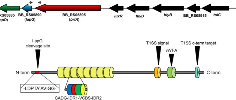
BB_RS05895 we found BB_RS05890 and BB_RS05885, which show sequence similarity to the Fig 1.B.bronchisepticaLapA domain organization.Diagram showing the organization of thelapD,lapGandbrtA genesinB.bronchiseptica(top) as well as the domain organization of the BrtA protein (bottom). The narrow arrows indicate the location of the primers used in the RT-PCR experiment. The thick, black arrow indicates the specific cleavage site for LapG. The type I secretion signal (T1SS) and T1SS target domains of BrtA are also shown (orange and cyan, respectively), as is the von Willebrand Factor A (vWFA) domain (light green). The repeat regions are indicated by the 8 yellow barrels. Domains found in repeated regions are also indicated: CADG domain (blue), VCBS domain (green) and IDR (interdomain region, red and yellow). The CADG and VCBS domain names are based on core conserved amino acid.
P.fluorescensLapG (48% identity) and LapD (30% identity) proteins, respectively. Based on their sequence as well as functional conservation to theP.fluorescensproteins (as described below), we decided to name BB_RS05890 and BB_RS05885 aslapGandlapD, respectively. We also identified a canonical predicted cleavage site for LapG (an Ala-Ala motif) in the N-termi-nal portion of BrtA (Fig 1) [21], indicating that BrtA cell surface localization is likely controlled by the LapG protease, a hypothesis we test further below.
The gene organization inB.bronchisepticasuggested thelapD,lapG and brtAgenes are part of an operon. Intergenic regions of 81 bp and 16 bp were found between the end of thebrtA
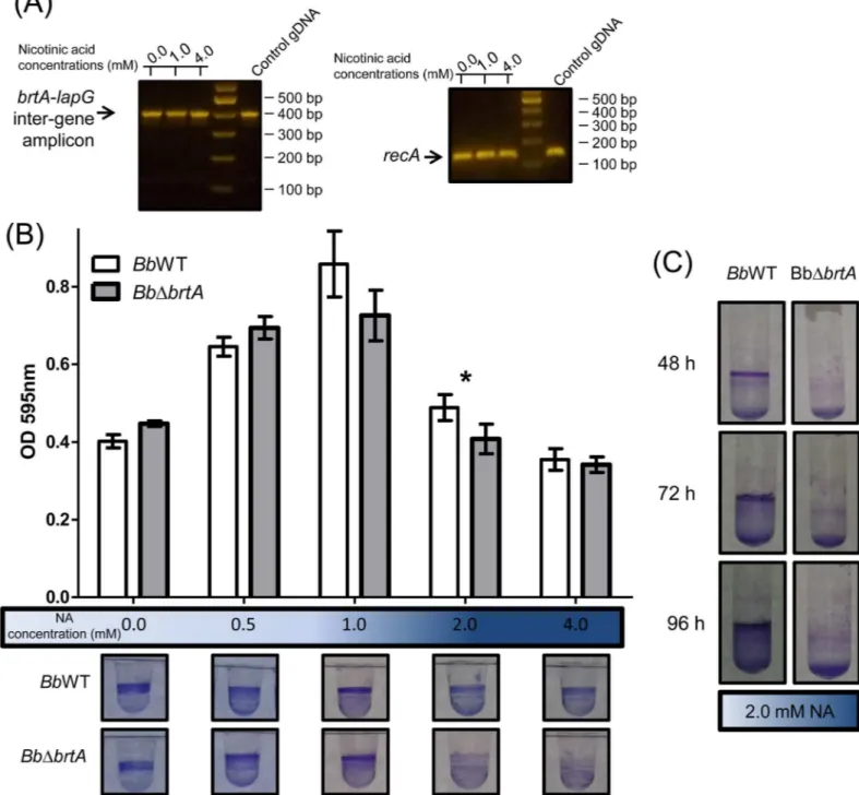
gene and start of thelapGgene and between thelapGandlapDgenes, respectively. To test whether these genes are co-transcribed, we performed RT-PCR using primers that amplified the intergenic region between thebrtAandlapGgenes (Fig 1). Results presented inFig 2A
show that thebrtAandlapGgenes appear to be co-transcribed. Moreover, these genes appear to be transcribed in both virulent and avirulent phases, as indicated by the detection of the PCR product across a range of nicotinic acid (NA) concentrations.
We compared the amino acid sequence of theB.bronchisepticaLapD- and LapG-like pro-teins. LapD ofB.bronchisepticahas cytosolic GGDEF and EAL domains that have been shown to be critical to the function of the LapD protein ofP.fluorescens[19]. TheB.bronchiseptica
LapG-like protein is 48% identical to itsP.fluorescenscounterpart; sequence analysis shows that all the amino acids described by Chatterjeeet al. as important for protease activity, as well as for binding to LapD and calcium [20] are conserved between the LapG proteins ofB. bronch-isepticaandP.fluorescens.
BrtA Is Important for Biofilm Formation in a Surface-Dependent Manner
Recently BrtA was described as an avirulence factor involved in biofilm formation inB. bronch-isepticaRB50 [16]. To confirm its role in biofilm formation, we deleted thebrtAgene ofB.
bronchiseptica9.73H+. The ability to form a biofilm was evaluated by the crystal violet (CV) biofilm assay using a 96-well polyvinylchloride (PVC) microtiter plates as described in the Material and Methods. We determined the amount of biofilm biomass formed as a function of NA concentration; this chemical serves as an environmental modulator of BvgAS activity [22]. As shown inFig 2B, we observed a modest but significant reduction in the amount of biofilm formed by a mutant lacking the BrtA protein (BbΔbrtA) at 2 mM NA. In contrast, biofilm for-mation by theBbΔbrtAstrain was substantially reduced at 2 mM NA when biofilm formation was assessed on a hydrophilic surface (glass,Fig 2C). Taken together, our data indicate that BrtA differentially contributes to biofilm formation on different surfaces.
B
.
bronchiseptica
LapG Is a Protease that Cleaves the N-Terminus of
BrtA
Newell and co-workers previously described theP.fluorescensmutantPfΔlapG, a strain with a clean deletion of thelapGgene and expressing a fully functional, HA-tagged variant of the LapA adhesin [20]. The lack of LapG inP.fluorescensresults in a strain that forms a hyper-bio-film due to the enhanced levels of LapA on the surface of the cell [20].
Fig 2. Loss of BrtA inB.bronchisepticanegatively impacts biofilm formationin vitro.(A) RT-PCR usingB.bronchisepticacDNA as template to amplify intergenic regions between thelapAandlapGgenes. TherecAamplicon was used as a positive control for the constitutively expressedrecA
gene. cDNA obtained from wild typeB.bronchisepticagrown in different NA concentrations was employed in these assays. Note: the RT-PCR assays used are not quantitative, but are useful for detecting the presence/absence of a transcript. (B) Biofilm development by theBbWT andBbΔbrtAstrains cultured in SS medium either alone or supplemented with NA at the indicated concentration. Biofilm formation was assessed by PVC microtiter plate assays as described in the Materials and Methods.*, indicates a significant difference between the wild type and mutant with same concentration of NA, p<0.01. After incubation in static conditions, the medium was removed, the bacteria were stained with CV, and the extent of biofilm formation quantified. (C) Biofilm formation by theBbWT andBbΔbrtAstrains assessed in SS supplemented with NA 2.0 mM in glass tubes.
the same mutant carrying pEmpty control vector (Fig 3A). Expression of theP.fluorescens
LapG protein in thePfΔlapGmutant served as a positive control for this assay.
To confirm that the reduction in biofilm formation caused by the expression of theB.
bronchisepticaLapG protein in thePfΔlapGmutant was due to a reduction in LapA on the cell Fig 3.B.bronchisepticaLapG and LapD complement their respectiveP.fluorescens mutations.(A) Biofilm formation by theP.fluorescensΔlapG
mutant (PfΔlapG) carrying plasmids expressing theB.bronchisepticaorP.fluorescens lapGgenes was determined by the CV biofilm assay. (B) Cell surface levels of LapA as measured by dot blot. Shown is a representative dot blot assay (top) and quantification of the pixel density (n = 6±SD; bottom) to assess the level of cell surface LapA of theP.fluorescensstrains expressing LapG or LapD, as well as indicated control strains.*,**or***indicate significant differences between strains, p<0.05, p<0.01 and p<0.005 respectively. (C) Biofilm formation by theP.fluorescensΔlapDmutant (PfΔlapD) carrying plasmids expressing theB.bronchisepticaorP.fluorescens lapDgenes was determined by the CV biofilm assay.*,**or***indicate significant differences between strains, p<0.05, p<0.01 and p<0.005 respectively.
surface, we performed the reported dot blot assay [19] to quantify cell-surface localized LapA protein inP.fluorescens. LapA was indeed reduced on the bacterial cell surface whenB. bronch-isepticaLapG was expressed in thePfΔlapGmutant (Fig 3B, first 4 bars), suggesting that LapG ofB.bronchisepticais indeed a protease that can targetP.fluorescensLapA as a substrate.
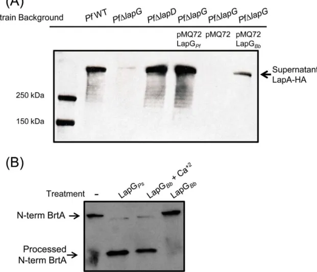
To confirm thatB.bronchisepticaLapG is a protease that cleaves BrtA we performed bio-chemical assays using two different approaches. In the experiment shown inFig 4A, the indi-catedP.fluorescensstrain was transformed with a vector control (pMQ72), a plasmid carrying the LapG protease fromP.fluorescens(LapGPf) as a positive control, orB.bronchiseptica
(LapGBb). In these assays, we assess LapG activity by measuring the amount of LapA protein of
P.fluorescensreleased into the supernatant; detecting supernatant LapA requires a functional LapG protease. The source of LapA in these studies is the chromosomally-encoded LapA modi-fied with an HA-tag to facilitate its detection. As expected, in the wild typeP.fluorescensstrain (PfWT,Fig 4A, lane 2), LapA protein can be detected in the supernatant but no LapA is detected in theP.fluorescensstrain lacking LapG (PfΔlapG,Fig 4A, lane 3). As an additional
Fig 4.B.bronchisepticaLapG cleavesP.fluorescens LapA andB.bronchisepticaBrtA.(A) Cleavage of LapA-HA from the surface ofP.fluorescenscells withB.bronchisepticaLapG. Western blot with anti-HA antibodies were used to detect LapA-HA ofP.fluorescensreleased into the supernatant fraction for each indicated strain. (B) Cleavage of N-terminal fragment of BrtA byB.bronchisepticaLapGin vitro. Samples correspond to reaction mixture ofE.colilysate expressing indicated protein or empty vector withE.colilysate expressing the N-terminus of BrtA (N-term BrtA) incubated at room temperature for 1h. Western blot with anti-HA antibodies was used to detect N-term BrtA.
control, we see abundant LapA in the supernatant in alapDmutant strain (PfΔlapD,Fig 4A, lane 4); alapDmutation results in high, unregulated LapG activity, and thus serves as an addi-tional positive control for LapA release into the supernatant.
Our data indicate thatB.bronchisepticaLapG can indeed substitute for the LapG protein of
P.fluorescensas indicated by its ability to release LapA into the supernatant (Fig 4A, lane 7), albeit to a lesser extent than theP.fluorescensLapG protein (Fig 4A, lane 5). In theP. fluores-censstrain lacking LapG (PfΔlapG) carrying the pMQ72 vector (lane 6), as expected, no LapA was detected in the supernatant. These data are consistent with the hypothesis thatB. bronchi-septicaLapG is a protease that can target cell-surface adhesins like LapA.
To extend the finding inFig 4Ato anin vitrosystem, anE.colistrain expressingB. bronchi-septicaLapG was grown, concentrated and sonicated to generate a crude cell-free extract, as described in the Materials and Methods. Crude cell free extracts ofE.colicontaining the HA-tagged, N-term fragment ofB.bronchisepticaBrtA (N-term BrtA, amino acids 1–330) were also generated. The crude extract containingB.bronchisepticaLapG was mixed in a 1:1 ratio with the extract containing the N-terminal fragment ofB.bronchisepticaBrtA, incubated at room temperature for 1 hr, then analyzed by Western blotting using an anti-HA-tag antibody to detect processing of BrtA. As a positive control, LapG fromP.fluorescenswas shown to pro-cess N-term BrtA, andB.bronchisepticaLapG was also able to cleave the N-terminal fragment ofB.bronchisepticaBrtA (Fig 4B). Interestingly,B.bronchisepticaLapG only cleaved BrtA in presence of 10 mM calcium. This finding is consistent with previous work showing that the LapG protein ofP.fluorescensis a calcium-dependent enzyme [23]. Interestingly, Nishikawa and colleagues observed that treatment ofB.bronchisepticawith EGTA, a calcium chelator, reduced biofilm formation; this reduction in biofilm formation may be due to the need for cal-cium to promote proper folding of some secreted proteins [24,25].
B
.
bronchiseptica
LapD Appears Partially Functional in
P
.
fluorescens
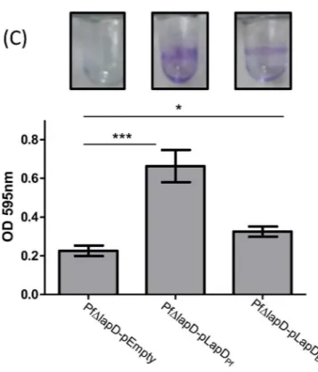
TheB.bronchiseptica lapD-like gene was cloned in a broad host vector to evaluate its ability to complement the phenotype of alapDmutant ofP.fluorescens(PfΔlapD). ThePfΔlapDstrain shows reduced biofilm formation and low LapA levels on the bacterial surface compared with the wild type [19]. When theB.bronchiseptica lapDgene was expressed in thePfΔlapDstrain, partial complementation was observed in CV biofilm assay (Fig 3C). The level of cell surface LapA in the strain carrying theB.bronchisepticaLapD was increased to an extent similar to that observed when theP.fluorescens lapDgene was introduced into this strain (Fig 3B). We do not understand why the increase in cell surface LapA to wild-type levels in the strain carry-ingB.bronchisepticaLapD does not also result in an increase in biofilm formation to levels equivalent to the wild-type strain.
The Deletion or Over-Expression of LapG Alters Biofilm Formation by
B
.
bronchiseptica
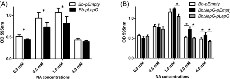
Previous studies ofP.fluorescensshowed that unregulated LapG expression results in increased cleavage of LapA and the release of this adhesin into the supernatant, results in reduced biofilm formation. In contrast, deleting thelapGgene ofP.fluorescensresults in enhanced levels of LapA on the cell surface and increased biofilm formation [20]. Thus, we assessed the effect of expressing LapG from a plasmid and deleting the gene from the chromosome on biofilm for-mation byB.bronchisepticaas a function of NA, anin vitroregulator of biofilm formation.
concentrations of NA added 0, 0.5 and 1 mM. At the highest concentration of NA tested (4.0 mM), where the extent of biofilm formation was the lowest, expression of theB.bronchiseptica
LapG did not further reduce biofilm formation.
Based on the work inP.fluorescens, we would expect aB.bronchisepticastrain with a dele-tion of thelapGgene to develop very robust biofilms compared to the wild type. As expected, theB.bronchisepticastrain lacking thelapGgene (BbΔlapG) showed more biofilm formation compared to wild type at the higher concentrations of NA tested, and this defect could be com-plementedin transby a wild-type copy of LapGBb(Fig 5B). This phenotype was observed on
either a PVC (Fig 5B) or glass surface (not shown).
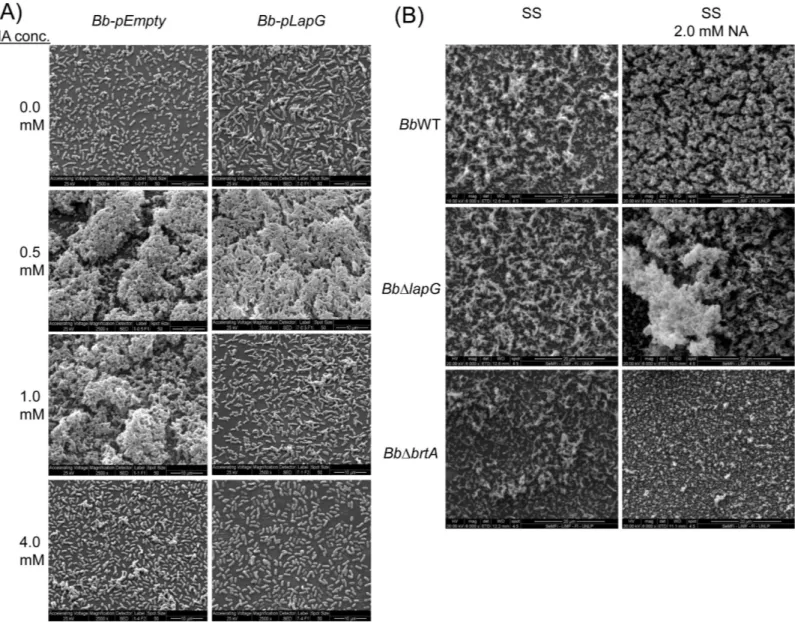
The observations inFig 5using the microtiter plate assay were confirmed by SEM analysis of biofilm structures formed in the air-liquid interface using glass as an abiotic surface.Fig 6
shows representative SEM images of the biofilms formed by strains over-expressing LapG com-pared to a vector control (Fig 6A) and in a strain lacking thelapGgene compared to the wild type (Fig 6B). As a control, deletion of thebrtAgene reduced (but did not eliminate) the bio-film formed under these conditions (Fig 6B). Similar results were observed when fluorescence microscopy was employed (S2 Fig).
LapG May Contribute to Infection
In Vivo
Biofilm formation on epithelial cells in the nose is thought to be a key early step inBordetella
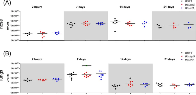
infection [9]. Given the role of BrtA and LapG forin vitrobiofilm formation, we hypothesized that these proteins might also play a role during colonizationin vivo. We intranasally inocu-lated suspensions of the wild type or mutantB.bronchisepticastrains into BALB/c mice, and the infection progress was evaluated for 21 days in the nose and lungs (Fig 7).
Mice were infected with wild typeB.bronchisepticaas previously described [4]. Wild type bacteria persisted in lung and nose mice after 21 days post infection. A typicalB.bronchiseptica
infection was observed; lung burden increased during the first 7 days and this microbe was gradually reduced across the remainder of the experiment. Our analysis indicates that BrtA Fig 5. LapG regulates biofilm formation byB.bronchiseptica.(A) Biofilm formation in different NA concentrations by the wild type carrying a vector control (Bb-pEmpty) or a plasmid over-expressingB.bronchisepticaLapG (Bb-lapG).*, indicates a significant difference versus theBb-pEmpty in same NA concentration, p<0.001. (B) Biofilm formation by the wild-type strain carrying a vector control (Bb-pEmpty), a strain with a deletion of thelapGgene carrying a vector control (BbΔlapG-pEmpty), and theBbΔlapGmutant complemented with a wild-type copy of theB.bronchiseptica lapGgene (BbΔlapG-pLapG). Biofilm formation was assessed using the microtiter plate assays as described in the Materials and Methods.*, indicates a significant difference in the indicated comparisons, p<0.001.
does not contribute to bacterial burden at any stage during infection in either body site evalu-ated (Fig 7). For the strain lacking thelapGgene (BbΔlapG), there was a small but statistically significant increase in bacterial burden at 14 days for those mice that survived until the end of the experiment, but no significant difference was detected at any other time point (Fig 7).
Interestingly, across all experiments, 25% ofBbΔlapGinfected mice presented symptoms of illness (indicated by ruffled fur, labored breathing, and diminished responsiveness) and were euthanized to avoid suffering (S3 Fig). Lungs of these euthanized mice carrying theBbΔlapG
strain had on average 3 x 10⁸CFU ml-1, a significant increase of 1–2 log over the wild type (7 x 10⁶CFU ml-1) and the mice carrying theBbΔlapGmutant that survived until the end of the experiment. These data indicated the possibility that loss of LapG function byB.bronchiseptica
could contribute to enhanced virulence, at least in some circumstances.
Fig 6. SEM images ofB.bronchisepticabiofilms.(A) The strainsBb-pEmpty (left column of panels) orBb-pLapG (right column of panels) were grown on vertically submerged coverslips in SS medium alone or supplemented with indicated NA concentrations. After 24 h of growth, biofilms formed at the air–liquid interface were visualized by SEM. (B) Indicated strains were grown on vertically submerged coverslips in SS medium alone (left column of panels) or SS supplemented with 2.0 mM NA (right column of panels).
Discussion
In this work we analyze the BrtA protein ofB.bronchiseptica, a predicted adhesin based on its similarity in domain structure to the LapA adhesin ofP.fluorescens. We confirm the recent finding of Nishikawa and colleagues [16] that BrtA participates in biofilm formation byB.
bronchiseptica, and build on this recent study by identifying and characterizing homologs of the LapD and LapG proteins ofP.fluorescens. InP.fluorescens, LapD and LapG participate in the regulation of cell surface localization of the LapA protein; we show that theB. bronchisep-ticaLapD and LapG proteins likely play a similar role for BrtA. We also demonstrate that loss of BrtA results in a mild biofilm defect on plastic, and a much more severe defect in biofilm for-mation on glass, a hydrophilic substrate. BrtA, while having limited sequence similarity to the LapA protein ofP.fluorescensshares a number of conserved features, including T1SS and vWFA domains, and the conserved Ala-Ala motif near its N-terminus that serves as the site of proteolysis in LapA for the LapG protein ofP.fluorescens.
InP.fluorescens, the localization of the LapA protein to the surface of the cell is postran-scriptionally regulated by LapG. LapG is a periplasmic protease that targets the N-terminus of LapA and cleaves this adhesin at a conserved Ala-Ala motif, resulting in release of LapA into the supernatant. Loss of LapA results in a reduction in biofilm formation byP. fluores-cens. The LapD protein ofP.fluorescens, in response to c-di-GMP levels in the cytoplasm, regulates LapG’s ability to target LapA by sequestering LapG via a protein-protein interac-tion mechanism [20,26]. The studies presented here indicate that the LapD and LapG pro-teins ofB.bronchisepticalikely play a similar role in the context of BrtA. Ourin vivoandin vitrostudies, including complementation assays, support the conclusion that LapG is a Fig 7.B.bronchisepticainfection in a mouse model.Comparison ofB.bronchisepticabacterial burden in the mouse nose (A) and lungs (B) were determined as described in the Materials and Methods after the indicated days post-infection. Bacteria were intranasally inoculated into external nares with an air displacement pipette (5 x 10⁵CFU in 50μl). Green dots correspond to CFU from euthanized mice infected withBb
ΔlapG.*indicates a significant difference compared to the CFU determined in mice infected with wild type strain, p<0.01.
protease that targets the N-terminal domain of BrtA for cleavage, thereby negatively impact-ing biofilm formation. Similarly, our studies implicate LapD in the control of LapG inB.
bronchiseptica.
An interesting observation from our SEM and fluorescence microscopy studies was that in a mutant lacking BrtA, biofilm formation was reduced but not eliminated. This observation sug-gested that there may be at least one other adhesin contributing to biofilm formation on abiotic surfaces. Previous reports implicated FHA and the adenylate cyclase enzyme as involved in bio-film formation byBordetella[7,11], further work should be directed at assessing the role of these proteins (or others) in the context of a strain lacking BrtA. Interestingly, BrtA appeared to play no detectable role in disease progression in a mouse model of infection; there was no change in bacterial burden in the nose or lungs of mice for up to 21 days post-infection in the
brtAmutant strain compared to the wild type. This finding is consistent with the work of Nishikawa and colleagues who showed that abrtAmutant ofB.bronchisepticashowed no defect in a model of rat nasal septum and tracheal colonization. Given that BrtA plays a role in biofilm formation on abiotic surfaces, we propose that a major role for this adhesion might be in environmental persistence as the microbe transits between hosts.
A strain ofB.bronchisepticalacking LapG function, which presumably has increased cell-surface BrtA based on ourin vitrostudies, when infecting mice, yielded two distinct pheno-types. In 75% of the animals, infection was maintained for 21 days with a bacterial burden simi-lar to the wild type, with the exception of a small but significant increase in bacterial numbers in lungs at day 14 post-infection. However, 25% of the mice infected with theB.bronchiseptica lapGmutant showed increased disease symptoms that resulted in the need to euthanize these animals typically between days 4–9. These data may indicate that BrtA, when present in larger levels on the cell surface can enhance virulence, at least in some animals. However, given the lack of impact upon loss of BrtA function in this infection model, it is also possible that LapG may have (at least) one other target inB.bronchiseptica. Distinguishing between these possibil-ities should be explored in future studies.
Overall, we have established that the LapD/LapG system of this pathogen likely functions in an analogous manner to the well-described system inP.fluorescens. InB.bronchiseptica, the target of the LapD/LapG system appears to be BrtA, a LapA-like protein that has a clear role in biofilm formation on abiotic surfaces. The role of the LapD/LapG system in a mouse model of infection is less clear—additional investigation will be required to dissect the role (if any) of BrtA and the LapD/LapG system in host-pathogen interactions.
Supporting Information
S1 Fig. Weblogo diagram of the repeated regions (CADG-IDR1-VCBS-IDR2) in BrtA.A consensus sequence among the eight repeat regions in BrtA is shown.
(TIF)
S2 Fig. Fluorescent microscopy ofB.bronchisepticabiofilm on glass surface.Bordetella
strains were cultured on glass coverslips as described for the SEM studies. After 12 hr of incu-bation the coverslips were washed with sterile PBS, and the bacteria were fixed with 4% formal-dehyde in PBS. The samples were observed in a Nikon Ti-U Fluorescence Microscope, and the images were obtained with a Nikon Digital Sight DS Ri1 camera using the Nis-Elements soft-ware. White bars indicate 100μm.
(TIF)
line). The difference between the survival curves was analyzed by the log-rank test (z = 1.98; p = 0.047 with 95% confidence).
(PDF)
S1 Table. Strains and plasmids used in this study.
(DOCX)
S2 Table. Oligonucleotides used in this study.
(DOCX)
S3 Table. Number of repeats domains in sequencedB.bronchisepticagenomes.
(PDF)
S1 Text. Detailed Materials and Methods.
(DOCX)
Acknowledgments
We thank Peggy Cotter for providing plasmids pGFLIP and pTNS3. We thank Paula Giménez, Silvana Tongiani, Luciana Cayuela, Juan Guzmán and Abel Bortolameotti for their excellent technical assistance. We thank to Ulises Mancini for technical assistance onS3 Table. We acknowledge the Laboratorio de Investigaciones de Metalurgia Física (LIMF) service (Facultad de Ingeniería–UNLP) and the Servicio de Microscopía Electrónica de Barrido (CINDECA-CONICET-UNLP) for SEM analysis.
Author Contributions
Conceived and designed the experiments: NA JF GAO FS. Performed the experiments: NA CDB JF FS. Analyzed the data: NA CDB JF GAO FS. Contributed reagents/materials/analysis tools: JF GAO FS. Wrote the paper: JF GAO FS.
References
1. Goodnow RA. Biology ofBordetella bronchiseptica. Microbiol Rev. 1980; 44: 722–738. PMID:7010115 2. Harvill ET, Cotter PA, Yuk MH, Miller JF. Probing the function ofBordetella bronchisepticaadenylate
cyclase toxin by manipulating host immunity. Infect Immun. 1999; 67: 1493–500. PMID:10024599 3. Harvill ET, Preston A, Cotter PA, Allen AG, Maskell DJ, Miller JF. Multiple roles forBordetella
lipopoly-saccharide molecules during respiratory tract infection. Infect Immun. 2000; 68: 6720–6728. PMID: 11083787
4. Sisti F, Fernández J, Rodríguez ME, Lagares A, Guiso N, Hozbor DF.In vitroandin vivo characteriza-tion of aBordetella bronchisepticamutant strain with a deep rough lipopolysaccharide structure. Infect Immun. 2002; 70: 1791–1798. PMID:11895940
5. Skinner JA, Reissinger A, Shen H, Yuk MH. Bordetella type III secretion and adenylate cyclase toxin synergize to drive dendritic cells into a semimature state. J Immunol. 2004; 173: 1934–1940. PMID: 15265927
6. Inatsuka CS, Julio SM, Cotter PA.Bordetellafilamentous hemagglutinin plays a critical role in immuno-modulation, suggesting a mechanism for host specificity. Proc Natl Acad Sci U S A. 2005; 102: 18578– 18583. doi:10.1073/pnas.0507910102PMID:16339899
7. Irie Y, Mattoo S, Yuk MH. The Bvg virulence control system regulates biofilm formation inBordetella bronchiseptica. J Bacteriol. 2004; 186: 5692–5698. doi:10.1128/JB.186.17.5692-5698.2004PMID: 15317773
8. Mishra M, Parise G, Jackson KD, Wozniak DJ, Deora R. The BvgAS signal transduction system regu-lates biofilm development inBordetella. J Bacteriol. 2005; 187: 1474–1484. doi:10.1128/JB.187.4. 1474-1484.2005PMID:15687212
10. Conover MS, Mishra M, Deora R. Extracellular DNA is essential for maintainingBordetellabiofilm integ-rity on abiotic surfaces and in the upper respiratory tract of mice. PLoS One. 2011; 6: e16861. doi:10. 1371/journal.pone.0016861PMID:21347299
11. Serra DO, Conover MS, Arnal L, Sloan GP, Rodriguez ME, Yantorno OM, et al. FHA-mediated cell-sub-strate and cell-cell adhesions are critical for Bordetella pertussis biofilm formation on abiotic surfaces and in the mouse nose and the trachea. PLoS One. 2011; 6: e28811. doi:10.1371/journal.pone. 0028811PMID:22216115
12. Sisti F, Ha D-G, O’Toole GA, Hozbor D, Fernández J. Cyclic-di-GMP signalling regulates motility and biofilm formation inBordetella bronchiseptica. Microbiology. 2013; 159: 869–79. doi:10.1099/mic.0. 064345-0PMID:23475948
13. Nicholson TL, Conover MS, Deora R. Transcriptome profiling reveals stage-specific production and requirement of flagella during biofilm development inBordetella bronchiseptica. PLoS One. 2012; 7: e49166. doi:10.1371/journal.pone.0049166PMID:23152870
14. Whiteley CG, Lee D-J. Bacterial diguanylate cyclases: structure, function and mechanism in exopoly-saccharide biofilm development. Biotechnol Adv. 33: 124–41. doi:10.1016/j.biotechadv.2014.11.010 PMID:25499693
15. Ryan RP. Cyclic di-GMP signalling and the regulation of bacterial virulence. Microbiology. 2013; 159: 1286–97. doi:10.1099/mic.0.068189-0PMID:23704785
16. Nishikawa S, Shinzawa N, Nakamura K, Ishigaki K, Abe H, Horiguchi Y. Thebvg-repressed genebrtA, encoding biofilm-associated surface adhesin, is expressed during host infection byBordetella bronchi-septica. Microbiol Immunol. 2016; 60: 93–105. doi:10.1111/1348-0421.12356PMID:26756546 17. Stainer DW, Scholte MJ. A simple chemically defined medium for the production of phase IBordetella
pertussis. J Gen Microbiol. 1970; 63: 211–220. doi:10.1099/00221287-63-2-211PMID:4324651 18. Bertani G. Studies on lysogenesis. I. The mode of phage liberation by lysogenicEscherichia coli. J
Bac-teriol. American Society for Microbiology (ASM); 1951; 62: 293–300.
19. Newell PD, Monds RD, O’Toole GA. LapD is a bis-(3’,5')-cyclic dimeric GMP-binding protein that regu-lates surface attachment byPseudomonas fluorescensPf0-1. Proc Natl Acad Sci U S A. 2009; 106: 3461–6. doi:10.1073/pnas.0808933106PMID:19218451
20. Newell PD, Boyd CD, Sondermann H, O’Toole GA. A c-di-GMP effector system controls cell adhesion by inside-out signaling and surface protein cleavage. PLoS Biol. 2011; 9: e1000587. doi:10.1371/ journal.pbio.1000587PMID:21304920
21. Chatterjee D, Boyd CD, O’Toole GA, Sondermann H. Structural characterization of a conserved, cal-cium-dependent periplasmic protease fromLegionella pneumophila. J Bacteriol. 2012; 194: 4415–25. doi:10.1128/JB.00640-12PMID:22707706
22. Akerley BJ, Monack DM, Falkow S, Miller JF. ThebvgASlocus negatively controls motility and synthe-sis of flagella inBordetella bronchiseptica. J Bacteriol. 1992; 174: 980–990. PMID:1370665
23. Boyd CD, Chatterjee D, Sondermann H, O’Toole GA. LapG, required for modulating biofilm formation byPseudomonas fluorescensPf0-1, is a calcium-dependent protease. J Bacteriol. 2012; 194: 4406– 14. doi:10.1128/JB.00642-12PMID:22707708
24. Bakás L, Veiga MP, Soloaga A, Ostolaza H, Goñi FM. Calcium-dependent conformation ofE.coli
alpha-haemolysin. Implications for the mechanism of membrane insertion and lysis. Biochim Biophys Acta. 1998; 1368: 225–34. PMID:9459600
25. Subrini O, Sotomayor-Pérez AC, Hessel A, Spiaczka-Karst J, Selwa E, Sapay N, et al. Characterization of a membrane-active peptide from theBordetella pertussisCyaA toxin. J Biol Chem. 2013; 288: 32585–32598. doi:10.1074/jbc.M113.508838PMID:24064217