1361
EFFECTS OF P22 BACTERIOPHAGE ON
SALMONELLA ENTERICA
SUBSP.
ENTERICA
SEROVAR TYPHIMURIUM DMC4 STRAIN BIOFILM FORMATION AND ERADICATION
Basar Karaca1,*, Nefise Akcelik2 and Mustafa Akcelik1
1 Department of Biology, Faculty of Science, Ankara University, Ankara, Turkey
2 Institute of Biotechnology, Ankara University, Ankara, Turkey
*Corresponding author: [email protected]
Abstract:Over the last decades, several antimicrobial agents have been made available. Due to increasing antimicrobial resistance, bacteriophages were rediscovered for their potential applications against bacterial infections. In the present study, biofilm inhibition and eradication of Salmonellaenterica subsp. enterica serovar Typhimurium DMC4 strain (S. Typhimurium) was evaluated with respect to different incubation periods at different P22 phage titrations. The efficacy of P22 phage on biofilm formation and eradication of S. Typhimurium DMC4 strain was screened in vitro on polystyrene and stainless steel surfaces. The biofilm forming capacity of S. Typhimurium was significantly reduced at higher phage titrations (106 pfu/mL ≤). All phage titers (104-108 pfu/mL) were found to be effective at the end of the 24 h-incubation period whereas higher phage titrations were found to be effective at the end of the 48 h and 72 h of incubation. P22 phage has less efficacy on already formed, especially mature biofilms (72 h-old biofilm). Notable results of P22 phage treatment on S. Typhimurium biofilm suggest that P22 phage has potential uses in food systems.
Key words: Salmonella;biofilm; bacterial adhesion; bacteriophage;eradication
Received: November 20, 2015; Revised: February 18, 2015; Accepted: February, 2015
INTRODUCTION
The enteric pathogen Salmonella Typhimurium causes gastroenteritis in humans and other mam-mals. S. Typhimurium serovar, along with Enteriti-dis, Virchow and Hadar serovars, is one of the most common serovars isolated from humans in develop-ing countries. It has a zoonotic origin and can exhibit resistance to commonly used antibiotics (Philips et al., 2004). Moreover, many Salmonella strains are able to form complex surface-associated communi-ties, called biofilms. Biofilms contribute to the resis-tance and persistence of Salmonella in both host and non-host environments and are especially important in food processing environments and poultry (Steen-ackers et al., 2012; Diez-Garcia et al., 2012). Biofilms are multicellular cell aggregates that are encased in a self-produced extracellular matrix (Branda et al., 2005; Hall-Stoodley and Stoodley, 2009). Further-more, it is known that approximately 80% of all
bac-terial infections are related to biofilms (Davies, 2003; Hall-Stoodley and Stoodley, 2009).
Many scientists in the last decades have focused their research on designing novel biofilm treatment strategies. Well-known antimicrobial treatment strat-egies may be mostly inefficient in eradicating biofilms (Davies, 2003). There are three major strategies to treat biofilms: prevention of biofilm formation, re-moval/killing of biofilm and weakening of biofilm (Alhede et al., 2011).
bacteria resistant to the most currently available an-timicrobial agents, bacteriophages were rediscovered for their potential applications in the eradication of bacterial infections (Sulakvelidze et al., 2001).
The utilization of bacteriophages to fight against bacterial biofilms has become a novel strategy. Bac-teriophages can be used efficiently against biofilm in-fections and in invitro biofilm setups. Many reports have shown that bacteriophages have a widespread use to reduce biofilm formation and to eradicate already formed biofilms. It has been also proved that bacterio-phages can reduce biofilm formation invivo. Invitro biofilms of Streptococcusepidermidis on silicone cathe-ters can be reduced effectively when the catheter is pre-treated with a lytic bacteriophage (Curtin and Donlan, 2006). Lytic bacteriophage treatments showed hopeful results on septicemic mouse infected with the multi-drug-resistant Pseudomonasaeruginosa and Klebsiella pneumonia (Vinodkumar et al., 2005; Vinodkumar et al., 2008). There are also several reports about phage and biofilm interactions for many bacteria such as Ba-cillussubtilis (O’Flaherty et al., 2009), Escherichiacoli (Corbin et al., 2001), Enterococcusfaecalis (Teng et al., 2009), Lactococcuslactis (Briandet et al., 2008), Listeria monocytogenes (Hibma et al., 1997), Pseudomonas fluo-rescens (Del Pozo and Patel, 2007) and Staphylococcus aureus (Resch et al., 2005).
P22 phage infects S. Typhimurium via binding to the O-antigen lipopolysaccharide on the surface of the host. The phage’s tail fiber protein has endorham-nosidase activity, which cleaves the O-antigen chain (Prevelige, 2006). P22 is a temperent phage and it can be used as a perfect transduction tool for genetic studies of S. Typhimurium. In this study, we aimed to evaluate the prevention and eradication potentials of P22 phage on S. Typhimurium biofilm.
MATERIALS AND METHODS
Bacterial strains and growth conditions
S. Typhimurium DMC4 strain isolated from a beef product in Turkey was obtained from the Ankara University Prokaryotic Genetics Laboratory culture
collection. The culture was inoculated into Luria-Ber-tani broth and incubated at 37°C for 18 h with shak-ing at 200 rpm. LB without NaCl (LBwo/NaCl; bacto-tryptone 10 g/L (Fluka, Taufkirchen, Germany), yeast extract 5 g/L (Merck, Darmstadt, Germany)) was used to maintain optimum conditions for biofilm as-says (Römling et al., 1998).
Preparation of P22 phage lysates
Preparation and titration of P22 phage were per-formed as described by Davis et al. (1980). Briefly, 1 mL overnight culture of S. Typhimurium DMC4 strain was added to 4 mL P22 transduction broth (transduction broth; 2 mL 50xE medium (sterilized with CHCl3), 1 mL sterile 20% glucose, 0.1 mL P22 lysate (sterilized with CHCl3); the final volume was adjusted to 100 mL by the addition of sterile LB broth. This suspension was incubated for 18 h at 37°C with shaking at 200 rpm. The culture was transferred into microfuge tube and centrifuged for 2 min at 12000 rpm to pellet the cell debris. Finally, several drops of CHCl3 (Merck, Darmstadt, Germany) were added to the lysate. The lysate was stored for a long time at 4°C. To check the titrations of phage lysates, LB soft agar was melted in a microwave and cooled in a 50°C heating block. Then 0.1 mL of overnight culture of S. Typhimurium DMC4 strain was added to melted soft agar and poured immediately onto LB agar plates. Finally, 20 µL of appropriate phage dilutions were spotted onto the culture lawn (from 104 to 109 phage dilutions in sterile 0.85% NaCl).
Biofilm sampling for microtiter plate assay and stainless steel application
the test broth only. Following the incubation, plates were washed twice with sterile 0.85% NaCl solution to remove planktonic cells. The plates were allowed to dry under a safety hood. Following this step, 130 µL of 98% methanol was added (Merck, Darmstadt, Ger-many) and the plates were incubated for 10 min. The methanol was removed and plates were dried again. One hundred thirty µL of 1% crystal violet (Merck, Darmstadt, Germany) was transferred into the wells and stained for 30 min. After staining, plates were washed under running tap water. Finally, 130 µL of 33% glacial acetic acid (Sigma-Aldrich, Taufkirchen, Germany) was added to each well and bound dye was solubilized. The OD of each well was measured at 595 nm (Bio-Rad, USA). The results for the tested strain were calculated by subtracting the median OD595nm of the three parallels of the control (LB without NaCl broth only) from the median OD595nm of the three par-allels of sample.
Stainless steel coupons (type 304) were chosen for screening biofilm production since stainless steel is fre-quently used for food-processing environments. The coupons (0.1, 0.8 and 2.5 cm) were prepared for treat-ment assays as previously described by Giaouris et al. (2005). Coupons were placed individually in 3.5 mL of LB without NaCl broth media, and test tubes with the coupons were autoclaved at 121°C for 15 min. Over-night cultures were adjusted to OD595nm of 0.2 (~108 CFU/mL), and 1 mL of these suspensions were inocu-lated into test tubes. The test tubes were incubated for 24, 48 and 72 h at 20°C. Following incubation, each coupon was taken with sterile forceps, transferred to an empty sterile tube and allowed to dry at room tempera-ture for 5 min. The coupons were rinsed twice with 5 mL of 0.85% sterile physiological saline solution to remove planktonic counterparts. After washing, each coupon was transferred to a new tube containing 4.5 mL of sterile physiological saline solution and five ster-ile glass beads (3 mm in diameter). The coupons were then vortexed for 1 min at maximum intensity (IKA Genius Vortex 3, Staufen, Germany).
Quantification of biofilm cells was performed using the agar plating method. Colonies on LB agar plates were counted after incubation at 37°C for 18 h.
The count of CFU (colony forming unit) per square centimeter was transformed to a logarithmic value.
Evaluation of P22 phage on microbial adhesion and biofilm formation (phage treatment)
S. Typhimurium DMC4 strain was cultured with P22 phage to assess the inhibition potential of P22 phage on biofilm formation. The 96-well polystyrene mi-crotiter plates were filled with the appropriate phage suspensions and control wells were filled with the test broth and inoculum only. Titration of phage suspen-sions were adjusted to 104-108 PFU/mL using test medium (LB without NaCl) to maintain the same ex-perimental conditions of biofilm sampling. The assay was performed as described above. The plates were incubated for 24, 48 and 72 at 20°C. The biofilm re-duction percentages were calculated according to the following formula [(C-B)-(T-B)]/ [(C-B)]x100 where C = control wells containing only strain inoculum, B = blank wells containing test medium and appropri-ate phage dilution without strain inoculum, and T = treatment wells containing strain inoculum and ap-propriate phage dilution.
To perform stainless steel application, test tubes containing sterile stainless steel coupons were filled with appropriate phage suspensions adjusted to 104 -108 PFU/mL in test medium. The control tubes were filled with only test medium. The test tubes were in-cubated for 24, 48 and 72 h at 20°C. The assay was performed as described previously. The logarithmic reduction of biofilm cells was calculated according to the following formula: [PK: (1-10–LR)x100%] where PK is the percentage killing, LR is the log reduction [log10(untreated viable cell density)-log10(treated vi-able cell density)].
Evaluation of P22 phage on already formed biofilms (phage post-treatment)
wells and stainless steel coupons were treated with appropriate phage dilutions (104-108 PFU/mL). The negative control samples were prepared only with test medium. Following incubation at 37°C for 24 h to maintain the optimum phage infection conditions, experimental data were obtained as described above. Finally, the values obtained from experiments were compared to evaluate the biofilm eradication poten-tial of P22 phage. The percentage of biofilm eradica-tion was calculated by the same formulas given above for both microtiter plate and stainless steel assays.
Statistical analysis
All statistical analyses were performed by Minitab statistics software, version 15 (USA). Experiments were performed in triplicate. Bars were given as mean standard deviation values. The one-way ANOVA test was preferred for all mean values associated with data from the microtiter plate and stainless steel assays. Pearson’s correlation was used to analyze the differ-ences in biofilm production on polystyrene and stain-less steel surfaces.
RESULTS
Effect of P22 phage on bacterial adhesion and biofilm forming (phage treatment)
To evaluate the efficacy of P22 phage on the inhibition of bacterial adhesion and biofilm formation, phage treatment assays were performed using S. Typhimuri-um DMC4 strain for different incubation periods (24, 48 and 72 h) and two types of abiotic surface (polysty-rene and stainless steel). Many of the S. Typhimurium strains were screened for their biofilm formation ca-pabilities, and the DMC4 strain was selected as a good biofilm producer in further experiments (data not shown). Ninety-six-well polystyrene microtiter plates and stainless steel surfaces to evaluate the biofilm-pro-ducing abilities of S. Typhimurium DMC4 strain were preferred due to their common usage in food manu-facturing and processing environments. To detect the effects of P22 phage on the biofilm-producing cells of S. Typhimurium DMC4 strain, microtiter plates
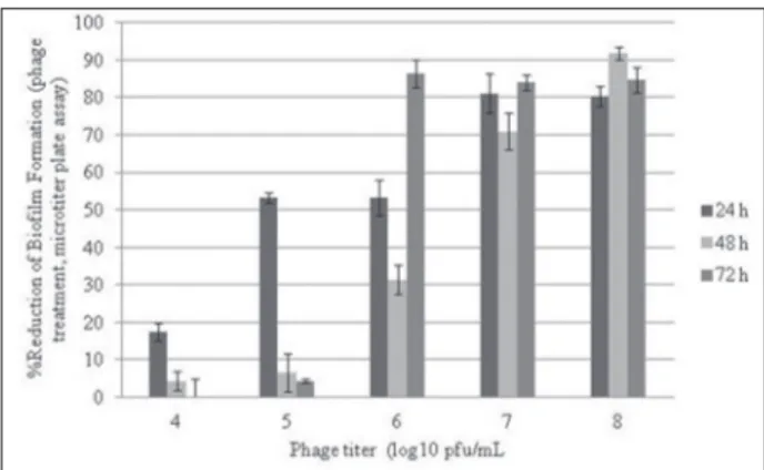
were incubated with appropriate phage dilutions for 24, 48 and 72 h. The effects of phage treatment on biofilm formation are summarized as a percentage of biofilm reduction in Fig. 1. All phage titrations (104 -108 PFU/mL) were found only to be effective at the end of the 24-h and 48-h incubation periods, whereas higher phage titrations were only found to be effective at the end of the 72-h incubation period (106 PFU/ mL ≤). Means of biofilm quantities were compared by one-way ANOVA; p <0.05. As regards the microtiter plate assay, the 48-h incubation period and the highest phage titrations (108 PFU/mL) were detected as the most effective conditions for the prevention of biofilm formation (Fig. 1).
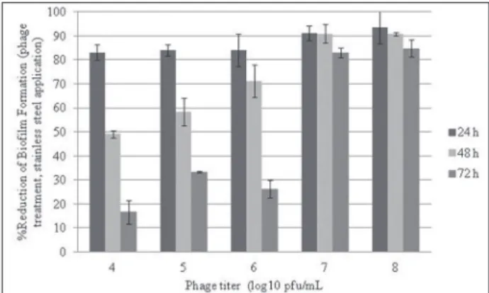
Results obtained from the stainless steel assay were not similar with microtiter plate assay results. Results are shown in Fig. 2. The effect of P22 phage on biofilm reduction of the tested strain on stainless steel surfaces was found to be much more effective in comparison to the microtiter plate assay results. A negative correlation was detected between the rates of biofilm quantity on polystyrene microtiter plates and stainless steel surfaces (r=-0.627 by Pearson correla-tion, p <0.01). However, due to the long incubation periods, the phage effect was significantly reduced, similarly to the microtiter plate assay. Significant bio-film reduction was detected only for higher phage ti-trations (107≤PFU/mL) at the end of 72-h incubation period. The values obtained from these experiments
were converted to log reduction percentage (Fig. 2). P22 phage could not easily prevent biofilm formation after the 24-h and 48-h-old incubation periods and at lower phage titration (Fig. 2).
Effect of P22 phage on already formed biofilms (phage post-treatment)
The removal activity of P22 phage on already formed 24-, 48-, and 72-h-old biofilms was evaluated by microtiter plate assay and stainless steel applica-tion. First the microtiter plate assay was performed to detect the removal of biomass. The 24-, 48-, and 72-h-old biofilms in plate wells were post-treated with appropriate phage dilutions (104-108 PFU/mL) and incubated at 37°C for 24 h. The results are given in Fig. 3. Biomass removal activities of 57.14% and 68.99% of P22 phage were detected for the 24-h-old biofilm at phage titrations of 107 and 108 PFU/mL, respectively. In the 48-h-old biofilms, the phage ti-trations of 107 and 108 PFU/mL resulted in 39.97% and 43.36% of biomass removal activity, respectively (one-way ANOVA, p <0.05). Apart from higher phage titrations such as 107 and 108 pfu/mL, the eradication capability of P22 phage was found to be less effec-tive on the 48-h-old biofilm structure (the samples on polystyrene surfaces) at lower phage titrations (Fig. 3). The least removal efficiency was detected for the 72-h-old biofilm structure by phage post-treatment (0.54%, 0.78%, 1.5%, 4.37% and 7.56% eradication for 104, 105, 106, 107 and 108 PFU/mL phage titrations, respectively) (one-way ANOVA, p <0.05).
P22 phage showed less eradication capacity against 24- and 48-h-old biofilms on stainless steel coupons compared to microtiter plate surfaces. Similar to mi-crotiter plate assay results, phage efficiency was dra-matically decreased for the 72-h-old biofilm samples (samples on stainless steel surfaces) (no eradication; 0% was detected for 104, 105, 106 PFU/mL titrations) (Fig. 4). Crystal violet staining was also performed for stainless steel surfaces to confirm the removal activ-ity of P22 phage. As a result, decreased crystal violet staining was observed on stainless steel coupons, indi-cating that P22 phage has biofilm eradication activity.
Fig. 2. Effect of phage treatment on biofilm formation during 24-, 48- and 72-h incubation periods and the reduction percentages of biofilm formation (stainless steel application) (Bars represent standard deviation).
Fig.3. Effect of phage post-treatment on already formed 24-, 48- and 72-h-old biofilm samples and the eradication percentages of biofilm (microtiter plate assay) (Bars represent standard devia-tion).
DISCUSSION
This study has shown that P22 phage is able to in-fect S. Typhimurium biofilm and has an eradication potential on already formed biofilm of tested S. Ty-phimurium strain. P22 infection begins via binding of the gp9 tail spike protein of phage to the O-antigen lipopolysaccharide exposed on the surface of the host. This tail fiber protein has endorhamnosidase activ-ity, which cleaves to the O-antigen chain (Prevelige, 2006). It is well known that Salmonella produces an O-antigen capsule coregulated with the fimbria and cellulose-associated extracellular matrix (Gibson et al., 2006). Gram-negative bacteria typically consist of surface exposed O-antigen, a core structure that is embedded in the outer membrane lipid bilayer. This study may reveal that the infection of S. Typhimuri-um biofilm by P22 phage begins via the trapping of phages by cells that are in the outer layers of the bio-film and also phages can infect planktonic bacteria that could go on to form a biofilm. The results of the present study suggest that although the phage treat-ment of two abiotic surfaces can effectively reduce the formation of biofilm, it is less effective in eradicating already formed biofilms. This phenomenon indicates that P22 phage may have a notable potential to inhibit S. Typhimurium biofilm formation, especially in food and clinical environments.
Many researches showed that significant biofilm reductions (ranging from 1 to 6 log), depend upon the constituents of the biofilm, biofilm age, phage selection and duration of treatment (Corbin et al., 2001; Sharma et al., 2005). It was found that treatment with P22 phage was much more effective in inhibit-ing biofilm formation (ranginhibit-ing from 0.08 to 1.2 log10) than eradicating post-treated mature biofilm (from 0 to 0.5 log10). The duration of the phage treatment and the age of biofilm are critical to the inhibition of biofilm formation and eradication of mature bio-films of S. Typhimurium. When phage treatment is extended, biofilm formation is completed in the late incubation period due to the decrement of phage ti-tration or a possible increasing of phage resistance. We also checked the effectiveness of phage infection capability on planktonic cultures of the strain in the
exponential phase. Therefore, phage resistance may increase or there might be a lack of phage penetration due to the long-term incubation periods and biofilm matrix (data not shown). This phenomenon should be examined to illuminate the phage resistance in S. Typhimuriumbiofilms for further studies. P22 phage has some potential to develop a control strategy for the biofilm formation of S. Typhimurium. However, it has less effectiveness in the eradication of mature biofilms. Hence, it might be used in future studies as a contributing agent for effective phage cocktails that can eradicate mixed biofilm formations and can be combined with different antimicrobial agents to treat biofilm formations.
There are also some reports showing that phage predation can enhance the formation of the biofilm. Hosseinidoust et al. (2013) discovered that single-species biofilm formation of Pseudomonas aerugi-nosa can be enhanced with the treatment of differ-ent phages. However, in our study, the induction of biofilm formation after the P22 phage challenge was not observed. Furthermore, P22 phage can be a good candidate to study phage and S. Typhimurium biofilm interactions. In this respect, phage-induced biofilm dispersion models should be studied to reveal the roles of lysogens in biofilms.
Salmonella strains can cause serious problems due to cross contamination in food environments. Discovering novel antimicrobial agents, such as bac-teriophages, for the foodborne pathogens, especially for disinfectant- and antibiotic-resistant strains, is a current topic. P22 phage could be a potential alterna-tive for eliminating S. Typhimurium biofilms in food systems, and it might be used as a supplement with other effective phage cocktails and disinfectants. The results of this study are also important for providing initial data to unveil P22 phage/S. Typhimurium bio-film interactions. Finally, it was concluded that P22 phage might have possible critical characteristics to control S. Typhimurium biofilms.
Akcelik and Mustafa Akcelik analyzed the data and pro-vided the reagents, material and all laboratory equipment.
Conflict of interest disclosure: The authors declare that they have no competing interests.
REFERENCES
Alhede, M., Kragh, K.N., Qvortrup, K., Holm, M.A., Van Gennip, M., Christensen, L.D., Jensen, P.O., Nielsen, A.K., Parsek, M., Wozniak, D., Molin, S., Tolker-Nielsen, T., Hoiby, N., Givskov, M. and T. Bjarnsholt (2011). Phenotypes of non-attached Pseudomonas aeruginosa aggregates resemble sur-face attached biofilm. PLoS ONE. 6, e27943.
Branda, S. S., Vik, A., Friedman, L. and R. Kolter (2005). Biofilms: the matrix revisited. Trends Microbiol.13, 20-26.
Briandet, R., Lacroix-Gueu, P., Renault, M., Lecart, S., Meylheuc, T., Bidnenko, E., Steenkeste, K., Bellon-Fontaine, M. N. and M. P. Fountaine-Aupart (2008). Fluorescence correlation spec-troscopy to study diffusion and reaction of bacteriophages inside biofilms. Appl. Environ. Microb.7, 2135-2143.
Corbin, B. D., Mclean, R. J. C. and G. M. Aron (2001). Bacterio-phage T4 multiplication in a glucose-limited Escherichia coli biofilm. Can. J. Microbiol.47, 680-684.
Curtin, J. J. and R. M. Donlan (2006). Using bacteriophages to reduce formation of catheter-associated biofilms by Staphy-lococcus epidermidis. Antimicrob. Agents Ch.50, 1268-1275.
Davies, D. (2003). Understanding biofilm resistance to antibacte-rial agents. Nat. Rev. Drug Discov.2, 114-122.
Davis, R. W., Botstein, D. and J. R. Roth (1980). Advanced Bacte-rial Genetics, Cold Spring Harbor Laboratory, 78-80. NY.
Del Pozo, J. L. and R. Patel (2007). The challenge of treating bio-film-associated bacterial infections. Clin. Pharmocol. Ther.
82, 204-209.
Diez-Garcia, M., Capita, R. and C. Alonso-Calleja (2012). Influ-ence of serotype on the growth kinetics and the ability to form biofilms of Salmonella isolates from poultry. Food Microbiol.31, 173-180.
Giaouris,E., Chorianoupoulos, N. and G. J. E. Nychas (2005). Effect of temperature, pH, and water activity on biofilm formation by Salmonella enterica Enteritidis PT4 on stainless steel sur-faces as indicated by the bead vortexing method and conduc-tance measurements. J. Food Prot.68, 2149-2154.
Gibson, D. L., White, A. P., Snyder, S. D., Martin, S., Heiss, C., Azadi, P., Surette, M. and W. W. Kay (2006). Salmonella
produces an O-antigen capsule regulated by AgfD and important for environmental persistence. J. Bacteriol.22,
7722-7730.
Hall-Stoodley, L. and P. Stoodley (2009). Evolving concepts in bio-film infections. Cell. Microbiol.11, 1034-1043.
Hibma, A. M., Jassim, S. A. A. and M. W. Griffiths (1997). Infec-tion and removal of L-forms of Listeria monocytogenes with bred bacteriophage. Int. J. Food Microbiol.34, 197-207.
Hosseinidoust, Z., Tufenkji, N. and T. G. M. van de Ven (2013). Predation in homogeneous and heterogeneous phage envi-ronments affects virulence determinants of Pseudomonas aeruginosa. Appl. Environ. Microbiol.79, 2862-2871.
O’Flaherty, S., Ross, R. P. and A. Coffey (2009). Bacteriophage and their lysins for elimination of infectious bacteria. FEMS Microbiol. Rev.33, 801-819.
Philips, S. J., Dud’ik, M. and R. E. Schapire (2004). A maximum entropy approach to species distribution modeling. In: Pro-ceedings of the 21st International Conference on Machine
Learning, 655-662. ACM Press, New York.
Prevelige, P. E. (2006). The Bacteriophages, Bacteriophage P22, Chapter 29, Oxford University Press, UK.
Resch, A., Fehrenbacher, B., Eisele, K., Schaller, M. and G. Friedrich
(2005). Phage release from biofilm and planktonic Staphy-lococcus aureus cells. FEMS Microbiol. Lett.252, 89-96.
Römling, U., Sierralta, W. D., Eriksson, K. and N. Staffan (1998). Multicellular and aggregative behaviour of Salmonella typhimurium strains is controlled by mutations in the agfD promoter. Mol. Microbiol.28, 249-264.
Sharma, M., Ryu, J. H. and L. R. Beuchat (2005). Inactivation of
Escherichia coli O157:H7 in biofilm on stainless steel by treatment with an alkaline cleaner and a bacteriophage. J. Appl. Microbiol.99, 449-459.
Steenackers, H., Hermans, K., Vanderleyden, J. and S. C. J. de Keersmacker (2012). Salmonella biofilms: An overview on occurrence, structure, regulation and eradication. Food Res. Int. 45, 502-531.
Stepanovic, S., Vukovic, D., Dakic, I., Savic, B. and M. Svabic-Vla-hovic (2000). A modified microtiter-plate test for quanti-fication of staphylococcal biofilm formation. J. Microbiol. Methods.40, 175-179.
Sulakvelidze, A., Alavidze,Z. and J. G. Morris Jr. (2001). Bacterio-phage therapy. Antimicrob. Agents Ch.45, 649-659.
Teng, F., Singh, K.V., Bourgogne, A., Zeng, J. and B.E. Murray
(2009). Further Characterization of the epa Gene Cluster and Epa Polysaccharides of Enterococcus faecalis. Infect. Immun.77, 3759-3767.
Vinodkumar, C. S., Neelagund, Y. F. and S. Kalsurmath (2005). Bacteriophage in the treatment of experimental septicemic mice from a clinical isolate of multidrug resistant Klebsiella pneumoniae. J. Commun. Dis.37, 18-29.
Vinodkumar, C. S., Neelagund, Y. F. and S. Kalsurmath (2008). Utility of lytic bacteriophage in the treatment of multidrug-resistant Pseudomonas aeruginosa septicemia in mice. Ind. J. Path. Microbiol.51, 360-366.
Woodward, M. J., Sojka, M., Sprigings K. A. and T. J. Humphrey