Two Major Vectors: A Comparison between a
Viscerotropic and a Dermotropic Strain
Carla Maia1,2, Veronika Seblova1, Jovana Sadlova1, Jan Votypka1, Petr Volf1*
1Department of Parasitology, Faculty of Sciences, Charles University, Prague, Czech Republic,2Unidade de Parasitologia Me´dica, Centro de Mala´ria e Doenc¸as Tropicais, Instituto de Higiene e Medicina Tropical, Universidade Nova de Lisboa, Lisboa, Portugal
Abstract
We quantifiedLeishmania infantum parasites transmitted by natural vectors for the first time. Both L. infantumstrains studied, dermotropic CUK3 and viscerotropic IMT373, developed well inPhlebotomus perniciosusandLutzomyia longipalpis. They produced heavy late-stage infection and colonized the stomodeal valve, which is a prerequisite for successful transmission. Infected sand fly females, and especially those that transmit parasites, feed significantly longer on the host (1.5–1.8 times) than non-transmitting females. Quantitative PCR revealed thatP. perniciosusharboured more CUK3 strain parasites, while inL. longipalpisthe intensity of infection was higher for the IMT373 strain. However, in both sand fly species the parasite load transmitted was higher for the strain with dermal tropism (CUK3). All but one sand fly female infected by the IMT373 strain transmitted less than 600 promastigotes; in contrast, 29% ofL. longipalpisand 14% of P. perniciosus infected with the CUK3 strain transmitted more than 1000 parasites. The parasite number transmitted by individual sand flies ranged from 4 up to 4.196104promastigotes; thus, the maximal natural dose found was still about 250 times lower
than the experimental challenge dose used in previous studies. This finding emphasizes the importance of determining the natural infective dose for the development of an accurate experimental model useful for the evaluation of new drugs and vaccines.
Citation:Maia C, Seblova V, Sadlova J, Votypka J, Volf P (2011) Experimental Transmission ofLeishmania infantumby Two Major Vectors: A Comparison between a Viscerotropic and a Dermotropic Strain. PLoS Negl Trop Dis 5(6): e1181. doi:10.1371/journal.pntd.0001181
Editor:Genevieve Milon, Institut Pasteur, France
ReceivedJanuary 6, 2011;AcceptedApril 8, 2011;PublishedJune 14, 2011
Copyright:ß2011 Maia et al. This is an open-access article distributed under the terms of the Creative Commons Attribution License, which permits unrestricted use, distribution, and reproduction in any medium, provided the original author and source are credited.
Funding:This work was supported by the Grant Agency of the Czech Republic (206/09/0777 and 206/09/H026) and Ministry of Education (MSM0021620828 and LC06009). C. Maia (SFRH/BPD/44082/2008) holds a fellowship from Fundac¸a˜o para a Cieˆncia e Tecnologia, Ministe´rio da Cieˆncia, Tecnologia e Ensino Superior, Portugal. The funders had no role in study design, data collection and analysis, decision to publish, or preparation of the manuscript.
Competing Interests:The authors have declared that no competing interests exist.
* E-mail: [email protected]
Introduction
Leishmania are intracellular protozoan parasites that establish infection in mammalian hosts following transmission through the bite of an infected phlebotomine sand fly. Visceral leishmaniasis, caused byLeishmania donovaniin the Old World andL. infantumin both the Old and New World, invariably leads to death if left untreated [1]. Despite the fact that parasites from theL. donovani
complex are mainly associated with disseminated infection of the spleen and liver, it has been shown thatL. infantumcan also cause cutaneous lesions [2–5]. A novel focus of cutaneous leishmaniasis caused by L. infantum was recently described in the Cukurova region in Turkey [6].
During the natural transmission ofLeishmaniainto the dermis, sand flies deposit pharmacologically active saliva [7] and egest parasite-released glycoconjugates, the promastigote secretory gel [8]. Both substances modulate the immune response of the bitten host and enhance the severity of infection (reviewed by [9]).
The ideal leishmaniasis model to test therapeutics and immunoprophylaxis candidates should reproduce the biological and immunological aspects of natural infection and disease. Different approaches regarding the parasite number and route of inoculation have been tested in order to develop an accurate experimental model for the L. donovani complex, most of them
using subcutaneous, intraperitoneal or intravenous injections of millions of axenic promastigotes or amastigotes [10–11]. Although in some studies up to 107parasites have been co-inoculated into the dermis with small amounts of sand fly saliva, is not clear how well these experiments mimic natural transmission [12–13].
The number of L. infantum parasites inoculated by infected vectors during natural transmission was not previously known, even though a determination of the natural infective dose is crucial for the development of an accurate experimental model to evaluate new drugs and vaccine candidates. In theL. major - P. duboscqi model, it was demonstrated that the number of pro-mastigotes inoculated by individual sand flies ranged between 10 and 16105 Leishmania [14]. The average number of L. infantum
Results
The following results summarize the data obtained in 15 and 10 independent experiments with both vectors andL. infantumstrain combinations: 9 with P. perniciosus-IMT373, 6 withP. perniciosus -CUK3, 6 with L. longipalpis-IMT373 and 4 with L. longipalpis -CUK3.
Experimental infections of sand flies: comparison of IMT373 and CUK3 strains
The L. infantum strains studied developed well in both P. perniciosus and L. longipalpis, producing heavy late-stage infection and colonizing the stomodeal valve of the vectors, which is a prerequisite for successful transmission. For both L. infantum
strains, the average parasite load in the sand fly midgut is summarized in Table 1. Quantitative PCR revealed that in P. perniciosusthe intensity of infection was higher for the CUK3 strain (p= 0.01) while L. longipalpis harboured more IMT373 parasites (p,0.001). However, in both sand fly species the number of parasites transmitted was higher for the dermotropic strain CUK3 (p,0.001); see below.
Transmission of the dermotropic strain CUK3
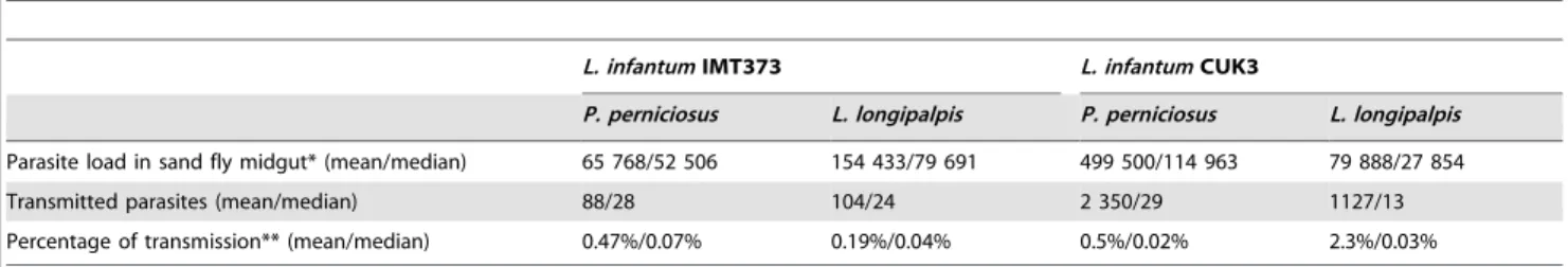
Out of 88P. perniciosus, females that bit mice, 62 (70.5%) were infected with CUK3; of these, 36 (58%) delivered parasites into the skin of the mice on days 10–14 post infective blood meal (Fig. 1a).
Out of 114 bitingL. longipalpisfemales, 86 (75.5%) were infected and 56 (65% of those infected) inoculated parasites into the mice on days 7–14 post infective blood meal (Fig. 1b).
Despite the fact that the intensity of infection was significantly higher inP. perniciosus(p,0.01), the percentage of transmission and number of inoculated parasites was comparable for both vectors. The parasite load delivered byP. perniciosusandL. longipalpisin the skin of mice ranged between 16 and 4.196104and between 4 and 1.116104, respectively. The average number of CUK3 parasites inoculated into the skin of mice and the percentages of transmission are summarized in Table 1.
InL. longipalpis, the feeding time was positively correlated with the number of CUK3 parasites delivered into host skin (p,0.05), while inP. perniciosusfemales no such correlation was observed. On the other hand, there was a significant correlation between the pre-feeding load inside both sand fly species’ midguts and the number of parasites transmitted (p= 0.0178 forL. longipalpis and
p,0.001 forP. perniciosus).
Transmission of the viscerotropic strain IMT373
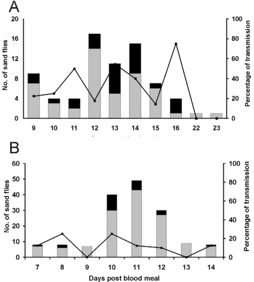
Out of 101P. pernicious females that bit mice, 73 (72%) were infected with IMT373, and of these 24 (33%) transmitted parasites into the mice’s skin.Leishmaniatransmission occurred between days 9 and 16 post infective bloodmeal (Fig. 2a). From 190 bitingL. longipalpis females, 159 (84%) were infected and 23 (14.5% on infected ones) inoculated parasites into the mice between days 7 and 14 post blood meal (Fig. 2b).
In contrast to above, the intensity of infection was significantly higher in L. longipalpis (p,0.001), but the transmission rate (i.e. percentage of transmitting females) and the number of parasites transmitted were significantly higher inP. perniciosus(p,0.01).
The number of parasites transmitted by P. perniciosus and
L. longipalpis ranged from 8 to 513 and between 7 and 1240 promastigotes, respectively. The median number of IMT373 transmitted is summarized in Table 1.
For both sand fly species, there was no correlation between feeding time and the number of IMT373 parasites in each female (p= 0.1594), or between the time to take a blood meal and the number of parasites transmitted (p= 0.6666). Moreover, no correlation was observed between the pre-feeding load in each sand fly species and the number ofLeishmaniadelivered (p= 0.1340 forP. perniciosus;p= 0.6473 forL. longipalpis).
Biting sites and feeding time of transmitting females
For all Leishmania-sand fly combinations, ears were the preferential biting place for sand flies transmitting the parasites, followed by the paws and tail. A few specimens that fed in the nose and eyes were also able to transmit parasites.
Table 2 summarizes the feeding times for both sand fly species:
L. longipalpistransmitting IMT373 completed their bloodmeals in
Table 1.Pre-feeding and transmitted parasite load forL. infantumstrains by both sand fly species.
L. infantumIMT373 L. infantumCUK3
P. perniciosus L. longipalpis P. perniciosus L. longipalpis
Parasite load in sand fly midgut* (mean/median) 65 768/52 506 154 433/79 691 499 500/114 963 79 888/27 854
Transmitted parasites (mean/median) 88/28 104/24 2 350/29 1127/13
Percentage of transmission** (mean/median) 0.47%/0.07% 0.19%/0.04% 0.5%/0.02% 2.3%/0.03%
*Parasite load was calculated as a sum of midgut parasites plus those transmitted by bite. **Percentage of parasite load transmitted by bite.
doi:10.1371/journal.pntd.0001181.t001
Author Summary
Leishmaniasis is a disease caused by protozoan parasites which are transmitted through the bites of infected insects called sand flies. The World Health Organization has estimated that leishmaniases cause 1.6 million new cases annually, of which an estimated 1.1 million are cutaneous or mucocutaneous, and 500,000 are visceral, the most severe form of the disease and fatal if left untreated. The development of a more natural model is crucial for the evaluation of new drugs or vaccine candidates against leishmaniases. The main aim of this study was to quantify the number ofLeishmania infantum parasites transmitted by a single sand fly female into the skin of a vertebrate host (mouse). TwoL. infantumstrains, viscerotropic IMT373 and dermotropic CUK3, were compared in two natural sand fly vectors: Phlebotomus perniciosus and Lutzomyia longipalpis. We found that the parasite number transmit-ted by individual sand flies ranged from 4 up to 4.196104.
times ranging from 2 to 27 minutes, while those transmitting CUK3 parasites needed between 3 to 55 minutes. The maximum and minimum feeding times forP. perniciosus transmitting CUK3 and IMT373 parasites ranged between 4–33 and 1–32 minutes, respectively. Infected sand flies transmitting CUK3 needed more time to feed than those that were infected but non-transmitting, while no differences in feeding time were observed between transmitting and non-transmitting females with IMT373 parasites.
Discussion
For the first time, we have quantified the number of parasites belonging to L. infantum dermotropic and viscerotropic strains transmitted to the dermis of experimental mice by individual sand fly females. The only previous attempt to calculate the number of transmittedL. infantumparasites was performed just recently [15], with the average number of promastigotes inoculated by 63 L. longipalpisinto culture medium through a chicken membrane skin
being 457 parasites, with 95% (431 promastigotes) of these corre-sponding to metacyclic parasites. However, these results do not allow us to take into consideration the individual variability of parasite transmission by a single specimen. The wide range of parasites inoculated per individual sand fly in our study (from 4 up to 4.196104 promastigotes) is in accordance to data previously obtained with other Leishmania-vector combinations [14,17], although the approach using microcapillaries as artificial feeding systems [17] could have interfered with the normal sand fly feeding behaviour.
In our study,Phlebotomus perniciosus harboured moreL. infantum
dermotropic parasites of the CUK3 strain, while inL. longipalpis
the intensity of infection was higher for the viscerotropic strain IMT373. However, in both sand fly species the parasite load transmitted was higher for the strain with dermal tropism. All but one sand fly female infected by IMT373 strain transmitted less than 600 promastigotes, the exception being aL. longipalpisfemale that inoculated 1240 parasites. On the other hand, 29% of L.
Figure 1. Transmission of Leishmania infantum CUK3. Percentage transmission of CUK3 strain by experimentally infected Phlebotomus perniciosus(A) andLutzomyia longipalpis(B). Black bars, infected females that transmitted by bite; grey bars, infected females that did not transmit; line, percentage of females that transmitted parasites.
longipalpisand 14% ofP. perniciosusinfected with the CUK3 strain transmitted more than 1000 parasites.
The majority of transmitting females inoculated less than 600 parasites. As most of these females were fully engorged by blood we may expect that their feeding pumps (the cibarial and pharyngeal pumps) and stomodeal valve were functioning
normally. On the other hand, in those transmitting more than 1000 parasites there was a significant correlation between the pre-feeding load and the number of parasites transmitted. We suggest that these females with high dose deliveries regurgitated parasites because of impaired stomodeal valve function [18]. This would be consistent with previous studies [19,20] which have demonstrated Figure 2. Transmission ofLeishmania infantumIMT373.Percentage transmission of IMT373 strain by experimentally infectedPhlebotomus perniciosus(A) andLutzomyia longipalpis(B). Black bars, infected females that transmitted by bite; grey bars, infected females that did not transmit; line, percentage of females that transmitted parasites.
doi:10.1371/journal.pntd.0001181.g002
Table 2.Average feeding time of infected and noninfectedP. perniciosusandL. longipalpis.
L. infantumIMT373 L. infantumCUK3
P. perniciosus L. longipalpis P. perniciosus L. longipalpis
Non-infected 10 11 8 10
Infected but without transmission 11 10 9 12
Infected and with transmission 12 10 12 18
an opened stomodeal valve due to the physical presence of a parasite plug and damage of the chitin layer of the valve by
Leishmaniachitinase.
Infected sand fly females, and especially those that transmit parasites, feed longer on hosts than non-transmitting ones do.
Lutzomyia longipalpisfemales transmitting dermotropic CUK3 strain parasites took an average of 1.5 times longer to complete a bloodmeal compared to specimens infected but not transmitting, and 1.8 times longer than uninfected females. Similarly,P. perniciosus
infected by CUK3 and IMT373 take 1.5 and 1.2 times more time for a blood meal. Most of the infected sand flies exposed to anaesthetized mice did not demonstrate increased probing, but rather remained feeding for longer periods until either they were fully or partially engorged. This is in agreement with data previously published on theL. longipalpis-L. mexicanacombination [21].
Although only one dermotropic and one viscerotropic L. infantum strains were evaluated, the significant variation in inoculum size between them allow us to hypothetise that the infectious dose delivered by vector sand flies may be an inherent character of eachLeishmaniastrain. Moreover, the infectious dose might be a determining factor in the outcome of Leishmania
infection. Local cutaneous lesions might result from a high-dose inoculum of dermotropic Leishmania resulting in a strong local immune response, whereas dissemination to internal organs might be the result of infected sand flies delivering a low number of parasites below the threshold required to produce/develop a localized and restraining immune response. This hypothesis corresponds with the data of Kimblin et al. [14] on theL. major-P. duboscqi combination. These authors evaluated the impact of inoculum size on infection outcome by comparing L. major
infections with high (56103) and low (16102) dose intradermally inoculated by needle in the ears of C57BL/6 mice, and observed the rapid development of large lesions in the ears of mice receiving the high-dose inoculum. In contrast, the low dose resulted in only minor pathology but a higher parasite titre during the chronic phase [14]. Nevertheless, it will be necessary to evaluate moreL. infantum strains with visceral and cutaneous tropism in order to determine if differences detected in our study were due to individual stock characteristics or if they are associated with parasite tropism in vertebrate hosts.
In conclusion, we have demonstrated that individual sand flies transmitLeishmaniaparasites in a wide dose range. However, the maximal natural dose found was still about 250 times lower than the challenge dose used for the L. donovani complex in most previous experimental works. This finding emphasizes the importance of determining the natural infective dose for the development of an accurate experimental model, which is crucial for the evaluation of new drugs and vaccine candidates against leishmaniasis.
Materials and Methods
Parasite strains
The viscerotropic Leishmania infantum strain IMT373 MON-1 (MCAN/PT/2005/IMT373) and the dermotropic L. infantum
strain CUK3 (ITOB/TR/2005/CUK3) were used in this study. CUK3 was isolated fromPhlebotomus tobbifrom a Cukurova focus of cutaneous leishmaniasis [6] while IMT373 was isolated from a dog with leishmaniasis and passaged through mice in order to keep its virulence [13,22]. Promastigotes (with less than 12 in vitro
passages since isolation) were cultured at+26uC in M199 medium (Sigma, USA) containing 10% heat-inactivated foetal calf serum (Gibco, USA), 50 mg/ml mikacin solution (Bristol-Myers Squibb, Czech Republic) and 1% sterile urine.
Sand flies and experimental infections
Lutzomyia longipalpis (originating from Jacobina, Brazil) and
Phlebotomus perniciosus (originating from Murcia, Spain) colonies were maintained in an insectary under standard conditions as described by Volf and Volfova [23]. Five to six-day old female flies (200 P. perniciosus and 150 L. longipalpisfemales per experiment, respectively) were fed on heat inactivated rabbit blood containing promastigotes (107parasites per ml of blood) through a chicken-skin membrane. Blood-engorged females were separated immedi-ately and maintained on a 50% sucrose diet in.70% relative humidity at+26uC.
One group of females was dissected to study the development and localization of infection in the sand fly midgut two and ten days post blood meal, i.e., during early and late stage infection, respectively. Individual midguts were placed into a drop of saline buffer, and parasite numbers were estimated under a light microscope at 200X and 400X magnifications by an experienced worker. Parasite loads were graded as previously described [24] into four categories: negative, 1–100, 100–1000, and .1000 parasites per gut. A second group of females from the same batch was used for transmission experiments and parasite quantification by Real-time PCR (see below). Nine and six independent experiments were performed with P. perniciosus-IMT373 and P. perniciosus-CUK3 combinations, respectively, while six and four artificial infections were done with L. longipalpis-IMT373 and L. longipalpis-CUK3 combinations.
Mice
One hundred and eight BALB/c mice (41 for P. perniciosus -IMT373, 28 mice forL. longipalpis-IMT373, 23 forP. perniciosus– CUK3 and 16 forL. longipalpis-CUK3 combinations) older than 8 weeks of age were purchased from AnLab (Czech Republic) and housed at Charles University, Prague, under stable climatic and dietary conditions. Experiments were approved by the institutional Ethical Committee and performed in accordance with national legislation for the care and use of animals for research purposes. Mice were anaesthetized intraperitoneally with ketamine (150 mg/ kg) and xylazine (15 mg/kg).
Transmission by bite and sample collection
Sand fly females were allowed to feed on whole body of anesthetised mice in a rectangular cage (20620 cm) for about one hour at various days post infective blood meal (7–14 days forL. longipalpisand 9–23 days forP. perniciosus). Each mouse was placed individually into a cage together with about 50P. perniciosusor 10
L. longipalpis females (the difference was due to the fact that L. longipalpiswere more aggressive and had higher feeding rate). Two people followed each experiment; one recorded biting place and feeding time while the second ensured that each sand fly female probed in different place and then collected engorged flies by an aspirator immediately after terminating their blood meal; the site of bite and time of feeding were recorded for each female. After exposure, mice were sacrificed, biting place was inspected under a stereoscope and excised. Both samples (skin biopsies and corresponding fed sand flies) were stored at 220uC until DNA extraction.
Real-time PCR (qPCR)
was performed in a Rotor-Gene 2000 from Corbett Research (St. Neots, UK) using the SYBR Green detection method (iQ SYBR Green Supermix, Bio-Rad, Hercules, CA). For adequate sensitiv-ity, kinetoplast DNA was chosen as the molecular target, with primers as previously described [25] (forward primer 59 -CTTTTCTGGTCCTCCGGGTAGG-39 and reverse primer 59-CCACCCGGCCCTATTTTACACCAA- 39). Two microliters of eluted DNA was used per individual reaction. PCR amplifica-tions were performed in duplicate wells using the condiamplifica-tions described previously [26]. Briefly, 3 min at 95uC followed by 45 cycles of: 10 s at 95uC, 10 s at 56uC, and 10 s at 72uC. Reaction specificities were checked for all samples by melting analysis. Quantitative results were expressed by interpolation with a standard curve included in each PCR run. Mass cultures of L. infantum promastigotes were used to construct a series of 10-fold dilutions ranging from 105to 1 parasite per PCR reaction. Diluted parasites were co-processed with mouse tissue or sand fly females for DNA extraction. DNA from uninfected sand flies and mice were used as a negative control.
For sand fly females transmitting promastigotes into mouse skin, the pre-feeding midgut load was calculated as the sum of parasites
in the midgut after feeding and the number of parasites transmitted.
Statistical analysis
Statistical analysis was performed using the software STATIS-TICA. For eachL. infantumstrain, a nonparametric Kruskal-Wallis test was used to compare: (i) the intensity of infection in P. perniciosus and L. longipalpis, and (ii) the number of parasites transmitted by each sand fly species into mice’s skin. Correlations between feeding time and the number of parasites (i) in each sand fly female and (ii) inoculated into the skin, as well as the correlation between pre-feeding load and the number of parasites transmitted, were determined by simple linear regression analysis. Differences were considered statistically significant forpvalues,0.05.
Author Contributions
Conceived and designed the experiments: CM VS JS JV PV. Performed the experiments: CM VS JS JV. Analyzed the data: CM JV PV. Wrote the paper: CM PV.
References
1. WHO (2010) Working to overcome the global impact of neglected tropical dis-eases. Available: http://www.who.int/neglected_diseases/2010report/NTD_2010 report_web.pdf. Accessed 2 November 2010.
2. Gradoni L, Gramiccia M (1994)Leishmania infantumtropism: strain genotype or host immune status? Parasitol Today 10: 264–267.
3. Campino L, Abranches P (2002) Cutaneous leishmaniasis. Unusual disease in Portugal? Acta Med Port 15: 387–390.
4. Garcı´a-Almagro D (2005) Cutaneous leishmaniasis. Actas Dermosifiliogr 96: 1–24.
5. Lima H, Rodrı´guez N, Feliciangeli M, Barrios M, Sosa A, et al. (2009) Cutaneous leishmaniasis due toLeishmania chagasi/Le. infantumin an endemic area of Guarico State, Venezuela. Trans R Soc Trop Med Hyg 103: 721–726. 6. Svobodova M, Alten B, Zidkova L, Dvorak V, Hlavackova J, et al. (2008)
Cutaneous leishmaniasis caused byLeishmania infantumtransmittedPhlebotomus tobbi.Int J Parasitol 39: 251–256.
7. Titus R, Ribeiro J (1988) Salivary gland lysates from the sand fly Lutzomyia longipalpis enhanceLeishmaniainfectivity. Science 239: 1306–1308.
8. Rogers M, Chance M, Bates P (2002) The role of promastigote secretory gel in the origin and transmission of the infective stage ofLeishmania mexicanaby the sandflyLutzomyia longipalpis.Parasitology 124: 495–507.
9. Rohousova I, Volf P (2006) Sandfly saliva: effects on host immune response and Leishmaniatransmission. Folia Parasitol 53: 161–171.
10. Melby P, Yang Y, Cheng J, Zhao W (1998) Regional differences in the cellular immune response to experimental cutaneous or visceral infection withLeishmania donovani. Infect Immun 66: 18–27.
11. Rola˜o N, Melo C, Campino L (2004) Influence of the inoculation route in BALB/c mice infected byLeishmania infantum. Acta Trop 90: 123–126. 12. Ahmed S, Colmenares M, Soong L, Goldsmith-Pestana K, Munstermann L,
et al. (2003) Intradermal infection model for pathogenesis and vaccine studies of murine visceral leishmaniasis. Infect Immun 71: 401–410.
13. Maia C, Rola˜o N, Nunes M, Campino L (2009) Infectivity ofLeishmania infantum treated with amphotericin B plusPhlebotomussalivary gland in BALB/c mice. Int J Integr Biol 6: 105–111.
14. Kimblin N, Peters N, Debrabant A, Secundino N, Egen J, et al. (2008) Quantification of the infectious dose ofLeishmania majortransmitted to the skin by single sand flies. Proc Natl Acad Sci USA 105: 10125–10130.
15. Rogers M, Corware K, Mu¨ller I, Bates P (2010)Leishmania infantum proteopho-sphoglycans regurgitated by the bite of its natural sand fly vector,Lutzomyia longipalpis, promote parasite establishment in mouse skin and skin-distant tissues. Microbes Infect 12: 875–879.
16. Killick-Kendrick R (1990) Phlebotomine vectors of the leishmaniases: a review. Med Vet Entomol 4: 1–24.
17. Warburg A, Schlein Y (1986) The effect of post-bloodmeal nutrition of Phlebotomus papatasion the transmission ofLeishmania major. Am J Trop Med Hyg 35: 926–930.
18. Schlein Y (1993)Leishmania and sandflies: interactions in the life cycle and transmission. Parasitol Today 9: 255–258.
19. Volf P, Hajmova M, Sadlova J, Votypka J (2004) Blocked stomodeal valve of the insect vector: Similar mechanism of transmission in two trypanosomatid models. Int J Parasitol 34: 1221–1227.
20. Rogers M, Hajmova´ M, Joshi M, Sadlova J, Dwyer D, et al. (2008)Leishmania chitinase facilitates colonization of sand fly vectors and enhances transmission to mice. Cell Microbiol 10: 1363–1372.
21. Rogers M, Bates P (2007)Leishmaniamanipulation of sand fly feeding behaviour results in enhanced transmission. PLoS Pathog 3(6): e91. doi:10.1371/journal.-ppat.0030091.
22. Maia C, Nunes M, Cristo´va˜o J, Campino L (2010) Experimental canine leishmaniasis: clinical, parasitological and serological follow-up. Acta Trop 116: 193–199.
23. Volf P, Volfova V (2011) Establishment and maintenance of sand fly colonies. J Vector Ecol. In press.
24. Cihakova J, Volf P (1997) Development of differentLeishmania majorstrains in the vector sandfliesPhlebotomus papatasiandP. duboscqi. Annals of tropical Medicine and Parasitology 91: 267–279.
25. Mary C, Faraut F, Lascombe L, Dumon H (2004) Quantification ofLeishmania infantumDNA by a realtime PCR assay with high sensitivity. J Clin Microbiol 42: 5249–5255.