75
NHL Journal of Medical Sciences/July 2013/Vol.2/Issue 2Research article
Sexing the human skull using the mastoid process.
Swati Shah*,Pratik Patel**
*Asst. Professor, Department of Anatomy,**Professor & head,Forensic medicine Smt. NHL Municipal Medical College, Ahmedabad
ABSTRACT
Sex determination of human or human skeletal remains is considered an initial step in its identification. In cases of fragmented or mutilated body, it is difficult to identify the body. This skillful process is carried out by forensic and anatomy experts.
In cases where intact skull is not found, mastoid play a vital role in sex determination as it is the most dimorphic bone of skull. The mastoid region, a fragmentary piece of skull is ideal for sex determination as it is resistant to damage due to its anatomical position at the base of skull and its toughness.
The skull measurements vary significantly in different ethnic groups of the world and number of research/studies are too few for Indian population. In present study, 100 adult human skulls of Gujarat population were studied to determine accuracy of mastoid process in sex determination. A quantitative blind study of the dimensions of the sides of the mastoid triangle was carried out. Results show significant craniometric difference between male and female mastoid triangles.
KEY WORDS
Asterion, craniometrical points, heron’s formula, mastoid triangle, mastoid, porion, sex determination.
INTRODUCTION
Identify means “determination of the individuality”. In number of civil and criminal matters identification of the individual, either living or dead, is required. The anatomical and medical features are the two major aspects to establish the identity. The establishment of identity is required from fresh intact corpses, decomposed corpses, mutilated and dismembered corpses or skeletalised material.1 Sex determination of human or human skeletal remains is considered vital step in identification and is crucial for further analysis.
When performing a forensic anthropological analysis for human identity, sex estimation of the individual is
one of the first and most important steps. In living, identification is possible with absolute fixation of the individuality but in case of advanced decomposed, mutilated or skeletalised body it is difficult to establish the identity of the dead body either partially or completely. In such circumstances the identification of human skeletal remains a critical issue. In present forensic scenario, dismemberment or mutilation of body has become the frequent method to conceal the identity of victim. In such instances the pelvis is considered the best bone to determine the sex of an individual2. When the pelvis is unavailable the skull is also widely considered the best indicator of sex. Krogman3 states that skull is the most dimorphic and easily sexed portion of skeleton after pelvis, providing up to 92% reliability. Bass4 states that the skull probably is the second best area of the skeleton to use for determining sex. Bass4, Byers5and Pickering and Bachman6 presented the idea that skull is the second best choice to estimate the sex of the dead body.
76
NHL Journal of Medical Sciences/July 2013/Vol.2/Issue 2sex in fragmented skull bone. Nagaoka T method is employed by some experts using two parameters on both sides of skull i.e. height and width of the mastoid7. This method has its own merits and significance. Paiva & Segre (2003) after studying a sample of 60 skulls introduced an easy technique for sex determination starting from the temporal bone. The technique is based on the triangular area calculation obtained between the points porion, mastoidale and asterion, measured from xerographic copy of skulls8. This technique has significant degree of predictability with a small observational error. Multiple measurements instead of single measurement of mastoid have increased its significant value.
AIMS AND OBJECTIVES
The objective of this study is to provide a new method for determination of sex of fragmentary human skeletal remains of natives of Gujarat state, India, using the three craniometric points of mastoid process i.e. the mastoidale, porion and asterion.
MATERIAL AND METHOD
100 dry skulls of known sex, 50 male and 50 female, from the department museum of anatomy of various medical colleges from Gujarat were analyzed for the present study after taking approval of Institutional Review Board of Smt NHL Municipal Medical College and written permission from the concerned department/ institute. Adult skulls of mature individuals, 18 or more years old, without destruction of the mastoid bone in the region of the craniometrical points, were chosen for the study. The skulls that present evidence of injury/fracture or deformity were excluded from this study. All the skulls were confirmed of the individual of native of Gujarat state, India from the concerned department record.
Three points on the mastoid portion on either side, porion [upper most lateral point of external acoustic meatus pore], Mastoidale [the most inferior point of mastoid process], Asterion [the meeting point of three posterior skull sutures i.e. lambdoid, occipitomastoid and parietomastoid] were selected as area of our study. These craniometrical points were marked by a single investigator. A triangle was prepared/drawn using these three points on skull bone
on both sides. Porion, asterion and mastoidale were referred as Po, As and Ma respectively. Measurements of the dimensions of the sides of the mastoid triangle were carried out using vernier caliper. Any of these three points if found damaged, the skull was excluded from the study.
The area (mm2) of the demarcated triangle for each side of the skull (right (R) and left (L) sides) was determined by Heron’s formula9
and the total value (T) of these measures of both sides was calculated as per method described by De Paiva and Segre8, Helmuth10, Demoulin11.
DATA MANAGEMENT AND ANALYSIS
Using appropriate analytical software, the data was analyzed. The descriptive statistics of the lineal dimensions and the mastoid triangle area were calculated.
DISCUSSION & CONCLUSION
The studies for sex determination of the bones are based on the dimorphism that is present in the majority of human bones. The present study was carried out using method applied by other researchers, based on craniometric dimensions of mastoid part of the skull.
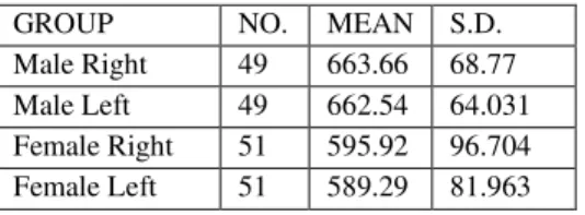
Total 100 dry skulls were measured. Out of these, 49 were male skulls and 51 were female skulls. The mean of Heron’s triangle was as follows.
Table 1: Male and female right and left side measurements
GROUP NO. MEAN S.D.
Male Right 49 663.66 68.77
Male Left 49 662.54 64.031
Female Right 51 595.92 96.704 Female Left 51 589.29 81.963
77
NHL Journal of Medical Sciences/July 2013/Vol.2/Issue 278
NHL Journal of Medical Sciences/July 2013/Vol.2/Issue 2Table 2: Comparison: male and female right and left side using Student t-test.
’t’Value Degre e of freedo m
P -value Level of Significance
Male Right side Vs Female Right side
4.023 98 <0.0001 Extremely significant
Male Left side Vs female Left side
4.966 98 <0.0001 Extremely significant
As per Table no. 2, it was found that the student t-test was extremely significant when male right side was compared with female right side as well as when male left side was compared with female left side indicating that the mastoid process measurements can be used as one of the bone determinant of sex.
As per the ANOVA test, for all four measurements, the test was statistically extremely significant. On comparing the male right and female right side by student t-test, the test was found to be extremely significant (P value < 0.0001).
Similarly, on comparing the male left and female left side by student t-test, the test was found to be extremely significant (P value < 0.0001) Area of triangle in male varies between -`600-700 mm. .in male and in female between 500-600 mm. in majority of cases in my study.
According to the study by Paiva LA and Segre12, male tringle area was significantly greater than female triangle area. Values calculated according to their study mean values was : in female : 570.9 mm2 and 575.1 mm2 in right and left sides. In male : 723.9 mm2 and 731.2 mm2 in right and left sides.
In our study male right :663.66mm2 ,male left : 662.54 mm2, female right : 595.92 mm2, female left : 589.29mm2 was found. Results of our study shows following P value.
MRT VS FRT P <0.001 MRT VS FLT P <0.001
FRT VS MLT P <0.001 MLT VS FLT P <0.001
MRT VS FRT unpaired student t-test P < 0.0001, extremely significant
‘t’ = 4.023, d.f. 98
MLT VS FLT unpaired student t-test P < 0.0001, extremely significant
‘t’ = 4.966, d.f. 98
As both the graph shows, when the measurement values are plotted in ascending order, the Heron’s triangle measurements are found more in males as compared to females.
The means of mastoid dimensions and mastoid triangular area in male are significantly larger than those of the female (p < 0.01). In the group of male skulls, we found the values in Table 1. In the group of female skulls we found the values in Table 2.
REFERENCES
1.Pekka Saukko, Bernard Knight, Knight’s Forensic pathology, 3rd edition, the establishment of identity of human remains: 98-99.
2.Byers SN, Introduction to forensic anthropology: a textbook, 3rd edition, 2002,pp176-186
3.W.M.Krogman, Mehmet Yasar Iscan, The human skeleton in Forensic medicine, 2nd edition, 191-194.
4.Bass WM : Human osteology : A Laboratory and field manual, 5th edn Columbia , MO : 2005
5.Byers S.N.Introduction to forensic anthropology , a textbook .Boston, A : Alyn and Bacon publishers,2002
6. Pickering RB, Bachman DC. The use of forensic anthropology. Boca Raton , FL : CRC Press,1997
7.Nagaoka T, Shizushima A, Sawada J, et al. Sex determination using mastoid process measurements: standards for Japanese human skeletons of the medieval and early modern periods. Anthropology Sic 2008;116:105-3
8.Paiva LA, and Segre M: sexing the human skulls through the mastoid process.Rev. Hosp. clin. Fac. Med.2003 Jan-Feb;58(1):15-20
9.Heron’s formula triangle area calculator
10. Helmuth H, Einige Mabe des processus mastoideus beim menschen und seine bedeutung für die geschlechtsbetimmung. Z Morphol Anthropology 1968; 60: 75-84.
11. Demoulin F, Importance de certaines mesures crâniennes (en particulier de la longueur sagittale de la mastoide) dans la determination sexuelle des crânes. Bull et Mém de la Soc
D’Anthropol 1972; 9: 259 – 64.