https://doi.org/10.1590/0004-282X20170082
ARTICLE
Middle cerebral artery aneurysms: aneurysm
angiographic morphology and its relation to
pre-operative and intra-operative rupture
Aneurismas da artéria cerebral média: morfologia angiográfica dos aneurismas e sua
relação com ruptura pré-operatória e intra-operatória
Iracema Araújo Estevão1, Bruno Camporeze1, Antonio Santos de Araujo Jr2, Breno Nery3,
Ápio Claudio Martins Antunes4,5, Timothy R. Smith6, Paulo Henrique Pires de Aguiar3,5,7
1Universidade São Francisco, Bragança Paulista SP, Brasil;
2Araujo e Fazzito Neurocirurgião e Neurologistas Associados, Departamento de Neurocirurgia, São Paulo SP, Brasil; 3Hospital Santa Paula, Departamento de Neurocirurgia, São Paulo SP, Brasil;
4Hospital de Clínicas de Porto Alegre, Departamento de Neurocirurgia, Porto Alegre RS, Brasil;
5Universidade Federal do Rio Grande do Sul, Departamento de Neurocirurgia, Divisão de Pós-Graduação, Porto Alegre, Brasil; 6Harvard University, Department of Neurosurgery, Massachusetts, USA;
7Pontifícia Universidade Católica de São Paulo, Departamento de Neurologia, Sorocaba SP, Brasil.
Correspondence: Iracema Araújo Estevão; Rua Ercílio Baratella, 334, Edifício Mônica; 12916-370 Bragança Paulista SP, Brasil; E-mail: iaestevão@gmail.com
Conflict of interest: There is no conlict of interest to declare. Received 20 January 2017; Accepted 30 March 2017.
ABSTRACT
Objective: Correlate the middle cerebral artery bifurcation aneurysm morphology with the pre-operative and intra-operative risk of
rupture. Methods: Forty patients with 46 middle cerebral artery bifurcation aneurysms were treated microsurgically by the same surgeon. Aneurysms were classiied according to shape and the Fisher test was applied to analyze the effect of morphology on the pre-operative and intra-operative rupture. Results: Pre-operative and intra-operative ruptures were observed in 8/46 patients (17.4%) and 14/46 patients (30.4%) respectively. Thirty-two cases (69.6%) had no symptoms postoperatively, modiied Rankin score (MRS) of 0; 6.5% had MRS of 1 (no signiicant disability); 13% had MRS of 2 (slight disability); 4.3% had moderately severe disability (MRS of 4); and there were 3 deaths (6.5%) post-operatively. The morphology was not directly related to the rupture rate. Conclusion: In general, ruptures are not affected by the morphology or the studied variables. Larger series are needed to validate these outcomes.
Keywords: aneurysm; subarachnoid hemorrhage; middle cerebral artery.
RESUMO
Objetivo: Correlacionar a morfologia do aneurisma da bifurcação da artéria cerebral média com o risco de ruptura pré-operatória e
intra-operatória. Métodos: 40 pacientes com 46 aneurismas de bifurcação da artéria cerebral média receberam tratamento microcirúrgico pelo mesmo cirurgião. Os aneurismas foram classiicados de acordo com a morfologia e o teste de Fisher foi aplicado para analisar o efeito da morfologia sobre a ruptura pré-operatória e intra-operatória. Resultados: As rupturas pré e intra-operatória foram observadas em 8/46 pacientes (17,4%) e 14/46 (30,4%) respectivamente. Trinta e dois casos (69,6%) não apresentaram sintomas pós-operatórios, pontuação de Rankin modiicada (MRS) de 0, 6,5% tinham MRS de 1 (sem incapacidade signiicativa), 13% tinham MRS de 2 (leve incapacidade), 4,3% moderadamente grave (MRS de 4) e houve 3 óbitos (6,5%) durante o pós-operatório. A morfologia não estava diretamente relacionada à taxa de ruptura pré-operatória ou intra-operatória. Conclusão: Em geral, as rupturas não são afetadas pela morfologia ou pelas variáveis estudadas. São necessárias séries maiores para validar esses resultados.
Palavras-chave: aneurisma; hemorragia subaracnoidea; artéria cerebral média.
Middle cerebral artery aneurysms are some of the most common vasculopathies in the anterior cerebral circula-tion, with an overall incidence of 18% to 20% of all aneurysms encountered1. Aneurysms in this location are typically
com-plex, multi-lobed, and incorporate eloquent vascular branches;
incorporation, and whether they are ruptured or not. he aneurysm neck is an important aspect when considering the endovascular treatment of the aneurysm. As opposed to endo-vascular therapies, neck-dome proportion is not as important when microsurgery is considered, since adequate neck dissec-tion and sometimes aneurysm dome dissecdissec-tion are pivotal in determining complete aneurysm obliteration and avoid-ance of eloquent incorporated cortical branches. In order to obtain complete comprehension of the relationship between the nearby vascular branches and perforating arteries, surgical dissection of the neck and dome are sometimes needed when microsurgical clipping is considered; this is especially true when we are dealing with middle cerebral artery bifurcation
aneurysm. he authors propose a simple morphological clas
-siication of middle cerebral artery bifurcation aneurysms in order to study the relationship of the aneurysm’s morphology and its tendency to rupture pre-operatively or intra-operatively.
he afore-mentioned morphological characteristics are partic
-ularly addressed by the authors, especially regarding the cur-rent technical repertoire and the surgeons’ expertise.
METHODS
From January 1998 to July 2015, 40 patients with 46 bifur-cation aneurysms of the middle cerebral artery were surgi-cally clipped by the senior author. Pre-operative historical and radiological records were reviewed in detail and mor-phological aspects charted. Middle cerebral artery
bifurca-tion aneurysms were classiied according to shape into glo
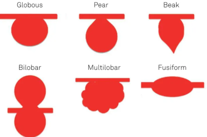
-boid, pear-shaped, bilobar, beak-shaped, multilobar, and fusiform (see classiication model in Figure 1).
here were 27 females and 13 male patients. Average age was 50.20 years old (min = 15 years old, max = 75 years old).
he same surgeon and surgical team operated all the aneu
-rysms. Categorical and scalar variables were described for each patient and Fisher’s test was applied to analyze the efect of the morphology on the pre-operative and intra-operative rupture rate. Fisher’s test was also applied to verify the degree of association between the interest categorical variables (right side, left side, gender, presence of associated aneurysms other than middle cerebral artery bifurcation aneurysms, anatomi-cal features such as an M1 segment of the middle cerebral artery larger than 3 cm, presence of trifurcated middle cerebral artery and aspect ratio higher than 1.5) and the rupture vari-ables. he Mann-Whitney test was applied to verify the degree of association between the scalar variables (age and size of the aneurysm) and the rupture variables.
Classification
We classiied middle cerebral artery aneurysms based on their shapes into globoid, pear-shaped, beak-shaped, bilobar, multilobar and associated with ibromuscular dysplasia ( fusiform), as shown in Figure 1.
here were 16 beak-shaped aneurysms (34.8% of all aneu
-rysms) with one pre-operative rupture in this category. Pear-shaped aneurysms were also seen in 14 of the aneurysms
(30.4%), with one ruptured aneurysm at presentation. A glo
-bous shape was encountered in nine cases (19.60% of the aneurysms), with one ruptured aneurysm at presentation (11.1%). A bilobar shape was seen in two cases (4.3%) with one ruptured aneurysm at presentation (50%). A multilobar shape was seen in three cases (6.5%) with two ruptured aneu-rysms at presentation (66.6%). A fusiform aneurysm was seen
in two cases (4.3%) and none of those were ruptured at pre
-sentation. All the categorical variables evaluated during data collection are described in Table 1. Examples of the morphol-ogy can be seen in Figures 2 and 3 (A to E).
Middle cerebral artery segment anatomic variations A long M1 segment was observed in 26%, M1 trifurcation was observed in approximately 11%, and M1 bifurcation was observed in 89% of the angiographies.
Associated aneurysms
Seven cases had one associated aneurysm other than the bifurcation of the middle cerebral artery on the right side and two patients had two associated aneurysms on the right side of the head. Four patients had one associated aneurysm on the left side other than the bifurcation of the middle cerebral artery (Table 1). Only one aneurysm was distal to the bifurca-tion of the middle cerebral artery. All the ruptures were related to the aneurysm of the bifurcation of the middle cerebral artery. Spearman’s analysis was done to verify the degree of relation-ship between the variables of interest (Table 2). he relationrelation-ship between the presence of associated aneurysms and middle cerebral artery bifurcation aneurysms, as well as the relation-ship between left side and right side of the associated aneu-rysms are weak; that is, nothing makes us believe that there is a
higher probability of inding an associated rupture due to previ
-ously-ruptured aneurysms in our series (Table 2). Figure 1. Classiication model of aneurysms according to shape into globoid, pear -shaped, bilobar, beak-shaped, multilobar and fusiform.
Globous Pear
Bilobar Multilobar Fusiform
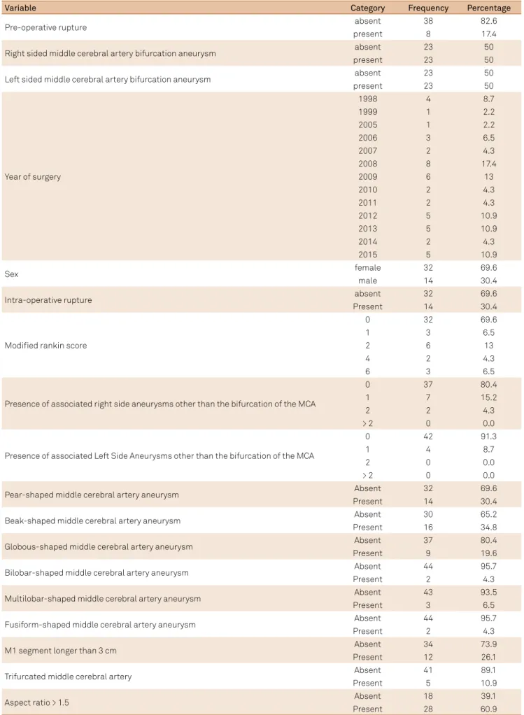
Table 1. Description of all the categorical variables evaluated during data collection.
Variable Category Frequency Percentage
Pre-operative rupture absent 38 82.6
present 8 17.4
Right sided middle cerebral artery bifurcation aneurysm absent 23 50
present 23 50
Left sided middle cerebral artery bifurcation aneurysm absent 23 50
present 23 50
Year of surgery
1998 4 8.7
1999 1 2.2
2005 1 2.2
2006 3 6.5
2007 2 4.3
2008 8 17.4
2009 6 13
2010 2 4.3
2011 2 4.3
2012 5 10.9
2013 5 10.9
2014 2 4.3
2015 5 10.9
Sex female 32 69.6
male 14 30.4
Intra-operative rupture absent 32 69.6
Present 14 30.4
Modiied rankin score
0 32 69.6
1 3 6.5
2 6 13
4 2 4.3
6 3 6.5
Presence of associated right side aneurysms other than the bifurcation of the MCA
0 37 80.4
1 7 15.2
2 2 4.3
> 2 0 0.0
Presence of associated Left Side Aneurysms other than the bifurcation of the MCA
0 42 91.3
1 4 8.7
2 0 0.0
> 2 0 0.0
Pear-shaped middle cerebral artery aneurysm Absent 32 69.6
Present 14 30.4
Beak-shaped middle cerebral artery aneurysm Absent 30 65.2
Present 16 34.8
Globous-shaped middle cerebral artery aneurysm Absent 37 80.4
Present 9 19.6
Bilobar-shaped middle cerebral artery aneurysm Absent 44 95.7
Present 2 4.3
Multilobar-shaped middle cerebral artery aneurysm Absent 43 93.5
Present 3 6.5
Fusiform-shaped middle cerebral artery aneurysm Absent 44 95.7
Present 2 4.3
M1 segment longer than 3 cm Absent 34 73.9
Present 12 26.1
Trifurcated middle cerebral artery Absent 41 89.1
Present 5 10.9
Aspect ratio > 1.5 Absent 18 39.1
Present 28 60.9
RESULTS
he average aneurysm size was 6.59 mm. Pre-operative and intra-operative ruptures were observed in 8/46 patients (17.4%) and 14/46 patients (30.4%) respectively. he average age was 50.2 years old (15–75), with a median age of 51.5 years of age
(Table 3). hirty-two cases (69.6%) had no symptoms postop
-eratively (modiied Rankin score (MRS) of 0, 6.5% had an MRS
of 1 (no signiicant disability), 13% had MRS of 2 (slight disabil -ity), 4.30% had moderately severe disability (MRS of 4) and there were 3 deaths (6.5%) post-operatively. Fourteen aneurysms
were classiied as pear shaped (30.4%), 16 aneurysms were clas
-siied as beak shaped (34.8%), 9 were considered to be globous shaped (19.6%), 2 patients (4.3%) had bilobar shaped aneu -rysms, 3 patients (6.5%) had multilobar shaped aneurysms and 2 (4.3%) had fusiform aneurysms (Table 1).
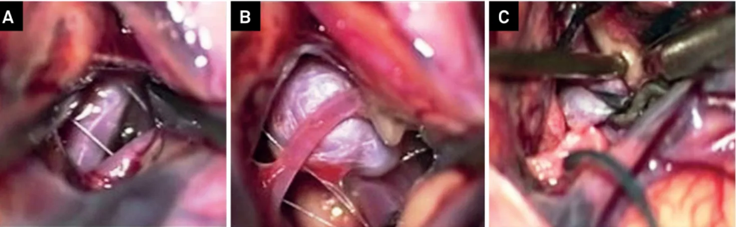
Figure 2. Intra-operative vision and clipping of middle cerebral artery aneurysms.
A
B
C
Figure 3. A. CT scan axial section of a patient with middle cerebral artery aneurysm. B. Angiogram of a patient with MCA aneurysm in M2 portion. C. Identiication of the aneurysm. D. E. F. Intra-operative vision of clipping of a middle cerebral artery aneurysm.
A
B
C
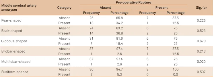
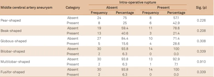
Fisher’s test was applied to analyze the efect of mor -phology on the pre-operative and intra-operative rupture (Tables 4 and 5, respectively). Statistical analysis showed that the morphology of the aneurysm does not afect the rate of pre-operative or intra-operative rupture. Fisher’s test was also applied to verify the degree of association between the categorical variables of interest (right side, left side, gender, presence of associated aneurysms other than middle cere-bral artery bifurcation aneurysms, anatomical features such as an M1 segment of the middle cerebral artery larger than 3 cm, the presence of a trifurcated middle cerebral artery and aspect ratio higher than 1.5) and the rupture variables
(Tables 6 and 7). he Mann-Whitney test was applied to ver
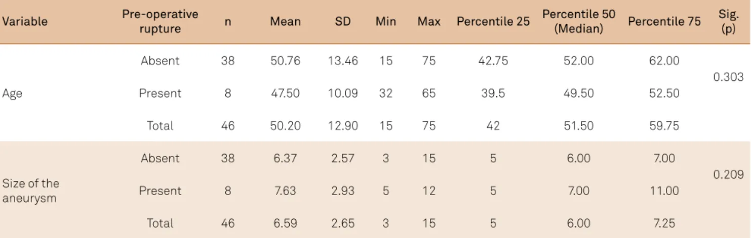
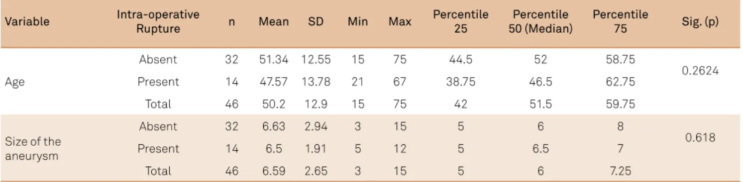
-ify the degree of association between the scalar variables (age and size of the aneurysm) and the rupture variables (Tables 8 and 9). According to the analyses, none of the
vari-ables seemed to afect the rate of pre-operative or intra-oper
-ative rupture. Spearman’s correlation analysis was applied to verify the degree of relationship between the morphology variables and the above-mentioned categorical variables of interest (Table 10). According to this analysis the relationship between those variables seemed to be weak, showing that they do not represent bias when correlating the morphology of the aneurysms and the risk of rupture.
Table 2. Spearman analysis was done to verify the degree of relationship between the variables of interest.
Variable Statistics
Right sided bifurcation middle cerebral artery
aneurysm
Left sided bifurcation middle cerebral artery
aneurysm
Presence of associated left side aneurysms other
than the bifurcation of the MCA
Left sided middle cerebral artery bifurcation aneurysm
Coef. Correl. (r) -1.000 — —
Sig. (p) < 0.001 — —
n 46 — —
Right sided middle cerebral artery bifurcation aneurysm
Coef. Correl. (r) +0.031 -0.031 —
Sig. (p) 0.839 0.839 —
n 46 46 —
Presence of associated left sided aneurysms other than the bifurcation of the MCA
Coef. Correl. (r) 0.000 0.000 -0.152
Sig. (p) > 0.999 > 0.999 0.315
n 46 46 46
MCA: Middle Cerebral Artery ;Coef. Correl.: Correlation Coeffcient ; Sig: statistical signiicance.
Table 3. Scalar variables (age and size of the operated aneurysms).
Variable n Min Max Average Standard
deviation Percentile 25
Percentile 50
(Median) Percentile 75
Age 46 15 75 50.2 12.9 42 51.5 59.75
Size of the aneurysm 46 3 15 6.59 2.65 5 6 7.25
Table 4. Fisher’s test to verify the association between the morphology of the aneurysms and the pre-operative rate of rupture.
Middle cerebral artery
aneurysm Category
Pre-operative Rupture
Sig. (p)
Absent Present
Frequency Percentage Frequency Percentage
Pear-shaped Absent 25 65.8 7 87.5 0.225
Present 13 34.2 1 12.5
Beak-shaped Absent 24 63.2 6 75 0.523
Present 14 36.8 2 25
Globous-shaped Absent 31 81.6 6 75 0.670
Present 7 18.4 2 25
Bilobar-shaped Absent 37 97.4 7 87.5 0.213
Present 1 2.6 1 12.5
Multilobar-shaped Absent 37 97.4 6 75 0.020
Present 1 2.6 2 25
Fusiform-shaped Absent 36 94.7 8 100 0.507
Table 5. Fisher’s test to verify the association between the morphology of the aneurysms and the intra-operative rate of rupture.
Middle cerebral artery aneurysm Category
Intra-operative rupture
Sig. (p)
Absent Present
Frequency Percentage Frequency Percentage
Pear-shaped Absent 24 75 8 57.1 0.226
Present 8 25 6 42.9
Beak-shaped Absent 19 59.4 11 78.6 0.208
Present 13 40.6 3 21.4
Globous-shaped Absent 27 84.4 10 71.4 0.308
Present 5 15.6 4 28.6
Bilobar-shaped Absent 30 93.8 14 100 0.339
Present 2 6.3 0 0.0
Multilobar-shaped Absent 30 93.8 13 92.9 0.910
Present 2 6.3 1 7.1
Fusifor-shaped Absent 30 93.8 14 100 0.339
Present 2 6.3 0 0.0
Table 6. Fisher’s test to verify the association between the variables of interest and pre-operative rupture.
Variable Category
Pre-operative Rupture
Sig. (p)
Absent Present
Frequency Percentage Frequency Percentage
Right sided middle cerebral artery bifurcation aneurysm
Absent 19 50 4 50
> 0.999
Present 19 50 4 50
Left sided middle cerebral artery bifurcation aneurysm
Absent 19 50 4 50
> 0.999
Present 19 50 4 50
Sex
Female 26 68.4 6 75
0.713
Male 12 31.6 2 25
Presence of associated right side aneurysms other than the bifurcation of the MCA
0 29 76.3 8 100
0.308
1 7 18.4 0 0.0
2 2 5.3 0 0.0
> 2 0 0,.0 0 0.0
Presence of associated left side aneurysms other than the bifurcation of the MCA
0 34 89.5 8 100
0.337
1 4 10.5 0 0.0
2 0 0.0 0 0.0
> 2 0 0.0 0 0.0
M1 segment > 3 cm
Absent 29 76.3 5 62.5
0.419
Present 9 23.7 3 37.5
Trifurcated middle cerebral artery
Absent 34 89.5 7 87.5
0.871
Present 4 10.5 1 12.5
Aspect ratio > 1.5
Absent 15 39.5 3 37.5
0.917
Present 23 60.5 5 62.5
Table 7. Fisher test to verify the association between the variables of interest and intra-operative rupture.
Variable Category
Pre-operative Rupture
Sig. (p)
Absent Present
Frequency Percentage Frequency Percentage
Right sided middle cerebral artery bifurcation aneurysm
Absent 19 50 4 50
> 0.999
Present 19 50 4 50
Left sided middle cerebral artery bifurcation aneurysm
Absent 19 50 4 50
> 0.999
Present 19 50 4 50
Sex
Female 26 68.4 6 75
0.713
Male 12 31.6 2 25
Presence of associated right side aneurysms other than the bifurcation of the MCA
0 29 76.3 8 100
0.308
1 7 18.4 0 0.0
2 2 5.3 0 0.0
> 2 0 0,.0 0 0.0
Presence of associated left side aneurysms other than the bifurcation of the MCA
0 34 89.5 8 100
0.337
1 4 10.5 0 0.0
2 0 0.0 0 0.0
> 2 0 0.0 0 0.0
M1 segment > 3 cm
Absent 29 76.3 5 62.5
0.419
Present 9 23.7 3 37.5
Trifurcated middle cerebral artery
Absent 34 89.5 7 87.5
0.871
Present 4 10.5 1 12.5
Aspect ratio > 1.5
Absent 15 39.5 3 37.5
0.917
Present 23 60.5 5 62.5
MCA: Middle Cerebral Artery ;M1: Segment M1 of Middle Cerebral Artery.
Table 8. Mann-Whitney’s test was applied to verify the degree of association between the scalar variables (age and size of the aneurysm) and pre-operative rupture.
Variable Pre-operative
rupture n Mean SD Min Max Percentile 25
Percentile 50
(Median) Percentile 75
Sig. (p)
Age
Absent 38 50.76 13.46 15 75 42.75 52.00 62.00
0.303
Present 8 47.50 10.09 32 65 39.5 49.50 52.50
Total 46 50.20 12.90 15 75 42 51.50 59.75
Size of the aneurysm
Absent 38 6.37 2.57 3 15 5 6.00 7.00
0.209
Present 8 7.63 2.93 5 12 5 7.00 11.00
Total 46 6.59 2.65 3 15 5 6.00 7.25
Table 9. Mann-Whitney’s test applied to verify the degree of association between the scalar variables (age and size of the aneurysm) and intra-operative rupture.
Variable Intra-operative
Rupture n Mean SD Min Max
Percentile 25
Percentile 50 (Median)
Percentile
75 Sig. (p)
Age
Absent 32 51.34 12.55 15 75 44.5 52 58.75
0.2624
Present 14 47.57 13.78 21 67 38.75 46.5 62.75
Total 46 50.2 12.9 15 75 42 51.5 59.75
Size of the aneurysm
Absent 32 6.63 2.94 3 15 5 6 8
0.618
Present 14 6.5 1.91 5 12 5 6.5 7
Total 46 6.59 2.65 3 15 5 6 7.25
SD: standard deviation.
Table 10. Spearman correlation analysis to verify the degree of relationship between the morphology variables (irst line above) and the categorical variables of interest (left column).
Variable Statistics Pear* Beak* Globous* Bilobar* Multilobar* Fusiform*
Right sided middle cerebral artery bifurcation aneurysm
Coef. Correl. (r) 0.000 0.365 -0.164 0.000 -0.264 -0.213
Sig. (p) > 0.999 0,013 0.275 > 0.999 0.076 0.155
n 46 46 46 46 46 46
Left sided middle cerebral artery bifurcation aneurysm
Coef. Correl. (r) 0.000 -0.365 0.164 0.000 0.264 0.213
Sig. (p) > 0.999 0.013 0.275 > 0.999 0.076 0.155
n 46 46 46 46 46 46
Age
Coef. Correl. (r) 0.153 0.002 -0.008 0.088 -0.100 -0.301
Sig. (p) 0.310 0.991 0.957 0.559 0.510 0.042
n 46 46 46 46 46 46
Sex
Coef. Correl. (r) 0.076 -0.086 -0.088 0.091 0.017 0.091
Sig. (p) 0.616 0.569 0.561 0.549 0.913 0.549
n 46 46 46 46 46 46
Modiied rankin score
Coef. Correl. (r) 0.042 -0.027 -0.043 -0.138 0.273 -0.138
Sig. (p) 0.784 0.856 0.776 0.359 0.066 0.359
n 46 46 46 46 46 46
Presence of associated right side aneurysms other than the bifurcation of the MCA
Coef. Correl. (r) 0.289 0.080 -0.242 -0.105 -0.130 -0.105
Sig. (p) 0.052 0.598 0.105 0.489 0.390 0.489
n 46 46 46 46 46 46
Presence of associated left side aneurysms other than the bifurcation of the MCA
Coef. Correl. (r) -0.036 0.099 0.042 -0.066 -0.082 -0.066
Sig. (p) 0.810 0.514 0.780 0.664 0.590 0.664
n 46 46 46 46 46 46
Size of the aneurysm
Coef. Correl. (r) -0.112 -0.505 0.448 0.057 0.128 0.348
Sig. (p) 0.457 < 0.001 0.002 0.705 0.395 0.018
n 46 46 46 46 46 46
M1 segment > 3 cm
Coef. Correl. (r) -0.178 -0.018 0.206 -0.127 -0.157 0.359
Sig. (p) 0.237 0.905 0.169 0.402 0.298 0.014
n 46 46 46 46 46 46
Trifurcated middle cerebral artery
Coef. Correl. (r) -0.079 0.038 0.004 0.268 -0.092 -0.074
Sig. (p) 0.601 0.801 0.980 0.072 0.542 0.623
n 46 46 46 46 46 46
Aspect ratio > 1,5
Coef. Correl. (r) -0.051 -0.163 0.283 -0.047 -0.149 0.171
Sig. (p) 0.739 0.280 0.057 0.754 0.323 0.256
n 46 46 46 46 46 46
DISCUSSION
Intracranial aneurysms occur in diverse shapes and forms.
However, shape is an independent factor since the inal con
-formation will depend on the aneurysm wall’s outer molecu-lar composition and the inluence of external factors such as underlying systemic hypertension, history of tobacco abuse, as well as local environmental changes such as shear stress forces and hemodynamic characteristics of such2. In reality,
according to classic studies by Pierre Lasjaunias and others, molecular wall weakening was likely the most important fac-tor linked to aneurysm formation, explaining multiple aneu-rysms, and mirror aneurysmal formation in humans. Several inheritable connective tissue disorders have been associated with intracranial aneurysms, such as autosomal-dominant polycystic kidney disease, Ehlers Danlos type 4 syndrome
and neuroibromatosis type 13,4,5. Despite the knowledge of
several syndromic features related to aneurysm formation, the true incidence of these alterations in the population is still unknown. One feasible hypothesis is that the genetic dis-orders related to aneurysm formation remain undiagnosed, because there is a high index of phenotypical expression, and a complete genetic evaluation is lacking in most cases. he understanding of the true pathobiology of the aneurysm for-mation is essential in developing strategies for the preven-tion, diagnosis and treatment of such a treacherous disease. It is also well known that hemodynamic alterations within the internal cerebral artery, from either anatomic/pathologic hypoplastic segments or iatrogenic occlusion, can directly
inluence the development of intracranial aneurysms6,7. here
is also evidence that de novo aneurysms can arise in the
ante-rior communicating artery or contralateral internal cerebral artery after coiling or clipping of intracranial aneurysms8,9. his
imbalance between local hemodynamic stress and arterial wall strength makes the arterial wall prone to aneurysm formation, and factors such as the above-mentioned make them prone to rupture. here is some evidence showing that biologic changes at the cellular and molecular levels are related to a higher risk of aneurysm formation10,11,12,13. Human samples and animal
mod-els show a common pathway for the intracranial birth, progres-sion and subsequent rupture, initiating with the endothelial
dysfunction and progressing with inlammation and degenera
-tive changes that culminate with aneurysm rupture11,12.
Aneurysm rupture risk is related to the aneurysm mor-phology, in association with the aneurysm location and local hemodynamic factors rather than size, as initially sug-gested14,15,16,17. here are signiicant controversial points in the
management of intracranial aneurysms and, in most circum-stances, small aneurysms will rupture in a not-insigniicant proportion in clinical practice and accurate answers on their treatment indications are lacking.
Since the middle cerebral artery is embryological and phy-logenetically diverse and has diferent developmental ages, anatomical variations and genetic compositions, aneurysm
formation processes in this topography are typically complex, frequently involving multiple branches and sometimes having an unfavorable neck for unassisted endovascular embolization,
which can make microsurgical clipping hazardous. he chal
-lenge of a perfect arterial dissection and identiication of crucial branches that cannot be sacriiced during clipping makes the microsurgical procedure even more diicult. In some instances, utilization of the STA-MCA bypass techniques18, ultrasound
low probe measurement of branch low patency19,20,21, and
ade-quate neurosurgical technique with proper temporary clipping application, will decrease morbidity and mortality associated with microsurgical approaches for middle cerebral artery
aneu-rysms. he improvement in imaging technology permits ade
-quate study of the vascular anatomy of the patients, directions of the vessels and detailed assessment of the three-dimensional morphology expected intra-operatively22.
As presented in our study, middle cerebral artery aneu-rysms can present with diverse morphological features. Our study showed that neither pre-operative rupture nor
intra-operative rupture are associated with a speciic morphol
-ogy in aneurysms of the bifurcation of the middle cerebral artery, but further studies with larger series are needed to reach a irm conclusion on this topic. Ruptured aneurysms are associated with higher morbidity and mortality and treat-ment after rupture will signiicantly afect outcome, since the chances of clinical cerebral vasospasm becomes higher, increasing the long-term morbidity and mortality associated with those23,24,25,26,27,28. In addition, aneurysm dissection after a rupture can be challenging, and correct neck identiication may not be optimal. Survival and outcomes will be directly linked to the postoperative neurological critical care.
In conclusion, we noticed that morphology does not seem to be directly related to an increased rate of pre-operative or intra-operative rupture when dealing with middle cerebral artery bifurcation aneurysms. Pre- or intra-operative ruptures were more likely to be related to higher morbidity and mor-tality, albeit directly dependent on how aggressive the neuro-logical critical care was carried out after surgery. Larger multi-center case series and uniform treatment paradigms will likely correlate with safety of microsurgical treatments, particularly when endovascular treatment is not indicated or with rup-tured aneurysms that cannot be coiled unassisted, i.e., without stent or balloon utilization. Medical treatments that inhibit the inlammatory cascade are likely to prevent progression and rupture of aneurysms12. Statins and anti-inlammatory
References
1. Heros RC, Fritsch MJ. Surgical management of middle cerebral artery aneurysms. Neurosurgery. 2001;48(4):780-5.
2. Krings T, Lasjaunias PL, Geibprasert S, Pereira V, Hans FJ. The aneurysmal wall. The key to a subclassiication of intracranial arterial aneurysm vasculopathies? Interv Neuroradiol. 2008;14(1 Suppl):39-47. https://doi.org/10.1177/15910199080140S107
3. Gibbs GF, Huston J 3rd, Qian Q, Kubly V, Harris PC, Brown RD Jr et al. Follow-up of intracranial aneurysms in autosomal-dominant polycystic kidney disease. Kidney Int. 2004;65(5):1621-7. https://doi.org/10.1111/j.1523-1755.2004.00572.x 4. Pepin M, Schwarze U, Superti-Furga A, Byers PH: Clinical
and genetic features of Ehlers-Danlos syndrome type IV, the vascular type. N Engl J Med. 2000;342(10):673-80. https://doi.org/10.1056/NEJM200003093421001 5. Ronkainen A, Hernesniemi J, Puranen M, Niemitukia
L, Vanninen R, Ryynänen M et al. Familial intracranial aneurysms. Lancet. 1997;349(9049):380-4. https://doi.org/10.1016/S0140-6736(97)80009-8
6. Gast AN, Sprengers ME, Rooij WJ, Lavini C, Sluzewski M, Majoie CB. Long-term 3T MR angiography follow-up after therapeutic occlusion of the internal carotid artery to detect possible de novo aneurysm formation. AJNR Am J Neuroradiol. 2007;28(3):508-10.
7. Jou LD, Lee DH, Morsi H, Mawad ME. Wall shear stress on ruptured and unruptured intracranial aneurysms at the internal carotid artery. AJNR Am J Neuroradiol. 2008;29(9):1761-7. https://doi.org/10.3174/ajnr.A1180
8. Ferns SP, Sprengers ME, Rooij WJ, Berg R, Velthuis BK, Kort GA et al. De novo aneurysm formation and growth of untreated aneurysms: a 5-year MRA follow-up in a large cohort of patients with coiled aneurysms and review of the literature. Stroke. 2011;42(2):313-8. https://doi.org/10.1161/STROKEAHA.110.591594
9. Miller CA, Hill SA, Hunt WE. “De novo” aneurysms: a clinical review. Surg Neurol. 1985;24(2):173-80. https://doi.org/10.1016/0090-3019(85)90181-8 10. Bourcier R, Redon R, Desal H. Genetic investigations on intracranial
aneurysm: update and perspectives. J Neuroradiol. 2015;42(2):67-71. https://doi.org/10.1016/j.neurad.2015.01.002
11. Fukuda M, Aoki T. Molecular basis for intracranial aneurysm formation. Acta Neurochir Suppl. 2015;20:13-5. https://doi.org/10.1007/978-3-319-04981-6_2.
12. Kataoka H. Molecular mechanisms of the formation and progression of intracranial aneurysms. Neurol Med Chir (Tokyo). 2015;55(3):214-29. https://doi.org/10.2176/nmc.ra.2014-0337 13. Sathyan S, Koshy LV, Balan S, Easwer HV, Premkumar S, Nair S et al.
Association of Versican (VCAN) gene polymorphisms rs251124 and rs2287926 (G428D), with intracranial aneurysm. Meta Gene. 2014;2:651-60. https://doi.org/10.1016/j.mgene.2014.07.001 14. Ausman JI, Roitberg B. A response from the ISUIA. Surg Neurol.
1999;52(4):428-30.
15. Marshman LA, Aspoas AR, Rai MS, Chawda SJ. The implications of ISAT and ISUIA for the management of cerebral aneurysms during pregnancy. Neurosurg Rev. 2007;30(3):177-80. https://doi.org/10.1007/s10143-007-0074-8 16. Raymond J, Guillemin F, Proust F, Molyneux AJ, Fox AJ,
Claiborne JS et al. Unruptured intracranial aneurysms: a critical review of the international study of unruptured intracranial aneurysms (ISUIA) and of appropriate methods to address
the clinical problem. Interv Neuroradiol. 2008;14(1):85-96. https://doi.org/10.1177/159101990801400111
17. Raymond J, Nguyen T, Chagnon M, Gevry G. Bsc, the trial on endovascular aneurysm management collaborative g: unruptured intracranial aneurysms. opinions of experts in endovascular treatment are coherent,weighted in favour of treatment, and incompatible with ISUIA. Interv Neuroradiol . 2007;13(3):225-37. https://doi.org/10.1177/159101990701300302
18. Inoue T, Yoshida H, Tamura A, Saito I. Clipping and STA-MCA bypass for unrupturedAcomA aneurysm associated with unilateral MCA occlusion. Neurosurg Focus. 2015;38:Video2. https://doi.org/10.3171/2015.V1.FOCUS14469
19. Amin-Hanjani S, Meglio G, Gatto R, Bauer A, Charbel FT. The utility of intraoperative blood low measurement during aneurysm surgery using an ultrasonic perivascular low probe. Neurosurgery. 2008;62(6 Suppl 3):1346-53. https://doi.org/10.1227/01.NEU.0000209339.47929.34 20. Charbel FT, Hoffman WE, Misra M, Hannigan K, Ausman JI.
Role of a perivascular ultrasonic micro-low probe in aneurysm surgery. Neurol Med Chir (Tokyo). 1998;38 Suppl:35-8. https://doi.org/10.2176/nmc.38.suppl_35
21. Fagundes-Pereyra WJ, Hoffman WE, Misra M, Charbel FT. Clip readjustment in aneurysm surgery after low evaluation using the ultrasonic perivascular probe: case report. Arq Neuropsiquiatr. 2005;63(2A):339-44. https://doi.org/10.1590/S0004-282X2005000200028 22. Zurada A, Gielecki J, Tubbs RS, Loukas M, Maksymowicz W,
Cohen-Gadol AA et al. Three-dimensional morphometrical analysis of the M1 segment of the middle cerebral artery: potential clinical and neurosurgical implications. Clin Anat. 2011;24(1):34-46. https://doi.org/10.1002/ca.21051
23. Carter BS. ISAT subgroups: is aneurysm clipping better for some patients? Neurosurgery. 2008;63(6):14. https://doi.org/10.1227/01.NEU.0000313633.74773.E7 24. Maurice-Williams RS. Aneurysm surgery after the International
Subarachnoid Aneurysm Trial (ISAT). J Neurol Neurosurg Psychiatry. 2004;75(6):807-8. https://doi.org/10.1136/jnnp.2004.036962 25. Molyneux A, Kerr R, Stratton I, Sandercock P, Clarke M, Shrimpton J et al.
International Subarachnoid Aneurysm Trial (ISAT) of neurosurgical clipping versus endovascular coiling in 2143 patients with ruptured intracranial aneurysms: a randomized trial. J Stroke Cerebrovasc Dis. 2002;11(6):304-14. https://doi.org/10.1053/jscd.2002.130390 26. Molyneux A, Kerr R, Stratton I, Sandercock P, Clarke M,
Shrimpton J et al. International Subarachnoid Aneurysm Trial (ISAT) of neurosurgical clipping versus endovascular coiling in 2143 patients with ruptured intracranial aneurysms: a randomised trial. Lancet. 2002;360(9342):1267-74. https://doi.org/10.1016/S0140-6736(02)11314-6 27. Risselada R, Lingsma HF, Bauer-Mehren A, Friedrich CM,
Molyneux AJ, Kerr RS et al. Prediction of 60 day case-fatality after aneurysmal subarachnoid haemorrhage: results from the International Subarachnoid Aneurysm Trial (ISAT). Eur J Epidemiol. 2010;25(4):261-6. https://doi.org/10.1007/s10654-010-9432-x 28. Sellar R, Molyneux A. ISAT: The International Subarachnoid Aneurysm