435
THE ROLE OF OMI/HTRA2 PROTEASE IN NEONATAL POSTASPHYXIAL SERUM-INDUCED APOPTOSIS IN HUMAN KIDNEY PROXIMAL TUBULE CELLS
YONG ZHANG1, WEN-BIN DONG1, QING-PING LI1, CHUN-LIANG DENG2
, TAO XIONG
1,
XIAO-PING LEI1 and LIN GUO1
1 Department of Newborn Medicine, Ailiated Hospital of Luzhou Medical College, Luzhou, 646000, Sichuan, PR China 2 Department of Infection, Ailiated Hospital of Luzhou Medical College, Luzhou, 646000, Sichuan, PR China
Abstract - Omi/HtrA2, a proapoptotic mitochondrial serine protease, is involved in both dependent and caspase-independent apoptosis. A growing body of evidence indicates that Omi/HtrA2 plays an important role in the pathogenesis of a variety of ischemia-reperfusion (I/R) injuries. However, the role of Omi/HtrA2 in renal injuries that occur in neonates with asphyxia remains unknown. he present study was designed to investigate whether Omi/HtrA2 plays an important role in the types of renal injuries that are induced by neonatal postasphyxial serum. Human renal proximal tubular cell line (HK-2) cells were used as targets. A 20% serum taken from neonates one day ater asphyxia was applied to target cells as an attacking factor. We initially included control and postasphyxial serum-attacked groups and later included a ucf-101 group in the study. In the postasphyxial serum-treated group, cytosolic Omi/HtrA2 and caspase-3 expression in HK-2 cells was signiicantly higher than in the control group. Moreover, the concentration of cytosolic caspase-3 was found to be markedly decreased in HK-2 cells in the ucf-101 group. Our results suggest both that postasphyxial serum has a potent apoptosis-inducing efect on HK-2 cells and that this efect can be partially blocked by ucf-101. Taken together, our re-sults demonstrate for the irst time that postasphyxial serum from neonates rere-sults in Omi/HtrA2 translocation from the mitochondria to the cytosol, where it promotes HK-2 cell apoptosis via a protease activity-dependent, caspase-mediated pathway.
Key words:Asphyxia, serum, apoptosis, Omi/HtrA2, renal injury
INTRODUCTION
Asphyxia is one of the major factors that can cause death in neonates during the perinatal period. he essence of asphyxia is hypoxia and ischemia, which can lead to damage in almost every organ in the body. Several studies have indicated that injury to the organs of postasphyxial neonates was associ-ated with ischemia/reperfusion. Neonatal sequelae, in many cases, may be accounted for by perinatal asphyxia damage to organs. As the kidneys are very sensitive to oxygen deprivation (renal injury may occur within 24 h of a hypoxic ischemic episode)
body (Rami et al., 2010). Omi/HtrA2 is formed as a precursor in the cytoplasm, where it is then trans-located to the mitochondria. In the mitochondria it is processed to its mature form via the removal of an amino-terminal domain (amino acids 1 to 133), which exposes the AVPS motif. Upon induc-tion of apoptosis by cellular stresses, mature Omi/ HtrA2 is released from the mitochondria into the cytoplasm, where it binds to the baculovirus IAP repeat domain of IAPs via its AVPS sequence mo-tif. Following a speciic cleavage and degradation of the IAPs, Omi/HtrA2 initiates caspase-dependent apoptosis. Omi/HtrA2 is also able to induce ap-optosis in human cells in a caspase-independent protease pathway (Verhagen et al., 2002). Previ-ous studies have demonstrated that apoptosis is in-volved in ischemia/reperfusion renal injuries (Zhao et al., 2009). Recent studies from several diferent researchers have independently demonstrated that Omi/HtrA2 plays an important role in ischemia/ reperfusion injuries.Althauset al. (2007) reported that focal cerebral ischemia/reperfusion results in Omi/HtrA2 translocation from the mitochondria to the cytosol, where it participates in neuronal cell death. Prior to ischemia treatment with ucf-101, the speciic inhibitor of Omi/HtrA2 reduced both the infarct size and the number of apoptotic cells in the focal cerebral ischemia/reperfusion in animal mod-els. Hui-Rong Liu and colleagues demonstrated that myocardial ischemia/reperfusionsigniicantly increased cytosolic Omi/HtrA2 content and mark-edlyincreased apoptosis (Liu et al., 2005). Omi promotes cardiomyocyte apoptosis via a protease activity-dependent, caspase-mediated pathway. Treatment with ucf-101 exerts signiicant cardio-protective efects. he aim of the present study was to investigate the efect ofOmi/HtrA2 on the renal injury that occurs in postasphyxial neonates and to explore further the mechanisms involved.
MATERIALS AND METHODS
Cell culture
Human kidney proximal tubular cell line (Ameri-can Type Culture Collection) HK-2 cells were
grown in DMEM (GIBCO, Carlsbad, CA) sup-plemented with 10% fetal bovine serum (FBS) (Hyclone, Logan,UT), 50 U/ml penicillin, 50 µg/ ml streptomycin(GIBCO), and 15 mM HEPES, at 37°C in an atmosphere of 95% air and 5% CO2. he cells were passaged weekly by trypsinization (0.25% trypsin, 0.02% EDTA) ater formation of a conluent monolayer. he cultured cells were placed in serum-free media 24 h before stimulation and were divided into a control group, a postasphyxial serum-attacking group, and a ucf-101 group. he cells of the control group were grown in a normal nutritive medium. he cells of the postasphyxial serum-attacking group were grown in a medium with 20% postasphyxial serum. he cells of the ucf-101 group were treated with ucf-ucf-101 (10 µmol/L) and grown in 20% postasphyxial serum. During the preliminary study, we only examined the con-trol group and the postasphyxial serum-attacking group to investigate whether Omi/HtrA2 was in-volved in postasphyxial serum-induced injury to the human kidney proximal tubular cell line HK-2 cells. In the next stage of our study, we included the ucf-101 group to determine the intracellular signal transduction pathway of Omi/HtrA2 in such injuries. Cells from all groups were harvested and assayed ater 24 h of treatment.
Preparation of postasphyxial serum in neonates
Determination of cell viability
Cell viability was determined by MTT assay. Expo-nentially growing HK-2 cells from each group were seeded in 96-well culture plates in serum-free me-dium at optimal density. Ater 24 h of incubation, the cells were treated as described above. Ater 24 h, 20 µl (5 g/L) MTT (Sigma) solution was added to each well. Ater 4 h of incubation, the supernatant was re-moved and 150 µl DMSO was added to each well and swung for 10 min. he optical density at 590 nm was determined with an enzyme linked immunosorbent assay (ELISA) reader. he results were calculated as mean values of eight wells per treatment group.
Detection of the expression of Omi/HtrA2 and caspase-3
he expression of Omi/HtrA2 and caspase-3 in cy-toplast was detected by SP immunocytochemical staining. Immunostaining was carried out accord-ing to the standard-procedure (SP) method and the manufacturer’s instructions. Cells were incubated overnight with the primary antibody at 4°C. he same process with PBS instead of primary antibody was used as a control. Ater incubation with the sec-ondary antibody at 37°C for 10 min and then DAB staining, cells were mounted and observed under a microscope. Five high-power ields were randomly selected. he positive cells appeared brownish-yel-low in the cytoplast area. he expression of Omi/ HtrA2 was determined by counting the number of positive cells among 200 cells. he expression of cas-pase-3 was analyzed with the Image-Pro Plus6.0 Im-age Analyzing System.
Confocal microscopy for Omi/HtrA2 translocation
Cells cultured on glass cover slips in oriice were incubated with MitoTracker Red 580 (1:50000 dilu-tion) for 20 min at 37°C in the dark. he cells were then ixed on cover slips with 4% paraformaldehyde (15 min, room temperature) and washed with PBS followed by a permeabilization step with 0.1% Triton X-100 in PBS for 15 min at room temperature. Ater several washes with PBS, the cover slips were
incu-bated sequentially with blocking bufer for 20 min at room temperature, an anti-human Omi/HtrA2 rab-bit polyclonal antibody for 1 h at 37°C (5µg/mL), and secondary antibody (goat anti-Rb IgG/FITC) diluted in blocking bufer (1:150) for 30 min at 37°C in the dark. he cover slips were washed with PBS several times and mounted onto glass slides (VWR) using glycerin. Omi/HtrA2 translocation was observed by confocal microscopy. he images were acquired in confocal microscope atexcitation and emission wavelengths of 579 nm and 495 nm, respectively.
Flow cytometric analysis of cell apoptosis
Cell apoptosis was determined by low cytometry af-ter staining with propidium iodide (PI). HK-2 cells (at least 1×10⁶ per sample) from each group were harvested by trypsinization (0.25% trypsin), washed by PBS, and ixed in 70% pre-cooled ethanol. he tubes containing the cells were stored at 4°C for 24 h. he cells were washed by centrifugation at 2000 rpm for 5 min in PBS and stained with PI solution (0.05g/L, 5g/L RNase A, 10g/L Triton X-100) in the dark for 40 min at 37°C. Cells were then resuspended in PBS and centrifuged at 2000 rpm for 5 min. before being resuspended in 500 μl PBS for low cytometric analysis.
Statistical analyses
All parameters were presented as mean±SEM (x±S). he translocation and expression of Omi/HtrA2 be-tween groups were analyzed via T test. he other data among groups were analyzed using a one-way ANO-VA followed by a mean comparison using a post hoc
LSD test. Probabilities of 0.05 or less were considered statistically signiicant. he statistical analyses were performed using SPSS 10.0 sotware.
RESULTS
Changes in morphology and viability of HK-2 cells
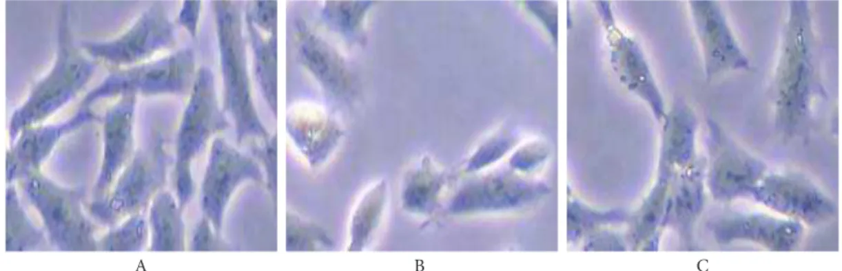
Com-pared to those of the control group, the cells from the postasphyxial serum-attacking group assumed an of-normal rounded or ellipsoid appearance. he refraction rate was lower and contour was enhanced. Vacuoles, lipid droplets, and granulation appeared in the cytoplasm. here was a great deal of cell debris in the accrescent intercellular space (Fig. 1B). Relative to those of the postasphyxial serum-attacking group, the changes in morphology of the HK-2 cells of the ucf-101-treated group were clearly improved (Fig. 1C).
Relative to the control group (0.47±0.02), the cell viability (optical density, OD) was signiicantly de-creased in the postasphyxial serum-attacking group (0.22±0.02). Relative to the postasphyxial serum-attacking group, cell viability was clearly increased in the ucf-101-treated group (0.36±0.02, P<0.05) but not to the same level as the control group (Fig. 2).
Efects of postasphyxial serum on Omi/HtrA2 expression in HK-2 cells
Several studies have reported that Omi/HtrA2 plays an important role in post-ischemic organ injury (Bhuiyan and Fukunaga, 2007; Saito et al., 2004). Fol-lowing ischemia/reperfusion in the kidney, the pro-teolytic activity of Omi/HtrA2 is markedly upregulat-ed (Faccio et al., 2000). To investigate whether Omi/ HtrA2 was involved in postasphyxial serum-induced injury to HK-2cells, we examined the level of expres-sion of Omi/HtrA2 in HK-2cells by SP
immunocy-tochemical staining. As shown in Fig. 2, Omi/HtrA2 was stained inaurate or yellow-brown and appeared localized in the cytoplasm. he percentage of control group cells expressing Omi/Htra2 was 9.00±2.50% (Fig. 3A). Ater postasphyxial serum challenge, the percentage of the postasphyxial serum-attacking-group Omi/Htra2-positive cells increased remark-ably (25.15±3.50%, P < 0.05) (Fig. 3B).
Efects of postasphyxial-serum on Omi/HtrA2 translocation from mitochondria to cytoplasm
he matureform of Omi/HtrA2 is localized in the mitochondria and may have a distinctfunction in-volved in the maintenance of mitochondrial home-ostasis (Jones et al., 2003). In the cytoplasm, mature Omi/HtrA2 can induce apoptosis in human cells,
Fig. 1. he efect of ucf-101 on the morphology of HK-2 cells treated with postasphyxial serum (inverted microscopy×400). A - control group; B - postasphyxial serum-attacking group; C - ucf-101-treated group.
A B C
either in a caspase-independent manner through its protease activity or in a caspase-dependent manner. he key step in this apoptotic pathway is the release of Omi/Htra2 from the mitochondrial intermem-brane space. To conirmthat the Omi/HtrA2 pro-tein is translocated from mitochondria to cytoplasm in HK-2cells treated with postasphyxial serum, we comparedthe luorescence-staining pattern of cells stained with Omi/HtrA2 antibodiesand MitoTrack-er Red 580. he results of the two staining pattMitoTrack-erns were almost identical in the control group,indicating that Omi/HtrA2 is localized predominantly in the mitochondria in normal HK-2 cells (Fig. 4C1). he two staining patterns were distinctive in the postas-phyxial serum-attacking group, indicating that Omi/ HtrA2 is translocated from the mitochondrial inter-membrane space to the cytoplasm (Fig. 4C2).
A1
A2
A B
B1
B2
C1
C2 Fig. 3. Postasphyxial serum-induced changes in expression of
Omi/HtrA2 in HK-2 cells. he level of expression of Omi/HtrA2 was detected by SP immunocytochemical staining. he percent-age of cells expressing Omi/HtrA2 is small in the control group (Fig. 3A). Ater postasphyxial serum challenge, the percentage of postasphyxial serum-attacking-group cells expressing Omi/ HtrA2 increased remarkably (Fig. 3B).
Efects of postasphyxial serum on apoptosis in HK-2 cells
Apoptosis plays an important role in the pathogen-esis of a variety of ischemia/reperfusion (I/R) inju-ries. We postulate that apoptosis is associated with postasphyxial serum-induced injury to HK-2cells. Apoptosis of HK-2cells was evaluated by quantita-tive determination of apoptosis using low cytomet-ric DNA analysis following propidium iodide (PI) staining. he data were presented as the percentage of apoptotic cells. As shown in Fig. 4, the proportion of cells undergoing apoptosis increased from fewer than 15% in the control group (Fig. 5A) to 36% af-ter being attacked by postasphyxial serum (Fig. 5B,
P<0.05). he proportion of cells undergoing apopto-sis decreased from 36% in the postasphyxial serum-attacking group to 26% ater ucf-101 treatment (Fig. 5C, P<0.05).
he efect of postasphyxial serum on caspase-3 expression in HK-2 cells
The caspase family is closely connected with many apoptotic processes. In the caspase family, caspase-3 is a common executer of apoptosis (horn-berry and Lazebnik, 1998). Multiple lines of evidence have demonstrated that caspase-3 plays a vital role in I/R injury. To conirmthat caspase-3 participates in postasphyxial serum-induced injury to HK-2,
we examined the level of expression of caspase-3 in HK-2cells. As shown in Fig. 5, caspase-3 was inau-rate or yellow-brown and appeared localized in the cytoplasm of HK-2 cells. Ater being attacked by postasphyxial serum, the level of expression of cas-pase-3 was signiicantly increased, as indicated by the stronger staining in the postasphyxial serum-at-tacking group (Fig. 6B) relative to the control group (Fig. 6A, P < 0.01). Ater treatment with ucf-101 (10 µmol/L), the expression of caspase-3 was signiicant-ly decreased (Fig. 6C) relative to the postasphyxial serum-attacking group (P<0.05).
DISCUSSION
It has been reported that ischemia/reperfusion induces apoptosis in many cells through different mechanisms. As a mitochondrial serine protein, mature Omi/HtrA2 is localized in mitochondria. Once released into the cytosol following apopto-sis stimuli, it promotes cell death by antagoniz-ing IAPs (in a caspase-dependent fashion) and via its proteolytic activity (in a caspase-independent fashion) (Saelens et al., 2004). Several studies have demonstrated that overexpression of Omi/HtrA2 markedly increases apoptosis (Kim et al., 2010; Tun et al., 2007). In this study, we demonstrated that postasphyxial serum incubation can signifi-cantly induce apoptosis in human HK-2 cells and that this effect is mediated by overexpression and
Fig. 5. he efect of postasphyxial serum on apoptosis in HK-2 cells. HK-2 cell apoptosis was detected by low cytometry. he proportion of cells undergoing apoptosis is small in the control group (Fig. 5A).he proportion of cells undergoing apoptosis increased signiicantly in the postasphyxial serum-attacking group (Fig. 5B). Relative to the postasphyxial serum-attacking group, the proportion of cells un-dergoing apoptosis decreased signiicantly in the group treated with ucf-101 (Fig. 5C).
mitochondrial release of the Omi/HtrA2 protein into cytoplasm.
Omi/HtrA2 is a member of the HtrA family of serine proteases, which shows extensive homology to the Escherichia coli HtrA genes that are essential for bacterial survival at high temperatures. he serine protease Omi/HtrA2 is synthesized as a precursor (premature Omi/HtrA2) and translocated into the mitochondria. Upon apoptotic stimuli, the N-termi-nal amino acids preceding alanine 134 are cleaved and the resulting mature Omi/HtrA2 is released from the mitochondria into the cytoplasm, where it induces apoptosis (Suzuki et al., 2004). Transcription of Omi/HtrA2 has been shown to be upregulated in ischemic human kidneys. he enzymatic activity of Omi/HtrA2 is substantially enhanced ischemic/ reperfusing mouse kidneys (Faccio et al., 2000). Here, we demonstrated that Omi/HtrA2 was overex-pressed in HK-2 cells ater these cells are attacked by postasphyxial serum. Arnold Levine and colleagues have identiied Omi/HtrA2 as a p53-targeted gene (Jin et al., 2003).Kelly et al. reported that p53 pro-tein levels increase signiicantly in the kidney over 24 h post-ischemia (Kelly et al., 2003). In postasphyxial serum-treated HK-2 cells, activation of the p53 pro-tein increases the transcription of the Omi/HtrA2 gene.
he key step in the role that Omi/HtrA2 plays in postasphyxial serum-induced HK-2 cell apoptosis is its release from the mitochondrial intermembrane space. Ater I/R injury, Ca2+ inlux through the L-type calcium channel triggers Ca2+ release from the InsP3R, which activates a cascade of Ca2+ release from the ER storage area (Wu et al., 2008). However, in ischemia/reperfusion injuries, severe depletion of ATP leads to failure of the pump-leak balance mechanism, leading to an inlux of Na+ that results in overload of Na+ in the cytosol. his increased Na+ level activates Na+-K+-ATPase and consumes ATP, which further activates nonselective Ca2+ channels, resulting in massive cytosolic Ca2+ accumulation. Increased cytoplasmic Ca2+ may activate calpain, and calpain can cleave Bid (Chen et al., 2001b). Af-ter cleavage, the truncated C-Af-terminal portion of Bid (tBid) translocates to the mitochondria and is in-serted into the outer membrane via its tail. It then binds to Bax, facilitating its insertion into the outer mitochondrial membrane and creates pores (Eskes et al., 2000). In addition, monomeric Bax and tBID to-gether can induce lipid remodeling following the per-meabilization of the outer mitochondrial membrane (Kuwana et al., 2002). Furthermore, full-length Bid may directly translocate to the mitochondria, where it may cause efects similar to those of Bax (Pei et al., 2007; Verhagen et al., 2002). As the permeability of
Fig. 6. he efect of postasphyxial serum on the expression of Omi/HtrA2 in HK-2 cells. he expression of caspase-3 was detected by SP immunocytochemical staining. he proportion of the cells expressing caspase-3 was small in the control group (Fig. 6A). Ater postas-phyxial serum challenge, the percentage of cells expressing caspase-3 increased remarkably, as shown in this image of the postaspostas-phyxial serum-attacking group (Fig. 6B). Ater ucf-101 treatment, the percentage of cells expressing caspase-3 decreased remarkably (Fig. 6C) but not to the level of the control group.
the outer mitochondrial membrane increases, Omi/ HtrA2 is released from the mitochondria to the cy-toplasm.
TNF-α may also play an important role in the translocation of Omi/HtrA2 from the mitochon-dria to the cytoplasm. In renal ischemia/reper-fusion, TNF-α gene transcription is primarily regulated by NF-κB activation (Donnahoo et al., 2000). Usually, NF-κB is localized in the cytoplasm in an inactive state due to its association with a class of inhibitory proteins termed inhibitory κB (IκB). During cellular ischemia/reperfusion injury, phosphorylation and subsequent ubiquitination of IκB leads to the release and nuclear translocation of NF-kB, where it promotes the transcription of genes such as TNF-α (Chen et al., 2001a). Recent studies have demonstrated that TNF-α involves a sequential signaling complex termed “complex I,” which contains TNFR-associated factor 2 (TRAF2) and promotes the activation of the cytoprotective transcription factor NF-κB (Micheau and Tschopp, 2003). When NF-κB is activated by complex I, a positive feedback for TNF-α gene transcription is formed. On one hand, TNF-α-induced oxidative stress alters redox homeostasis by impairing the MPTP protein adenine nucleotide translocator and voltage-dependent anion channel, thereby result-ing in the openresult-ing of pores and inducresult-ing the re-lease of Omi/HtrA2 (Mariappan et al., 2007). On the other hand, the increased TNF-α level in the cytosol activates caspase-8, a proximal efector protein from the tumor necrosis factor receptor family. Even small amounts of activated caspase-8 are able to cleave Bid eiciently, the truncated form of which translocates to mitochondria and induces the release of Omi/HtrA2 through the mechanism described above.
Ater being released from the mitochondria, Omi/HtrA2 may play an important role in apoptosis by means of binding and cleaving IAP proteins and relieving their inhibitory efect on caspases (Verhagen et al., 2007; Yang et al., 2003). he protease activity of Omi/HtrA2 is central to its function. Ucf-101, a speciic Omi/HtrA2 inhibitor, is able to inhibit the
protease activity of Omi/HtrA2 (Kim et al., 2010). Here we report that HK-2 cells treated with ucf-101 are resistant to postasphyxial serum-induced cell ap-optosis. Our data demonstrate that Omi/HtrA2 plays a signiicant role in postasphyxial serum-induced HK-2 cell apoptosis and that its serine protease ac-tivity is necessary and essential for its proapoptotic function in this system. he role of Omi/HtrA2 in the cytoplasm is associated with caspase-3. Many studies have demonstrated that caspase-3 is upregulated in organs afected by ischemia/reperfusion injury (Li et al., 2008; Teruya et al., 2008). Our results demonstrate that the level of expression of caspase-3 is upregu-lated in HK-2 cells attacked by postasphyxial serum. Caspase-3 can also cleave and activate Bid ater the onset of apoptosis as part of a positive feedback loop (Slee et al., 2000). HK-2 cells treated with ucf-101 are resistant to postasphyxial serum-induced caspase-3 expression. Our indings suggest that Omi/HtrA2 is associated with the expression of caspase-3 in HK-2 cells treated with postasphyxial serum.
Our studies suggest that Omi/HtrA2 plays an important role in neonatal postasphyxial serum-induced injury in renal tubular cells. Ater HK-2 cells are attacked by neonatal postasphyxial serum, Omi/HtrA2 was released from the mitochondria to the cytoplasm where it induced caspase-dependent apoptosis in HK-2 cells through its proteolytic ac-tivity. Our indings point to a novel way of relieving postasphyxial serum injuries to human kidney cells through inhibiting the proteolytic viability of Omi/ HtrA2.
Acknowledgments - his research was supported by Sichuan
Youth Science and Technology Foundation (04ZQ026-033) to W.-B.D. he authors are also grateful for support provided by the confocal microscope facility at the Institute of Myocar-dium Electrophysiology of Luzhou Medical College.
REFERENCES
Althaus J., Siegelin M.D., Dehghani F., Cilenti L., Zervos A.S.,
and A. Rami (2007) he serine protease Omi/HtrA2 is
involved in XIAP cleavage and in neuronal cell death
fol-lowing focal cerebral ischemia/reperfusion. Neurochem
Bhuiyan M.S., and K. Fukunaga (2007) Inhibition of HtrA2/Omi ameliorates heart dysfunction following
ischemia/reper-fusion injury in rat heart in vivo. Eur J Pharmacol557,
168-77.
Chen F., Castranova V., and X. Shi (2001a) New insights into the
role of nuclear factor-kappaB in cell growth regulation.
Am J Pathol 159, 387-97.
Chen M., He H., Zhan S., Krajewski S., Reed J.C., and R.A.
Gott-lieb (2001b) Bid is cleaved by calpain to an active fragment
in vitro and during myocardial ischemia/reperfusion. J
Biol Chem 276, 30724-8.
Donnahoo K.K., Meldrum D.R., Shenkar R., Chung C.S., Abraham
E., and A.H. Harken (2000) Early renal ischemia, with or
without reperfusion, activates NFkappaB and increases
TNF-alpha bioactivity in the kidney. J Urol 163, 1328-32.
Eskes R., Desagher S., Antonsson B., and J.C. Martinou (2000) Bid
induces the oligomerization and insertion of Bax into the
outer mitochondrial membrane. Mol Cell Biol 20, 929-35.
Faccio L., Fusco C., Chen A., Martinotti S., Bonventre J.V., and
A.S. Zervos (2000) Characterization of a novel human
ser-ine protease that has extensive homology to bacterial heat shock endoprotease HtrA and is regulated by kidney
isch-emia. J Biol Chem 275, 2581-8.
Gupta B.D., Sharma P., Bagla J., Parakh M., and J.P. Soni (2005)
Renal failure in asphyxiated neonates. Indian Pediatr 42,
928-34.
Jin S., Kalkum M., Overholtzer M., Stofel A., Chait B.T., and A.J.
Levine (2003) CIAP1 and the serine protease HTRA2 are
involved in a novel p53-dependent apoptosis pathway in
mammals. Genes Dev 17, 359-67.
Jones J.M., Datta P., Srinivasula S.M., Ji W., Gupta S., Zhang Z., Davies E., Hajnoczky G., Saunders T.L., Van Keuren M.L.,
Fernandes-Alnemri T., Meisler M.H., and E.S. Alnemri
(2003) Loss of Omi mitochondrial protease activity causes
the neuromuscular disorder of mnd2 mutant mice. Nature
425, 721-7.
Kelly K.J., Plotkin Z., Vulgamott S.L., and P.C. Dagher (2003) P53
mediates the apoptotic response to GTP depletion ater re-nal ischemia-reperfusion: protective role of a p53
inhibi-tor. J Am Soc Nephrol 14, 128-38.
Kim J., Kim D.S., Park M.J., Cho H.J., Zervos A.S., Bonventre J.V.,
and K.M. Park (2010) Omi/HtrA2 protease is associated
with tubular cell apoptosis and ibrosis induced by
unilat-eral uretunilat-eral obstruction. Am J Physiol Renal Physiol 298,
F1332-40.
Kuwana T., Mackey M.R., Perkins G., Ellisman M.H., Latterich
M., Schneiter R., Green D.R., and D.D. Newmeyer (2002)
Bid, Bax, and lipids cooperate to form supramolecular
openings in the outer mitochondrial membrane. Cell 111,
331-42.
Li H.Z., Han L.P., Jiang C.M., Li H., Zhao Y.J., Gao J., Lin Y., Ma
S.X., Tian Y., Yang B.F., and C.Q. Xu (2008) Efect of
do-pamine receptor 1 on apoptosis of cultured neonatal rat
cardiomyocytes in simulated ischaemia/reperfusion. Basic
Clin Pharmacol Toxicol 102, 329-36.
Liu H.R., Gao E., Hu A., Tao L., Qu Y., Most P., Koch W.J.,
Christopher T.A., LopezB.L., Alnemri E.S., Zervos A.S.,
and X.L. Ma (2005) Role of Omi/HtrA2 in apoptotic cell
death ater myocardial ischemia and reperfusion.
Circu-lation 111, 90-6.
Mariappan N., Soorappan R.N., Haque M., Sriramula S., and J.
Francis (2007) TNF-alpha-induced mitochondrial
oxida-tive stress and cardiac dysfunction: restoration by
super-oxide dismutase mimetic Tempol. Am J Physiol Heart Circ
Physiol 293, H2726-37.
Micheau O., and J. Tschopp (2003) Induction of TNF receptor
I-mediated apoptosis via two sequential signaling
com-plexes. Cell 114, 181-90.
Pei Y., Xing D., Gao X., Liu L., and T. Chen (2007) Real-time monitoring full length bid interacting with Bax during
TNF-alpha-induced apoptosis. Apoptosis 12, 1681-90.
Rami A., Kim M., and J. Niquet (2010) Translocation of the serine
protease Omi/HtrA2 from mitochondria into the cytosol upon seizure-induced hippocampal injury in the neonatal
rat brain. Neurochem Res 35, 2199-207.
Saelens X., Festjens N., Vande Walle L., van Gurp M., van Loo G.,
and P. Vandenabeele (2004) Toxic proteins released from
mitochondria in cell death. Oncogene 23, 2861-74.
Saito A., Hayashi T., Okuno S., Nishi T., and P.H. Chan (2004) Modulation of the Omi/HtrA2 signaling pathway ater transient focal cerebral ischemia in mouse brains that
overexpress SOD1. Brain Res Mol Brain Res 127, 89-95.
Slee E.A., Keogh S.A., and S.J. Martin (2000) Cleavage of BID
during cytotoxic drug and UV radiation-induced apop-tosis occurs downstream of the point of Bcl-2 action and is catalysed by caspase-3: a potential feedback loop for ampliication of apoptosis-associated mitochondrial
cyto-chrome c release. Cell Death Difer 7, 556-65.
Suzuki Y., Takahashi-Niki K., Akagi T., Hashikawa T., and R.
Takahashi (2004) Mitochondrial protease Omi/HtrA2
en-hances caspase activation through multiple pathways. Cell
Death Difer 11, 208-16.
Teruya R., Fagundes D.J., Oshima C.T., Brasileiro J.L., Marks G.,
Ynouye C.M., and M.J. Simoes (2008) he efects of
pen-toxifylline into the kidneys of rats in a model of unilateral
hindlimb ischemia/reperfusion injury. Acta Cir Bras23,
hornberry N.A., and Y. Lazebnik (1998) Caspases: enemies
within. Science 281, 1312-6.
Tun C., Guo W., Nguyen H., Yun B., Libby R.T., Morrison R.S.,
and G.A. Garden (2007) Activation of the extrinsic
cas-pase pathway in cultured cortical neurons requires p53-mediated down-regulation of the X-linked inhibitor of
apoptosis protein to induce apoptosis. J Neurochem 102,
1206-19.
Verhagen A.M., Kratina T.K., Hawkins C.J., Silke J., Ekert P.G.,
and D.L. Vaux (2007) Identiication of mammalian
mito-chondrial proteins that interact with IAPs via N-terminal
IAP binding motifs. Cell Death Difer 14, 348-57.
Verhagen A.M., Silke J., Ekert P.G., Pakusch M., Kaufmann H., Connolly L.M., Day C.L., Tikoo A., Burke R., Wrobel C.,
Moritz R.L., Simpson R.J., and D.L. Vaux (2002) HtrA2
promotes cell death through its serine protease activity and its ability to antagonize inhibitor of apoptosis
pro-teins. J Biol Chem 277, 445-54.
Wu D., Chen X., Ding R., Qiao X., Shi S., Xie Y., Hong Q., and
Z. Feng (2008) Ischemia/reperfusion induce renal tubule
apoptosis by inositol 1,4,5-trisphosphate receptor and
L-type Ca2+ channel opening. Am J Nephrol 28, 487-99.
Yang Q.H., Church-Hajduk R., Ren J., Newton M.L., and C. Du
(2003) Omi/HtrA2 catalytic cleavage of inhibitor of apop-tosis (IAP) irreversibly inactivates IAPs and facilitates
cas-pase activity in apoptosis. Genes Dev 17, 1487-96.
Zhao J., Dong W.B., Li P.Y., and C.L. Deng (2009) Mechanism
of intracellular signal transduction during injury of renal tubular cells induced by postasphyxial serum in neonates