Phosphotyrosine-Independent Interaction with DLC-1
Kun Dai1., Shanhui Liao1., Jiahai Zhang1
, Xuecheng Zhang2, Xiaoming Tu1*
1Hefei National Laboratory for Physical Sciences at Microscale, School of Life Sciences, University of Science and Technology of China, Hefei, Anhui, People’s Republic of China,2School of Life Sciences, Anhui University, Hefei, Anhui, People’s Republic of China
Abstract
Background:Src homology 2 (SH2) domain is a conserved module involved in various biological processes. Tensin family member was reported to be involved in tumor suppression by interacting with DLC-1 (deleted-in-liver-cancer-1) via its SH2 domain. We explore here the important questions that what the structure of tensin2 SH2 domain is, and how it binds to DLC-1, which might reveal a novel binding mode.
Principal Findings:Tensin2 SH2 domain adopts a conserved SH2 fold that mainly consists of fiveb-strands flanked by two a-helices. Most SH2 domains recognize phosphorylated ligands specifically. However, tensin2 SH2 domain was identified to interact with nonphosphorylated ligand (DLC-1) as well as phosphorylated ligand.
Conclusions: We determined the solution structure of tensin2 SH2 domain using NMR spectroscopy, and revealed the interactions between tensin2 SH2 domain and its ligands in a phosphotyrosine-independent manner.
Citation:Dai K, Liao S, Zhang J, Zhang X, Tu X (2011) Solution Structure of Tensin2 SH2 Domain and Its Phosphotyrosine-Independent Interaction with DLC-1. PLoS ONE 6(7): e21965. doi:10.1371/journal.pone.0021965
Editor:Dong-Yan Jin, University of Hong Kong, Hong Kong
ReceivedJanuary 26, 2011;AcceptedJune 15, 2011;PublishedJuly 12, 2011
Copyright:ß2011 Dai et al. This is an open-access article distributed under the terms of the Creative Commons Attribution License, which permits unrestricted use, distribution, and reproduction in any medium, provided the original author and source are credited.
Funding:This work was supported by the Knowledge Innovation Program of the Chinese Academy of Science, Grant KSCX2-EW-Q-4, the National High-Tech R&D Program, Grant 2006AA02A315 and the National Basic Research Program of China (973 Program), Grants 2007CB914503 and 2009CB918804. The funders had no role in study design, data collection and analysis, decision to publish, or preparation of the manuscript.
Competing Interests:The authors have declared that no competing interests exist. * E-mail: xmtu@ustc.edu.cn
.These authors contributed equally to this work.
Introduction
Src homology 2 (SH2) domain was firstly identified from N-terminal noncatalytic region of fujinami sarcoma virus P130gag-fps. It is a conserved domain of approximately 100 residues and shared by a number of cytoplasmic tyrosine kinase [1]. Few years after its identification, the SH2 domain was found in various proteins other than tyrosine kinases, such as PLCc1, RasGAP, Grb2, suggesting its role as an interaction module [2]. Meanwhile, the works on GAP and EGF receptor verified the interactions between the SH2 domain and phosphorylated tyrosine (pTyr) containing protein [3–5].
Post-translational modification by phosphate group is a highly dynamic process, which is critical for regulation of various cellular events. Especially, phosphorylation/dephosphorylation of tyrosine residues of intracellular proteins is a critical signal for multiple biological events, e.g. DNA replication and repair, chromosome recombination and segregation, motility, metabolism, and gene transcription [6]. The diverse functions of SH2 domain containing proteins require broad but specific recognition of pTyr-ligands. Structural analyses of different SH2 domains revealed some common features of their structures and the binding between the SH2 domains and their ligands: (1) The SH2 domain mainly contains five anti-parallelb-strands flanked by twoa-helices [7]. (2) The SH2 domain has two binding regions: one is the pTyr binding site, a deep positive-charged pocket [8], the other is the so-called ‘‘specificity determine region’’, which includes residues close
to the pTyr binding site [7–10]. It is revealed that the C-terminal three residues adjacent to pTyr are critical for the affinity and specificity of SH2 domain-ligand interaction [9,10]. These provide SH2 domain containing proteins with properties of precise recognition of ligands and efficient regulation of signaling pathway. On the other hand, the SH2 domains binding pTyr through variable C-termini may facilitate SH2 containing proteins interacting with various ligands [7]. In most cases, SH2 domain-ligand binding is dependent on phosphorylated tyrosine.
A previous work has revealed a relationship between tensin3 and tumorigenesis by showing a role of tensin3 in maintaining the transformed properties of cell lines from advanced cancers [14]. More recently, cten was considered to be a potential tumor suppressor by interacting with DLC-1 (deleted-in-liver-cancer-1), a member of RhoGTPase activating protein (GAP) family known to have suppressive activities in tumorigenicity and cancer metastasis [12,13,15–18]. This interaction was mediated by the SH2 domain of cten and the fragment ‘‘CSRLSIY442DNVPG’’ of DLC-1 [13]. Further experiments revealed that the interactions between cten SH2 domain and DLC-1 are phosphortyrosine independent [13]. This phosphorylation independent manner has also been observed for the SH2 domain-ligand binding of SAP, Grb7, and Vav1 [7,13,18–20], which might indicate a new binding mode of the SH2 domain.
In this study, we identified the interactions between the SH2 domain of tensin2, one member of tensin family, and the fragment ‘‘CSRLSIY442
DNVPG’’ of DLC-1. Similar to cten, the interac-tions between the SH2 domain of tensin2 and DLC-1 were phosphortyrosine independent. The solution structure of tensin2 SH2 domain was determined by Nuclear Magnetic Resonance (NMR) spectroscopy. Tensin2 SH2 domain adopted a typical SH2 fold. Furthermore, chemical shift perturbation indicated a similar manner of tensin2 SH2 domain in binding to nonphosphorylated and phosphorylated ligands.
Materials and Methods
Sequence analysis
Sequence alignments were performed using ClustalW2 [21]. Further processing of the alignment files was carried out using ESPript 2.2 [22]. The existence of the SH2 domain in tensin2 was determined using SMART (http://smart.embl-heidelberg.de/). Accession number of the proteins used for alignments are listed as follows: Swiss-Prot, O60880, Homo sapiensSH2_SAP; Swiss-Port, P16277,Mus musculusSH2_BIK; Swiss-Port, Q56XZ1,Arabidopsis thaliana SH2_SHB; Swiss-Prot, P23615.1, Saccharomyces cerevisiae
SH2_SPT6; Swiss-Prot, Q63HR2.2, Homo sapiens SH2_tensin2; Swiss-Prot, Q9HBL0, Homo sapiens SH2_tensin1; Swisst-Prot, Q68CZ2, Homo sapiens SH2_tensin3; and Swiss-Prot, Q8IZW8,
Homo sapiensSH2_cten.
Protein expression and purification
Sequence encoding the SH2 domain of human tensin2 was amplified and cloned into pET-22b (Novagen). Following primers were used for polymerase chain reaction (PCR): forward primer 59 -CCGCATATGGATACATCCAAGTTCTGGTAC-39and reverse primer 59 -TTCTCGAGTTTGCTGGGAATGCGCAGGCAGC-AfoG-39(restriction endonuclease sites underlined). The recombinant tensin2 SH2 domain contained a C-terminal tag of six histidine residues. The recombinant protein was expressed inEscherichia coli
strain BL21 Gold (DE3) and purified according to previous procedure [23]. 15N-labeled and 13C,15N-labeled tensin2 SH2 domain were prepared in the same way, except that super broth was replaced by M9 medium containing 0.5 g/L15N-labeled ammonium chloride and 2.5 g/L 13C-labeled glucose as the sole nitrogen source and carbon source, respectively. The purified protein was dialyzed with buffer containing 20 mM NaH2PO4, 150 mM NaCl, 50 mM
arginine, 50 mM glutamine and 2 mM EDTA, pH 6.8, and concentrated to 0.6 mM for further NMR experiments.
Site-directed mutagenesis
The site-directed mutagenesis (Y41S) was performed by PCR with the previousely constructed plasmid containing gene of
Tensin2 SH2. The following primer sets were used: forward primer, 59- CATTCATTCCAAGGAGCTTCTGGGCTGGCC-CTCAAG -39, reverse primer, 59- GCCACCTTGAGGGCCAG-CCCAGAAGCTCCTTGGAATG -39. The PCR products were digested with DpnI (TaKaRa) for 10 hours, and then transformed into E. coli BL21. The plasmids containing mutated gene were sequenced to confirm the Y41S mutagenesis. The tensin2 SH2 (Y41S) were prepared as described above for further study.
Peptide synthesis
Peptides ‘‘CSRLSIY442DNVPG’’ and ‘‘CSRLSI pY442DNVPG’’ (residues 436–447 of DLC-1 with Y442 nonphosphorylated/phos-phorylated) were synthesized by GL Biochem (shanghai). The peptides were purified by HPLC (purity of peptides.95%), and molecular weight of peptides were confirmed by MS analysis.
Surface plasmon resonance (SPR)
Real-time interactions between tyrosine-phosphorylated or nonphosphorylated peptide and tensin2 SH2 domain or mutated tensin2 SH2 domain (Y41S) were measured by surface plasmon resonance (SPR) on a Biacore 3000 (Biacore Inc.). SPR experiments were performed on sensor chips CM5 (carboxy-methylated dextran, BiacoreAB). Chip was treated as described previously [23]. Purified tensin2 SH2 domain and mutated tensin2 SH2 domain were diluted with running buffer (20 mM NaH2PO4,
150 mM NaCl, 50 mM arginine, 50 mM glutamine and 2 mM EDTA, pH 6.8). The kinetic analysis of the interactions between phosphorylated peptide and tensin2 SH2 domain was performed at 6-step concentration of tensin2 SH2 domain (0.18mM, 0.38mM, 0.75mM, 1.50mM, 3.00mM, and 6.00mM) at a flow rate of 30mL/min for 2 min, whereas the kinetic analysis of the
interactions between nonphosphorylated peptide and tensin2 SH2 domain was performed at 6-step concentration of tensin2 SH2 domain (0.41mM, 0.81mM, 1.63mM, 3.25mM, 6.50mM, and
13.00mM). The kinetic analysis of the interactions between
phosphorylated peptide and mutated tensin2 SH2 (Y41S) domain was performed at 6-step concentration of mutated tensin2 SH2 (Y41S) domain (0.50mM, 1.00mM, 2.00mM, 4.00mM, 8.00mM, and 16.00mM) at a flow rate of 30mL/min for 2 min, whereas the kinetic analysis of the interactions between nonphosphorylated peptide and mutated tensin2 SH2 domain (Y41S) was performed at 6-step concentration of mutated tensin2 SH2 domain (Y41S) (3.13mM, 6.25mM, 12.50mM, 25.00mM, 50.00mM, and 100.00mM). All analyses were performed three times. To
overcome refractive index changes, data of control surface were subtracted from that of peptide-immobilized surface. Interactions between peptide and Albumin Bovine V (BSA, Sigma, 8 mg/mL) were measured as a negative control. Kinetic analyses of SPR data were performed using BIAevaluation 4.1 (Biacore Inc.). Curves were fitted with 1:1 (langmuir) binding model. The equilibrium dissociation constant (KD) was derived from kinetic analysis.
Circular dichroism (CD) spectroscopy
CD experiments were performed on a Jasco-810 spectropho-tometer (JASCO, Japan) over the wavelength range from 200 to 260 nm at 20uC. Tensin2 SH2 domain was dissolved in buffer (20 mM NaH2PO4, 150 mM NaCl, 50 mM arginine, 50 mM
glutamine and 2 mM EDTA, pH 6.8) to a concentration of
,0.6 mg/mL. Different molar ratios of SH2/peptide (1:1, 1:2 and
successive scans were recorded and averaged. The analyses of CD spectra were performed using Jascow32 software (JASCO, Japan).
NMR spectroscopy, data processing and structure calculation
NMR samples were dissolved in buffer containing 20 mM NaH2PO4, 150 mM NaCl, 50 mM arginine, 50 mM glutamine,
2 mM EDTA, 90% H2O/10% D2O and pH 6.8. All NMR
measurements were performed at 293 K on a Bruker DMX500 spectrometer. The following spectra were recorded to obtain backbone and side chain resonance assignments:1H-15N HSQC, HNCO, HN(CA)CO, CBCA(CO)NH, CBCANH, H(CC)ONH, HBHA(CO)NH, HC(CO)NH. 3D 15N-edited and 13C-edited NOESY were acquired with mixing times of 100 and 130 ms, respectively. To identify the slowly exchanging amides, a 15 N-labeled sample was lyophilized and dissolved in 99.96% D2O.
Immediately, a series of1H-15N HSQC were recorded to monitor the attenuation of NH signals. NMR data were processed using NMRPipe and NMRDraw [24], and then analyzed using Sparky 3 [25]. All software was run on a Linux system.
3D 15N-edited and 13C-edited NOESY spectra were used to determine the NMR distance restraints for structure calculations. Backbone torsion angle restraints were predicted from chemical shifts of five types of nuclei:13Ca,13Cb,13CO,1Ha, and15NH by using TALOS+[26]. Hydrogen bond restraints were obtained by identifying slow-exchange amide protons after 12 hours’ incuba-tion following solvent exchange. For hydrogen bonds, two distance restraints were used: 2.0 A˚ for H-O and 3.0 A˚ for N-O. Structures
were calculated using the program CYANA 3.0 [27]. A total of 200 conformers were independently calculated, and 20 lowest-energy structures were selected and analyzed by MOLMOL [28] and PROCHECK [29] online (http://nihserver.mbi.ucla.edu/ SAVES_3/).
Chemical shift perturbation
To identify the residues responsible for binding to tyrosine-phosphorylated or nontyrosine-phosphorylated DLC-1 peptide in tensin2 SH2 domain, 0.5 mM 15N-labeled protein were titrated with unlabeled tyrosine-phosphorylated or nonphosphorylated peptide to different molar ratios (1:0, 1:1, 1:2 and 1:3, SH2/peptide).
1
H-15N HSQC spectrum was acquired for analysis. Peptide stock solutions in identical buffer were titrated stepwise with a sample dilution of less then 10%. Combined chemical shift pertur-fobation was calculated using the following equation: Ddppm~
ffiffiffiffiffiffiffiffiffiffiffiffiffiffiffiffiffiffiffiffiffiffiffiffiffiffiffiffiffiffiffiffiffiffiffiffiffiffiffiffiffiffiffiffi
(DdHN)2z(0:17DdN)
2
q
. DdNH and DdN are the chemical shift
variation in the proton and nitrogen dimensions, respectively. The threshold values used as a criterion for significance were 60% for intensity reduction and 0.1 ppm for chemical shift perturbation.
Results
Comparison of the primary sequence of the SH2 domain between tensins and other proteins
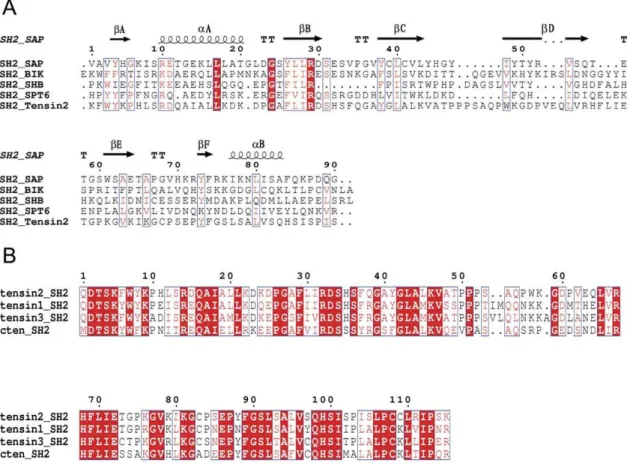
The SH2 domains have been found in hundreds of proteins involved in a variety of cellular processes. The SH2 domains from different proteins share low sequence identity (Figure 1A). For
Figure 1. Multiple sequence alignments between different SH2 domains of proteins from different species. Alignments were performed using ClustalW2 and ESPript 2.2. Identical residues were shaded in red box. A. SH2 domains share low primary sequence similarity, but similar secondary structure. B. SH2 domains of tensin family members share high primary sequence similarity.
example, the SH2 domain of SAP which is related to SLAM-induced signal-transduction events in T lymphocytes shares only about 24% sequence identity with tensin2 SH2 domain. The sequence diversity of the SH2 domains may reflect their ability of binding various ligands in different cellular processes. Although the SH2 domains vary dramatically in sequence and ligand recognition, the SH2 domains from different tensin family members display 70%–80% of sequence identity between each other (Figure 1B), which may imply their common properties not only in structure but also in ligand recognition.
Interactions between tensin2 SH2 domain and DLC-1 peptides
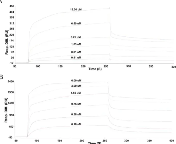
Recently, the SH2 domain of tensin family protein cten that is able to bind DLC-1 peptides in a pTyr independent manner has been reported [12,13]. To understand whether the SH2 domain of another tensin family member, tensin2, binds to the same ligand and whether this binding is also phosphorylation-independent, we investigated the interactions between tensin2 SH2 domain and non/phosphorylated DLC-1 peptides by SPR. The results indicated that tensin2 SH2 domain bound specifically to both phosphorylated and nonphosphorylated peptides. The equilibrium
dissociation constant (KD) derived from kinetic analyses was about 248.0068.82 nM for SH2-nonphosphorylated peptide interaction (Figure 2A) and 31.0060.50 nM for SH2-phosphorylated peptide interaction (Figure 2B). It was surprising that phosphorylation of DLC-1 peptide increased its affinity to tensin2 SH2 domain by only eight folds approximately under the condition for SPR experiments, whereas for typical SH2 domain, the affinity of tyrosine-phosphorylated peptide to the SH2 domain is about four orders of magnitude greater than that of nonphosphorylated counterpart [7,30]. This result indicated that the peptide recognition of tensin2 SH2 domain was relatively phosphorylation independent.
Secondary structures of tensin2 SH2 domain in free form and in complex with DLC-1 peptides
To investigate the secondary structure of tensin2 SH2 domain and its change upon binding to ligand, CD experiments were performed. The CD spectra of tensin2 SH2 domain in complex with non/phosphorylated DLC-1 peptides in different ratios indicated that there was no significant change in the secondary structure of tensin2 SH2 domain after binding to the ligands (Figure 3A and Figure 3B).
Figure 2. Kinetic analyses of interactions between tensin2 SH2 domain and nonphosphorylated/phosphorylated peptides by SPR. Kinetic analyses of interactions between peptides and tensin2 SH2 domain were performed at 6 steps of concentration of recombinant SH2 domain at a flow rate of 30mL/min for 2 mins. A. SPR spectra of the SH2 domain binding to nonphosphorylated peptide. B. SPR spectra of the SH2 domain
binding to phosphorylated peptide. Analyses were performed three times at each step of concentration. KD of the SH2 domain binding different peptide was derived from kinetic analysis.
Solution structure of tensin2 SH2 domain
Tensin2 SH2 domain, including residues from 1135 to 1248 of the protein, was recombinantly expressed and purified. The recombinant product exhibited good solubility and stability. The solution structure of tensin2 SH2 domain was calculated based on a series of NMR spectra. The NMR data used for structure
calculations are summarized in Table 1. The chemical shift assignments of tensin2 SH2 domain have been deposited in the Biological Magnetic Resonance Data Bank (accession number 17314). The assembly of the 20 lowest-energy structures is shown in Figure 4A. Structures of tensin2 SH2 domain have been deposited in Protein Data Bank (PDB ID code 2l6k). The statistical
Figure 3. CD spectra of tensin2 SH2 domain free and in complex with peptides. A. CD spectra of tensin2 SH2 domain with nonphosphorylated peptide in different molar ratios (blue, green, red and cyan represent SH2/peptide ratios of 1:0, 1:1, 1:2 and 1:3, respectively). B. CD spectra of the SH2 domain with phosphorylated peptide in different molar ratios (blue, green, red and cyan represent SH2/peptide ratios of 1:0, 1:1, 1:2 and 1:3, respectively).
parameters in Table 1 indicate a high-quality NMR structure of tensin2 SH2 domain. Calculated structure of tensin2 SH2 domain shows a characteristic SH2 fold, which includes the two maina -helices packing against either side of a centralbsheet. The lowest-energy structure was shown in Figure 4B. The centralb sheet is composed of three long antiparallel b strands (bB, bC, bD) followed by one short antiparallelbstrand (bE) and one shorterb -sheet-like structure (bF). The two maina-helices (aB andaC) flank either side of the central b sheet. The residues composing secondary structures are listed as follows: residue 4 to 7 (aA), residue 8 to 9 (bA), residue 13 to 22 (aB), residue 29 to 33 (bB), residue 40 to 46 (bC), residue 66 to 72 (bD), residue 78 to 81 (bE), residue 87 to 89 (bF) and residue 91 to 98 (aC), from N-terminus to C-terminus.
Structural comparison with other SH2 domains
As the SH2 domains of tensin2 and SAP both interact with ligands in a pTyr-independent way, the structure of free SAP was used to perform structural compare. The alignment of SAP and the lowest-energy structure of tensin2 SH2 domain was shown in Figure 5. Although the two SH2 domains share low sequence
identity, both structures show typical SH2 fold, with a central five
b strands flanked by two a helices at either side, which demonstrated that the SH2 domain is a structurally conserved. A DALI [31] search using tensin2 SH2 domain as query sequence indicates that SH2 domains share high similarity in 3D-structure. The RMSD and Z-score between tensin2 SH2 domain and SAP are 2.4 A˚ and 10.8, respectively. The structural difference between these two SH2 domains lies in the loop between bC and bD. Tensin2 SH2 domain has a longer loop than SAP, implying that the former structure might be more flexible than that of the latter in this region.
Identification of the residues of tensin2 SH2 domain involved in binding to DLC-1 peptides
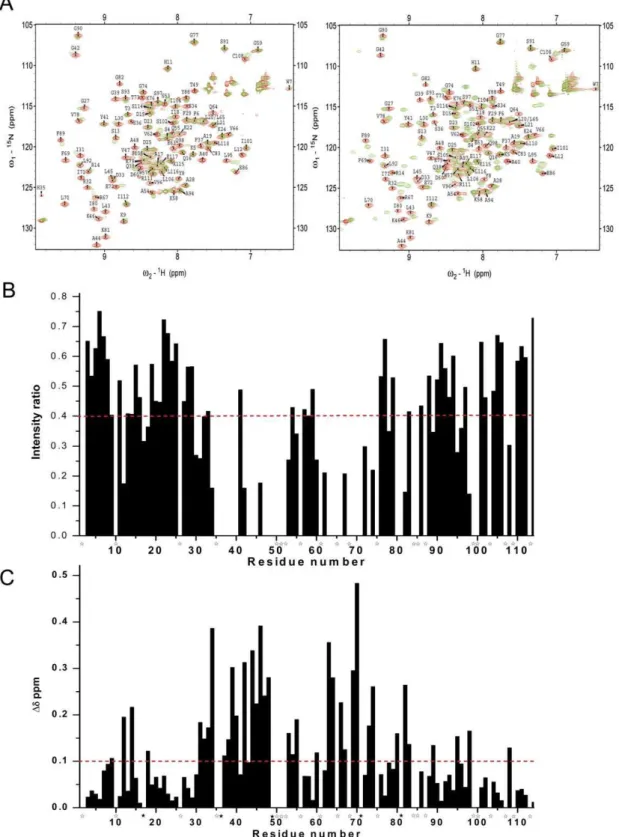
To determine the residues in tensin2 SH2 domain involved in recognition of non/phosphorylated DLC-1 peptides, chemical shift perturbation experiments were performed. 1H-15N HSQC experiments were recorded for15N-labeled tensin2 SH2 domain before and after addition unlabeled nonphosphorylated or phosphorylated peptides to different molar ratios of SH2/peptide. The spectral changes that occurred after peptide addition were characterized by resonance intensity reductions and chemical shift changes. The residues of tensin2 SH2 domain interacting with nonphosphorylated ligand were signified by the obviously reduced peak intensity. Compared with it, the residues of tensin2 SH2 domain interacting with phosphorylated ligand were signified by the substantial resonance shift (Figure 6A to 6C). The amino acids affected in the presence of the nonphosphorylated ligand were residues Leu12, Ala17, Ile18 (belong toaB), Leu30-Arg32 (belong tobB), Ser34, Ser36-Gly39 (belong to BC loop), Ala40, Gly42-Lys46 (belong tobC), Val47-Thr49, Ser53, Gln55, Asp60, Val62-Gln64 (belong to CD loop), Val66, Arg67, Phe69-Glu72 (belong to
bD), Thr73, Gly74 (belong to DE loop), Val78, Ile80, Lys81 (belong tobE), Gly82 (belongs to EF loop), Phe89 (belongs tobF), Leu95, Val96, Gln98 (belong toaC) and Cys108 (belongs to C-terminal coil) (Figure 6B). The amino acids affected in the presence of phosphorylated ligand were residues Lys9 (belongs to
bA), Leu12, Arg14, Ala17, Ile18 (belong to aB), Ile31-Asp33 (belong tobB), Ser34, Ser36-Gly39 (belong to BC loop), Ala40, Gly42-Lys46 (belong tobC), Val47-Thr49, Ser53-Gln55, Asp60, Glu63, Gln64 (belong to CD loop), Val66, Arg67, Phe69-Ile71 (belong tobD), Thr73, Gly74 (belong to DE loop), Ile80, Lys81 (belong tobE), Gly82, Cys83 (belongs to EF loop), Phe89 (belongs tobF), Leu95, Gln98 (belong toaC) and Cys108 (belongs to C-terminal coil) (Figure 6C). These residues might be involved in the interactions between tensin2 SH2 domain and the ligands. The distribution of the residues which showed intensity reductions in the presence of nonphosphorylated ligand was almost identical to that of residues which showed chemical shift perturbations in the presence of phosphorylated ligand, which might reflect the similar binding pattern of tensin2 SH2 domain to peptides with different phosphorylation status.
Tyr41 of tensin2 SH2 domain was shown to be affected both in the presence of phosphorylated and nonphosphorylated ligands in chemical shift perturbation experiments. Meanwhile, it has been reported previously that tensin3 SH2 domain’s Tyr1206, which was the counterpart of Tyr41 in tensin2 SH2 domain, is important for ligand binding [14]. Therefore, we mutated the Tyr41 residue to Ser residue and tested how this mutation affected their interactions. The equilibrium dissociation constant (KD) derived from SPR experiment was about 16.0060.20mM for the Y41S mutant-nonphosphorylated peptide interactions (Figure S1A) and 5.0060.04mM for the Y41S mutant-phosphorylated peptide
interactions (Figure S1B). The KD value of Y41S mutant-ligands
Table 1.NMR structural statistics.
NMR restraints in the structure calculation
Intraresidue 333
Sequential (|i - j| = 1) 447 Medium-range (|i - j|,5) 252 Long-range (|i - j|./ = 5) 369
Hydrogen bonds 66
Total distance restraints 1580 Dihedral angle restraints 113 Residual violations
CYANA target functions, A˚ 1.4360.24 NOE upper distance constrain violation
Maximum, A˚ 0.2060.04
Number.0.2 A˚ 061
Dihedral angle constrain violations
Maximum,u 3.2360.72
Number.5u 060
Vander Waals violations
Maximum, A˚ 0.3060.00
Number.0.2 A˚ 361
Average structural rmsd to the mean coordinates, A˚
Secondary structure backbonea 0.31
Secondary structure heavy atomsa 0.80
All backbone atomsb 1.30
All heavy atomsb 1.79
Ramachandran statistics, %of all residues
Most favored regions 81.5
Additional allowed regions 18.5 Generously allowed regions 0
Disallowed regions 0
aIncludes residues in secondary structure.
bObtained for residues T3-R111 since no long-range NOEs were identified for
reduced by two orders compared with that of tensin2 SH2 domain-ligands, which suggested an important role of Tyr41 of tensin2 SH2 domain both in the recognition of the nonpho-sphorylated and phononpho-sphorylated ligands.
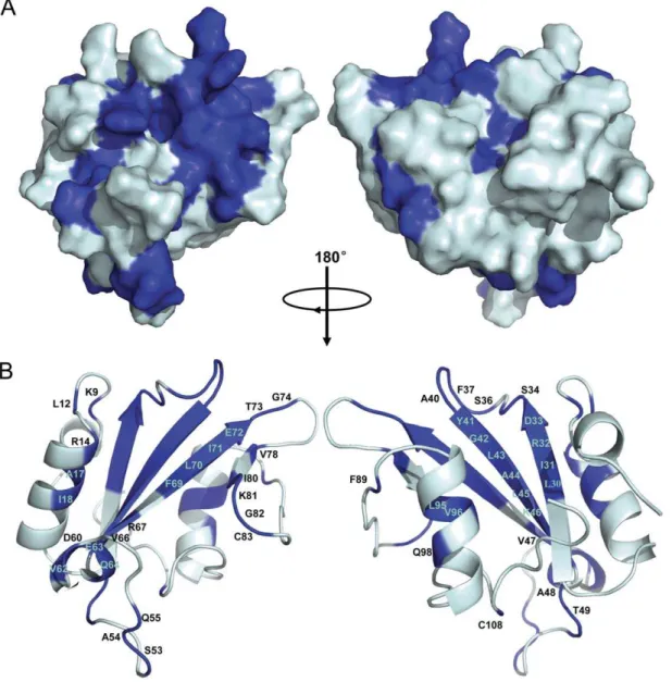
Based on chemical shift perturbation, the structure of tensin2 SH2 domain was shown with residues marked to display the ones involved in peptide interaction (Figure 7). It is remarkable that the majority of significantly affected residues, such as Leu30, Ile31,
Figure 4. The NMR structure of tensin2 SH2 domain.A. Superposition of 20 lowest-energy NMR structures. B. The lowest-energy conformation used for ribbon representation on highlighting the sixb-strands and two maina-helices that are identified in tensin2 SH2 domain.
Ser36 to Ala40, Gly42 to Thr49, Val62 to Gln64, Arg69 to Gly74 whose intensity reductions were higher than 70% even to 100% and Ser34, Gly39, Ala44, Lys46, Glu63 whose chemical shift perturbations were bigger than 0.3 ppm, were located in the central anti-parallel b-sheet and the loops between b strands. These sites are the conserved binding sites for typical SH2 domains recognizing their ligands. In addition, residues Lys9 (belongs tobA), Leu12, Arg14, Ala17 and Ile18 (belong toaB), which are located outside the central region, displayed discernable changes during titration as well. These residues may also participate in the interactions between tensin2 SH2 domain and its ligands.
Discussion
Tensin is a family containing four members that are localized to the cytoplasmic side of focal adhesions and play essential roles in extracellular matrix based signaling pathway. Previous studies have suggested cten, a tensin family member, may function as a tumor suppressor, through the interaction with DLC-1 [13] that has been found to be absent or suppressed in many cancers such as hepatocellular carcinoma (HCC). However, more recent studies revealed other roles of cten as an oncogene in colorectal cancer through enhancing the colony formation, anchorage-independent growth, cell migration and invasion [32,33]. Mutations in DLC-1 which disrupt its interaction with cten SH2 domain will abolish its role as a tumor suppressor [34]. Thus, cten is important for the functions of DLC-1 in tumor suppression. In addition, tensin3 has also been reported to be involved in tumorigenesis [14]. In this study, we indicate that another member of tensin family, tensin2, is
able to interact through its SH2 domain with DLC-1 as well; and the interaction is pTyr-independent, similar to cten SH2 domain. The interaction of tensin2 SH2 domain with tumor suppressor DLC-1 might reflect the general roles of tensin family members in preventing tumor development.
So far, no structure has been reported for the SH2 domains of tensin family proteins. Though the sequence similarity between tensin members is low, their SH2 domains share high sequence identity, which implies they might possess conserved structure. In this study, we reported the NMR solution structure of tensin2 SH2 domain. The structure is mainly composed of five b-strands flanked by twoa-helices at either side, which is a characteristic of typical SH2 domain, suggesting that SH2 domain family is highly conserved in structure.
Although tensin2 SH2 domain adopts a similar structure to other SH2 domains, it recognizes its ligand in a pTyr-independent instead of pTyr-dependent manner. SAP, which has only one SH2 domain, was the first protein reported to interact with ligand in a pTyr-independent manner. The interaction mechanism has been dis-closed by analyzing its free structure and complex structures with phosphorylated/nonphosphorylated ligands. A theory called ‘‘three-pronged plug and socket’’ was proposed to illustrate the pTyr-independent interaction. This theory suggests three distinct regions on SAP mediate specific interaction with N-terminus, residue Tyr, and C-terminus of ligand, respectively [35]. Ligands of SAP share a motif of ‘‘T/S-I-Y-X-X-V/I’’ (X represents any residue) [35]. Although the SH2 domain of tensin2 shares only about 24% of the primary sequence identity with that of SAP, the ligand of tensin2 SH2 domain, DLC-1, contains the motif of ‘‘T/S-I-Y-X-X-V/I’’ as well, within the sequence of ‘‘CSRLSIYDNVPG’’. The same motif
Figure 5. Structural comparison of tensin2 and SAP SH2 domains.The structure of SAP SH2 domain was shown in limon. The lowest-energy conformation of tensin2 SH2 domain was shown in cyan. The alignment was performed by using Pymol.
Figure 6. Identification of residues of tensin2 SH2 domain for binding to nonphosphorylated/phosphorylated ligands.A. The1H-15N HSQC spectra of the15N-labeled tensin2 SH2 domain free (red) and titrated with its ligand (left: nonphosphorylated peptide; right: phosphorylated
peptide) (green) were overlaid. B. The ratio of peak intensity after compared with before the addition of nonphosphorylated peptide. C. The chemical shift changes after the addition of phosphorylated peptide. All the comparison was performed at the SH2/peptide molar ratio of 1:3. The broken lines represent the significance level of intensity reductions.60% (intensity ratios,40%) and chemical shift changes.0.1 ppm, respectively. The residues that had significant changes and were untraceable in1H-15N HSQC spectra after the addition of phosphorylated peptide were labeled withw; the
recognized by SAP and tensin2 SH2 domains suggests that they may interact with ligands in a similar way.
Classical SH2 domains bind to phosphorylated peptides through a conventional phosphotyrosine-binding pocket. In our study, chem-ical shift perturbation and structural comparison indicate that tensin2 SH2 domain maintains this general binding feature. The two conserved binding regions existing in tensin2 SH2 domain are as follows: (1) Residues binding to DLC-1 ligands regardless of the phosphorylation status are located in the central anti-parallel b -sheets and the loops betweenbB andbC, including Leu30, Ile31, Arg32, Asp33, Ser34, Ser36, Phe37, Gln38, Gly39, Ala40, Tyr41, Gly42, Leu43, Ala44, Leu45 and Lys46. These residues might form the pTyr or Tyr binding site. Notably, Arg32, inbB strand of tensin2 SH2 domain, which is also present in other SH2 domains, is essential for recognition of the Tyr residue of ligand. (2) Residues (including Ile80, Lys81, Gly82, Phe89 and Cys108) located inbE,bF, the loop betweenbE andbF, and C-terminal coiled coil that correspond to
so-called ‘‘specificity determine region’’ might be responsible for the interactions between tensin2 SH2 domain and C-terminus of ligand. Additionally, the third group of residues, located in bA and aB, including Lys9, Leu12, Arg14, Ala17 and Ile18, were identified. Similar as those of SAP SH2 domain, these residues may participate in recognizing the N-terminus of ligand and be required for its specific phosphorylation-independent binding.
In summary, we determined the solution structure of tensin2 SH2 domain and identified the pTyr-independent interactions between tensin2 SH2 domain and DLC-1 fragment. Our data imply the SH2 domains of tensin family might have conserved structure, common mechanism of ligand recognition in a pTyr-independent way.
Supporting Information
Figure S1 Kinetic analyses of interactions between mutated tensin2 SH2 domain and nonphosphorylated/
Figure 7. Molecular surface and ribbon representation of tensin2 SH2 domain displaying residues involved in its interaction with nonphosphorylated/phosphorylated ligand.A. Molecular surface of tensin2 SH2 domain. B. Ribbon representation of tensin2 SH2 domain. Molecular surface and ribbon representation of tensin2 SH2 domain were rotated by 180uhorizontally. Residues marked in blue corresponded to the ones involved in the interactions with both nonphosphorylated and phosphorylated ligands.
phosphorylated peptides by SPR. Kinetic analyses of interactions between peptides and mutated tensin2 SH2 domain were performed at 6 steps of concentration of recombinant SH2 domain at a flow rate of 30mL/min for 2 mins. A. SPR spectra of the mutated SH2 domain (Y41S) binding to nonphosphorylated peptide. B. SPR spectra of the mutated SH2 domain (Y41S) binding to phosphorylated peptide. Analyses were performed three times at each step of concentration. KD of the SH2 domain binding different peptide was derived from kinetic analysis. (TIF)
Acknowledgments
We thank F. Delaglio and A. Bax for providing NMRPipe and NMRDraw, T. D. Goddard and D. Kneller for Sparky, and R. Koradi and K. Wuthrich for MOLMOL. We also thank Zhaofeng Luo for SPR analyses.
Author Contributions
Conceived and designed the experiments: XZ XT. Performed the experiments: KD SL JZ. Analyzed the data: KD SL XZ XT. Contributed reagents/materials/analysis tools: KD SL JZ. Wrote the paper: KD SL XZ XT. Prepared the sample: KD SL. Collected the data: JZ.
References
1. Sadowski I, Ston, JC, Pawson T (1986) A noncatalytic domain conserved among cytoplasmic protein-tyrosine kinases modifies the kinase function and trans-forming activity of Fujinami sarcoma virus P130gag-fps. Mol Cell Biol 6(12): 4396–4408.
2. Beattie J (1996) SH2 domain protein interaction and possibilities for pharmacological intervention. Cell Signal 8(2): 75–86.
3. Ellis C, Moran M, McCormick F, Pawson T (1990) Phosphorylation of GAP and GAP-associated proteins by transforming and mitogenic tyrosine kinases. Nature 343: 377–381.
4. Margolis B, Bellot F, Honegger AM, Ullrich A, Schlessinger J, et al. (1990) Tyrosine kinase activity is essential for the association of phospholipase C-gamma with the epidermal growth factor receptor. Mol Cell Biol 10(2): 435–441. 5. Morrison DK, Kaplan DR, Rhee SG, Williams LT (1990) Platelet-derived growth factor (PDGF)-dependent association of phospholipase C-gamma with the PDGF receptor signaling complex. Mol Cell Biol 10(5): 2359–2366. 6. Sawyer TK (1998) Src homology-2 domains: structure, mechanisms, and drug
discovery. Biopolymers 47(3): 243–261.
7. Machida K, Mayer BJ (2005) The SH2 domain: versatile signaling module and pharmaceutical target. Biochim Biophys Acta 1747(1): 1–25.
8. Waksman G, Kumaran S, Lubman O (2004) SH2 domains: role, structure and implications for molecular medicine. Expert Rev Mol Med 6(3): 1–18. 9. Songyang Z, Shoelson SE, Chaudhuri M, Gish G, Pawson T, et al. (1993) SH2
domains recognize specific phosphopeptide sequences. Cell 72(5): 767–778. 10. Songyang Z, Shoelson SE, McGlade J, Olivier P, Pawson T, et al. (1994) Specific
motifs recognized by the SH2 domains of Csk, 3BP2, fps/fes, GRB-2, HCP, SHC, Syk, and Vav. Mol Cell Biol 14(4): 2777–2785.
11. Lo SH (2004) Tensin. Int J Biochem Cell Biol 36(1): 31–34.
12. Chan LK, Ko FC, Ng IO, Yam JW (2009) Deleted in liver cancer 1 (DLC1) utilizes a novel binding site for Tensin2 PTB domain interaction and is required for tumor-suppressive function. PLoS One 4(5): e5572.
13. Liao YC, Si L, deVere White RW, Lo SH (2007) The phosphotyrosine-independent interaction of DLC-1 and the SH2 domain of cten regulates focal adhesion localization and growth suppression activity of DLC-1. J Cell Biol 176(1): 43–49.
14. Qian X, Li G, Vass WC, Papageorge A, Walker RC, et al. (2009) The Tensin-3 Protein, Including its SH2 Domain, Is Phosphorylated by Src and Contributes to Tumorigenesis and Metastasis. Cancer Cell 16: 246–258.
15. Martuszewska D, Ljungberg B, Johansson M, Landberg G, Oslakovic C, et al. (2009) Tensin3 is a negative regulator of cell migration and all four Tensin family members are downregulated in human kidney cancer. PLoS One 4(2): e4350.
16. Yam JW, Ko FC, Chan CY, Jin DY, Ng IO (2006) Interaction of deleted in liver cancer 1 with tensin2 in caveolae and implications in tumor suppression. Cancer Res 66(17): 8367–8372.
17. Qian X, Li G, Asmussen HK, Asnaghi L, Vass WC, et al. (2007) Oncogenic inhibition by a deleted in liver cancer gene requires cooperation between tensin binding and Rho-specific GTPase-activating protein activities. Proc Natl Acad Sci U S A 104(21): 9012–9017.
18. Sayos J, Wu C, Morra M, Wang N, Zhang X, et al. (1998) The X-linked lymphoproliferative-disease gene product SAP regulates signals induced through the co-receptor SLAM. Nature 395(6701): 462–469.
19. Pero SC, Oligino L, Daly RJ, Soden AL, Liu C, et al. (2002) Identification of novel non-phosphorylated ligands, which bind selectively to the SH2 domain of Grb7. J Biol Chem 277(14): 11918–11926.
20. Mahajan NP, Earp HS (2003) An SH2 domain-dependent, phosphotyrosine-independent interaction between Vav1 and the Mer receptor tyrosine kinase: a mechanism for localizing guanine nucleotide-exchange factor action. J Biol Chem 278(43): 42596–42603.
21. Larkin MA, Blackshields G, Brown NP, Chenna R, McGettigan PA, et al. (2007) Clustal W and Clustal X version 2.0. Bioinformatics 23(21): 2947–2948. 22. Gouet P, Courcelle E, Stuart DI, Me´toz F (1999) ESPript: analysis of multiple
sequence alignments in PostScript. Bioinformatics 15(4): 305–308.
23. Liao S, Shang Q, Zhang X, Zhang J, Xu C, et al. (2009) Pup, a prokaryotic ubiquitin-like protein, is an intrinsically disordered protein. Biochem J 422(2): 207–15.
24. Delaglio F, Grzesiek S, Vuister GW, Zhu G, Pfeifer J, et al. (1995) NMRPipe: a multidimensional spectral processing system based on UNIX pipes. J Biomol NMR 6(3): 277–293.
25. Goddard TD, Kneller DG SPARKY 3, University of California, San Francisco. 26. Shen Y, Delaglio F, Cornilescu G, BAx A (2009) TALOS+: a hybrid method for predicting protein backbone torsion angles from NMR chemical shifts. J Biomol NMR 44(4): 213–223.
27. Gu¨ntert P, Mumenthaler C, Wu¨thrich K (1997) Torsion angle dynamics for NMR structure calculation with new program DYANA. J Mol Biol 273(1): 283–298.
28. Koradi R, Billeter M, Wu¨thrich K (1996) MOLMOL: a program for display and analysis of macromolecular structures. J Mol Graph 14(1): 29–32.
29. Laskowski R A, MacArthur M W, Moss D S, Thornton J M (1993) PROCHECK - a program to check the stereochemical quality of protein structures. J App Cryst 26: 283–291.
30. Ladbury JE, Lemmon MA, Zhou M, Green J, Botfield MC, et al. (1995) Measurement of the binding of tyrosyl phosphopeptides to SH2 domains: a reappraisal. Proc Natl Acad Sci U S A 92(8): 3199–3203.
31. Holm L, Rosenstro¨m P (2010) Dali server: conservation mapping in 3D. Nucl Acids Res 38: W545–549.
32. Liao YC, Chen NT, Shih YP, Dong Y, Lo SH (2009) Up-regulation of C-terminal tensin-like molecule promotes the tumorigenicity of colon cancer throughb-catenin. Cancer Res 69(11): 4563–4566.
33. Albasri A, Seth R, Jackson D, Benhasouna A, Crook S, et al. (2009) C-terminal Tensin-like (CTEN) is an oncogene which alters cell motility possibly through repression of E-cadherin in colorectal cancer. J Pathol 218(1): 57–65. 34. Durkin ME, Yuan BZ, Zhou X, Zimonjic DB, Lowy DR, et al. (2007) DLC-1: a
Rho GTPase-activating protein and tumour suppressor. J Cell Mol Med 11(5): 1185–1207.