I
LEUCOCYTE VERSUS ADVANCED-PLATELET RICH
FIBRIN MEMBRANES RESISTANCE TO TRACTION: A
COMPARATIVE STUDY
Dissertação apresentada à Universidade Católica Portuguesa
para obtenção do grau de mestre em Medicina Dentária
Por:
Martim de Almeida Nóbrega Correia Pascoal
III
LEUCOCYTE VERSUS ADVANCED-PLATELET RICH
FIBRIN MEMBRANES RESISTANCE TO TRACTION: A
COMPARATIVE STUDY
Dissertação apresentada à Universidade Católica Portuguesa
para obtenção do grau de Mestre em Medicina Dentária
Por:
Martim de Almeida Nóbrega Correia Pascoal
Orientador: Professor Doutor Gustavo V. O. Fernandes
Coorientador: Mestre Nuno Bernardo Malta dos Santos
V
Epígrafe
VII
Dedicatórias
À minha mãe pelo apoio incessante e incondicional que me deu ao longo destes 5 anos. Ao meu pai por me mostrar sempre o caminho correto. Ao meu irmão por ser sempre um exemplo a seguir.
IX
Agradecimentos
Aos meus pais por me terem dado a oportunidade de perseguir os meus sonhos. E a toda a minha família por me ter apoiado nesta jornada.
Aos meus orientadores Professor Doutor Gustavo Fernandes e Doutor Nuno Malta por todo o apoio, disponibilidade e amizade que demonstraram durante o decorrer deste projeto.
À Universidade Católica Portuguesa, a todo o corpo docente e funcionários pelo ensino de excelente qualidade que recebi ao longo destes 5 anos.
À Faculdade de Medicina da Universidade de Coimbra em especial ao Departamento de Medicina Dentária, Estomatologia e Cirurgia Maxilofacial, incluindo todos os seus docentes e funcionários, pela oportunidade que me deram de aprender além do que conhecia e pela maneira como me acolheram e com amabilidade me fizeram sentir um Estudante de Coimbra.
A todos os meus colegas e amigos pelo apoio demonstrado em todos os momentos.
À Avelab - Laboratórios Médicos de Análises Clínicas em Aveiro pelo apoio disponibilizado na colheita de sangue.
Ao laboratório TEMA da Universidade de Aveiro em especial ao Professor Doutor António Completo e ao Doutor Nuno Almeida por toda a disponibilidade que demonstraram neste projeto.
XI
Resumo
Objetivos: Este estudo teve por objectivo fazer uma comparação directa da
resistência à tração entre membranas produzidas com o protocolo L-PRF (Leucocyte-Platelet Rich Fibrin) e A-PRF (Advanced-Platelet Rich Fibrin).
Materiais e Métodos: Após a colheita de sangue de uma pessoa saudável e
sem histórico de toma de anticoagulantes, procedeu-se à produção de membranas segundo os protocolos de L-PRF e A-PRF, previamente descritos na literatura. De seguida, as membranas (13 para cada protocolo) foram submetidas a um teste de tração, para os quais foram obtidos valores de tração máxima e de tração média. A análise estatística dos dados foi feita com recurso a teste t não pareado.
Resultados: Relativamente à tração média o protocolo A-PRF obteve 0.0288
N.mm-2 e o L-PRF 0.0192 N.mm-2 (p<0.05 utilizando teste-t não pareado; n=13). Para a tração máxima registou-se para o protocolo A-PRF o valor de 0.0752 N.mm-2 e L-PRF 0.0425 N.mm-2 (p<0.05 utilizando teste t não pareado; n=13).
Conclusão: Com este estudo foi possível concluir que o protocolo A-PRF
permite obter membranas com valores de tração máxima e tração média mais elevados do que os obtidos para o protocolo L-PRF, apontando assim para uma maior resistência quando duas forças opostas são aplicadas sobre a membrana. Este facto, associado à otimização das suas propriedades celulares e biológicas, fazem do protocolo A-PRF uma escolha melhor em detrimento do L-PRF.
Palavras-chaves: “Fibrina”, “Rutura”, “Fibrina Rica em Plaquetas”, “Teste de
XIII
Abstract
Purpose: This study aimed at comparing the resistance traction between
membranes produced with the protocol L-PRF (Leucocyte-Platelet Rich Fibrin) versus the protocol A-PRF (Advanced-Platelet Rich Fibrin).
Materials and Methods: After blood collection of a healthy individual with no
history of anticoagulant usage, we produced fibrin membranes according to the protocols L-PRF and A-PRF, previously described in the literature. Afterwards the membranes (13 for each condition) were submitted to a traction test, assessing the maximal traction and the average traction obtained for each membrane. The data was analyzed using unpaired t-test.
Results: Regarding average traction, the A-PRF protocol obtained a value of
0.0288 N.mm-2 and L-PRF 0.0192 N.mm-2 (p<0.05 using unpaired t-test; n=13). For maximal traction A-PRF obtained 0.0752 N.mm-2 and L-PRF 0.0425 N.mm-2 (p<0.05 using unpaired t-test; n=13).
Conclusion: With this study, we conclude that the A-PRF protocol generates
membranes with higher maximal traction average traction scores, which indicates an increased resistance when two opposing forces are applied to it. This fact, associated with the optimization of the cellular and biological properties, make A-PRF a better protocol for the preparation of fibrin membranes.
XV
Table of Contents
1- Introduction ... 1
1.1- Blood Concentrates - Historic introduction ... 3
1.2- Leucocyte-Platelet Rich Fibrin (L-PRF) ... 6
1.3- Advanced Platelet Rich Fibrin (A-PRF)... 7
1.4- Purpose of this study ... 7
2- Materials and Methods ... 9
2.1- Blood collection and Membrane Preparation ... 11
2.2- Traction test ... 12
2.3- Data and statistical analysis ... 12
3 – Results ... 15
4 – Discussion ... 21
5 – Conclusion ... 25
1
3
1.1- Blood Concentrates - Historic introduction
Various materials, some of them foreign, such as allografts and xenografts, have been used in several medical procedures to promote wound healing in an attempt to repair and regenerate damaged tissue. In dentistry, specifically in periodontal surgery, these materials have been used with the aim of restoring function and to rehabilitate edentulous regions of the mouth.
Platelet concentrates were traditionally used in transfusions aiding in the control of hemorrhage caused by severe thrombocytopenia, often associated with multiple blood illnesses or as consequence of blood loss during long surgeries.(1) These concentrates were always considered very interesting, since platelets have high quantities of growth factors such as the TGF (transforming growth factor) super family, PDGF (platelet derived growth factor) and many others, all associated with the promotion of healing (2,2–4). Researchers and companies tried to develop a biomaterial that would incorporate platelets and such growth factors (GF) to provide a more efficient heal and thus a better quality of tissue through regeneration. Therefore, blood-derived products were first used as a support in the regeneration of tissue over fifty years ago. Fibrin glues were used to seal wounds and promote healing and were composed of concentrated fibrinogen whose polymerization was induced by adding calcium chloride or bovine thrombin (1,5). Currently, fibrin glues are mainly autologous in order to prevent contamination risk.
The platelet gel was first described by Whitman et al. as a substitute for fibrin glues (6). The main difference is the way they were produced, since platelet gel was prepared with preoperative collection of blood, fibrin glue required pre donated autologous blood or homologous blood increasing the risk for cross infection (6). Although many companies developed their own way to produce PRP, most of them had some points in common: blood was collected and anticoagulant was added just before, or during the surgery, then the blood went through a first centrifugation designed to separate the blood in its components. Three layers were formed, a bottom one composed of red blood cells (RBC), in the middle a “buffy coat” where platelets are concentrated, and in the top layer a supernatant composed mainly of platelet-poor plasma (PPP). The following steps
4
are different depending on the adopted protocol, but they all aim at discarding the RBC layer and the supernatant in an attempt to collect only the “buffy coat” layer. In the end, the product is applied via syringe with thrombin and calcium chloride (in some cases) in order to trigger platelet activation and the polymerization of fibrin(1). This process usually takes about an hour. The first automated methods to produce PRP used a concept called plasmapheresis: a patient was connected to a device called cell separator filtering the blood and collecting platelets until the right quantity was achieved. Alternatively, the process could start from a bag of previously collected blood with anticoagulant(1). Various studies were performed with this material, including the one by Robert E. Marx and colleagues. Their research showed the potential of PRP in bone grafts through the release of growth factors such as TGF-b1, TGF-b2 and PDGF, all linked to the process of bone regeneration, especially TGF, Transforming growth factor beta superfamily which includes the bone morphogenetic protein (BMP). This superfamily is involved in the process of chemotaxis and mitogenesis of osteoblast precursor cells and the deposition of collagen in the bone matrix by osteoblasts. Furthermore, BMP is responsible for the differentiation of stem cells into active osteoblast capable of producing bone matrix (4). Also, the authors reported that the growth factors found to be present in PRP were responsible for an acceleration in the rate of bone formation and in the quantity of bone formed in the grafts they were applied to, in comparison to the ones with no added PRP. Therefore, by implementing the technique of PRP, the clinician would be augmenting the concentration of this growth factors into the surgical site or wound, possibly achieving a higher bone regeneration rate (4). However, PRP was difficult to use: it was mostly a liquid substance which made handling it very cumbersome and it also required specific machinery in order to process the blood, being unsuitable for most of the situations and for most of the patients.
Anitua et al. introduced a new type of PC (platelet concentrate), the Plasma-rich in Growth Factors (PRGF). This autologous blood derived material was produced in order to further enhance the action of the growth factors by adding not only the anticoagulants to the blood, but also calcium chloride, which would activate platelets and lead to the release of growth factors (7). Produced as a manual protocol, venous blood was collected into small tubes and centrifuged to obtain the three layers. The top layer was discarded using a pipette
5
and measured optically by the operator (Figure 1). The rest of the plasma (PRGF) was collected into a collection tube through a process of continuous pipetting, leading to possible errors in the method (1).
Figure 1-Step by step how to make PGRF. (Adapted from David M. Dohan
Ehrenfest et al. (1))
This protocol proved to be problematic due to its lack of reproducibility and the possibility of collecting leucocytes, which could, according to the author, trigger pro-inflammatory processes (2,8,9).
PRGF complex was expected to enhance wound healing, through angiogenesis and the formation of extracellular matrix (2,10). It can be used as a clot, fibrin membrane or as a liquid, depending on which fraction of the plasma column is collected after the centrifugation (7). This product has been used in multiple surgical situations, throughout medical fields, being known for its
6
significant positive outcomes in contributing to the repair of muscle, bone, nerve, cartilage and tendon.
1.2- Leucocyte-Platelet Rich Fibrin (L-PRF)
The work of Joseph Choukroun et al. was the first to develop a new platelet concentrate with a smaller cost, easier to manipulate and to produce and better success rates in some cases (11–15). Leucocyte-Platelet Rich Fibrin (L-PRF) was produced with a simpler process which involved withdrawing blood, and centrifuging it in a specific but simple protocol. Following this procedure, the clot is taken from the tube and compressed into a membrane followed by application in the surgical site (16). Being from an autologous source there was no risk of cross-infection (11,17). The fibrin matrix created a scaffold containing a larger number of platelets, cytokines, growth factors and leukocytes in a structure promoting cell migration. In vivo PRF is absorbed at a slower rate than PRP: similar to a natural blood clot it is slowly remodeled. This three dimensional scaffold allows for a continuous release of growth factors and cytokines over a period of almost 10 days (9,18).
L-PRF has shown promising results with soft tissue regeneration in non-healing diabetic venous and arterial ulcers, pressure ulcers or other complex wounds. A study by Nelson R. Pinto et al. showed the potential for full closure by applying L-PRF membranes weekly to chronic wounds for a period of at least 15 weeks. This study showed a 100% wound closure for ulcers with ≤10 cm2 and at least 10 out of 15 large wounds achieved full closure (19). This treatment was applied where the standard treatment for chronic ulcers has failed and this extensive process could include debridement of necrotic tissue, infection control through high doses of antibiotics, revascularization surgery, mechanical compression and management of blood glucose levels. This technique appeared very promising in the coverage of wounds and showed a very positive outcome (19).
7
1.3- Advanced Platelet Rich Fibrin (A-PRF)
In recent years, a new PRF was developed: the Advanced Platelet Rich Fibrin (A-PRF). This protocol aimed at optimizing the properties of the clot produced with L-PRF in order to achieve a more appropriated scaffold with an even population of cells (9). For that, Choukron et al. introduced the concept of Low-speed centrifugation, which was proven to deliver membranes more porous, with a larger interfibrous space in comparison to L-PRF (18,20). Furthermore, with the work of Ghanaati et al, we know that the membranes produced with the A-PRF protocol have a higher concentration of neutrophils and macrophages. B- and T-lymphocytes are also present in this membrane, which we know to be responsible for key factors in tissue injury (20). We also know that A-PRF clots display a higher concentration of growth factors which will induce a more significant effect on angiogenesis (18,21). All these characteristics will surely deliver a different resistance to the membrane (18).
1.4- Purpose of this study
In collaboration with the Centre for Mechanical Technology and Automation (TEMA) of the University of Aveiro, this research aims to evaluate the mechanical resistance to tension of platelet rich fibrin membranes produced with the protocols of L-PRF and A-PRF. As objectives we expect to be able to produce each of the membranes according to the protocols, determine the tensile strength of each protocol with success and establish a comparison between the values found. We expect to arrive to the conclusion that one protocol can produce a membrane with better mechanical properties and more able to withstand the stress when applied in the oral cavity.
Despite existing some research on this topic like the study by Hooman Khorshidi et al. testing the mechanical properties of early L-PRF versus PRGF/Endoret Membranes, there is no direct comparison of the properties of L-PRF and A-L-PRF (22). And by the same author the study where silver nanoparticles was added into L-PRF in a way to improve its mechanical properties.(23) To the best of the authors knowledge, there is no direct comparison on the literature of these two protocols and how they’re products
8
behave mechanically, so we believe this to be a tool to further extend the knowledge on how this kind of materials will function when applied in a surgical site, and to understand what kind of resistance a clinician can expect to achieve with this technique.
PRF is a material that has proven to be valuable in wound healing and bone regeneration, being applied by several authors in implant sites, bone defects, periodontal defects or sinus lift procedures with high efficiency and good results, achieving higher bone regeneration rate, better quantity and quality of bone, lower cost to produce and ease in application (9,24–26).
9
11
2.1-
Blood collection and Membrane Preparation
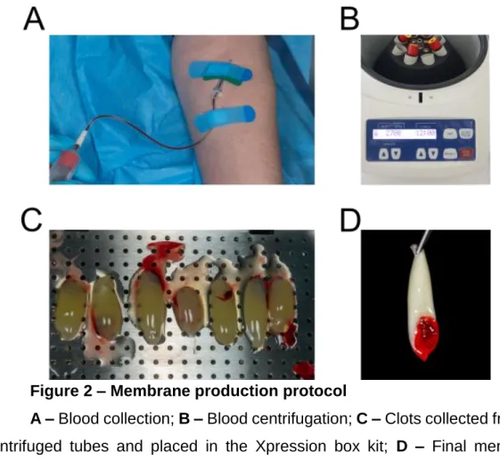
Blood was collected from a healthy person, with no history of anticoagulant usage, into 9 mL sterile glass coated plastic tubes (IntraLock; U.S.A.) (Figure
2A). 8 tubes will be prepared at each time. L-PRF will be prepared according to
Nelson R. Pinto et al. guidelines, centrifugation at 2700 rpm for 12 minutes with the IntraSpin centrifuge by IntraLock, the only CE and FDA approved system for PRF production.(16,27) (Figure 2B). After the first two tubes of blood are collected, they were immediately placed into the IntraSpin centrifuge, opposite to each other to ensure the centrifuge is properly balanced. The cover was closed and the timer set to one minute. We allowed the centrifuge to run for one minute. While it was spinning for 1 minute we collected the third and fourth tubes of blood from the patient, and repeated the procedure for the other tubes. Centrifugation should be at 2700 rpm using the IntraSpin centrifuge, for at least 12 minutes (start timing after loading the centrifuge with last 2 tubes).• After ≥ 12 minutes centrifugation, L-PRF clots are ready. The fibrin clots were taken out of the tubes and separated from the red blood cells. For A-PRF membrane preparation the only difference was the centrifugation speed and time: 1500 rpm for 14 minutes (18,20). Following membrane preparation fibrin clots were placed in the Xpression box (IntraLock) for gentle compression by gravity (Figure 2C). 5 minutes later the L-PRF and A-PRF membranes are ready for use (Figure 2D).
12
Figure 2 – Membrane production protocol
A – Blood collection; B – Blood centrifugation; C – Clots collected from the
centrifuged tubes and placed in the Xpression box kit; D – Final membrane obtained.
2.2
- Traction test
Before traction test membranes were measured using a WHO Periodontal probe and cut in a rectangular shape in which the short ends measured 5mm each. Traction test was performed using the Shimadzu MMT-101N (Shimadzu Corporation; Japan) equipment. By applying divergent forces (1mm spacing between the claws of the equipment) the maximum traction was measured in 13 membranes for each protocol, until rupture. The maximum value for traction using this equipment is set to 12mm
2.3-
Data and statistical analysis
The force applied to the membrane per area of section in the claws (N.mm -2), and the traction of the membrane (percentage of deformation in comparison to the initial spacing between the claws, 1mm) were plotted for each membrane.
13
This allowed us to construct a graph in order to determine the maximum force that was applied, when the membrane ruptured, giving us the maximum tensile strength. Data was collected in Microsoft Excel and GraphPad Prism. All statistical analyses were performed with GraphPad Prism software. Values are presented as mean ± s.e.m. in figure legends. Statistical comparisons included two-sided unpaired t-test.
15
17
The traction evaluation is based on the quantification of the average traction obtained for each membrane tested and the maximum value detected upon rupture of each membrane. This proved important since we wanted to discover the maximum resistance of the membranes but also if it would represent an actual statistically different average resistance. From the traction evaluation of 13 A-PRF and 13 L-PRF membranes, we found that there was significant statistical difference in the maximum traction with rupture and in the average traction between the A-PRF and L-PRF protocols. The traction test results had some variability within and between groups (Figure 3A-B). In reference to the average traction A-PRF obtained a value of 0.0288 N.mm-2 and L-PRF 0.0192 N.mm-2 (p<0.05 using unpaired t-test; n=13). For maximal traction A-PRF obtained 0.0752 N.mm-2 and L-PRF 0.0425 N.mm-2 (p<0.001 using unpaired t-test; n=13) (Figure 3C-D). From this evaluation we concluded that A-PRF obtained both a maximum traction value and an average traction value much higher than L-PRF.
19
Figure 3 – A-PRF has higher average and maximal traction values.
A – Representative traction profile with maximal traction with rupture of
membrane and average traction identified by arrows; B – Individual values of each membrane tested for each protocol with average values of traction, and maximal value for traction measured; C – Average traction difference between L-PRF and A-L-PRF protocol (*p<0.05 using unpaired t-test n=13); D – Maximal traction difference between L-PRF and A-PRF protocol (***p<0.001 using unpaired t-test n=13).
21
23
With this study we intended to evaluate if there was any difference in mechanical properties between L-PRF and A-PRF. We consider this to be a vital question, since these techniques are being more and more used throughout many fields. In dentistry, periodontal surgery is starting to question the use of PRF membranes opposed to a subepithelial connective tissue graft. We know that in terms of root coverage the result will not be the same. However, the use of PRF avoids a donor site which greatly decreases the postoperative discomfort (28).
Through the low-speed centrifugation concept researchers found that it was possible to reduce the cell pull-down by the g forces applied in the centrifugation(18), which increases the quantity of cells within the top layer of the fibrin matrix. This surely modifies the properties of A-PRF when compared to L-PRF, which suffers much higher forces during the centrifugation, concentrating almost all cellular content in the bottom of the clot (18).
With the results we obtained, it is now possible to state that A-PRF has a higher resistance to traction then L-PRF. A-PRF scored 0.0752 N.mm-2 and L-PRF 0.0425 N.mm-2 (p<0.05 using unpaired t-test; n=13). Concerning average traction A-PRF scored 0.0288 N.mm-2 and L-PRF 0.0192 N.mm-2 (p<0.05 using unpaired t-test; n=13). We noticed that in the A-PRF protocol some membranes scored the same in regard to the maximal traction, however the average traction was different, indicating that the structure of each membrane is different. In the L-PRF protocol the same happened but with slightly different values. This indicates that there is a large variation to be expected when producing PRF membranes, and one could not expect to achieve the highest traction possible.
We knew that A-PRF had a higher concentration of growth factors within its fibrin matrix, increasing the tissues regeneration rate when applied in a surgical wound. This fact, allied to the higher maximal traction and average traction, appears to make it a more suitable material for regeneration then L-PRF(20).
As objectives we wanted to be able to produce membranes with the L-PRF and A-PRF protocols which we did and also to find some difference in the mechanical properties of both protocols. Which we as well were able to find, proving that indeed the low-speed centrifugation concept produces membranes with a more even distribution of cells throughout the clot and also a more
24
mechanically resistant membrane. This reveals that when applied in multiple situations A-PRF should be more effective. With this study we can now advise clinicians to use A-PRF membranes instead of L-PRF in order to achieve a possibly better result.
However we know that there are some factors that can influence our study. As stated before, through the centrifugation process most of the cellular content is pulled to the bottom of the clot, and so when cutting the membrane macroscopically for the traction test we need to be sure to use the bottom part in order to use the most resistant part of the membrane. Also the variety in results of maximal traction could indicate a small sample of membranes was used in this study.
Our research team is developing a new study which will focus on a new type of PRF released recently, with still very little research. And we wish to compare its resistance to our findings. Also we think that by increasing the resistance of the membrane we will be producing a more fibrous matrix which will result in a longer absorption time. Prolonging the effects of the release of growth factors and leukocytes in the wound that the membrane has been applied to. This question is yet to be answered, however our team is already tackling this issue. .
25
27
Through this study we can now state that there is a significantly higher resistance to traction in membranes produced with the A-PRF protocol. These results show a promising future in the field of applications for PRF membranes. As shown, PRF membranes show a better capability to withstand traction forces. For the above-mentioned reason, we believe that the use of PRF membranes would be beneficial in newer techniques for periodontal surgery like the Vestibular incision subperiostal tunnel access (VISTA) technique, for example. The newer techniques for periodontal surgery require a material with a high traction resistance to be pulled using sutures. Also, as stated before, we believe the higher resistance of the matrix has a positive influence in the absorption time of the membrane, which would be beneficial when applying it to a surgical wound with the intent of raising the regeneration rate of the tissues.
29
31
1. Dohan Ehrenfest DM, Rasmusson L, Albrektsson T. Classification of platelet concentrates: from pure platelet-rich plasma (P-PRP) to leucocyte- and platelet-rich fibrin (L-PRF). Trends in Biotechnology. Março de 2009;27(3):158–67.
2. Anitua E, Aguirre JJ, Algorta J, Ayerdi E, Cabezas AI, Orive G, et al. Effectiveness of autologous preparation rich in growth factors for the treatment of chronic cutaneous ulcers. Journal of Biomedical Materials Research Part B: Applied Biomaterials. Fevereiro de 2008;84B(2):415–21. 3. Steenvoorde P, van Doorn LP, Naves C, Oskam J. Use of autologous
platelet-rich fibrin on hard-to-heal wounds. Journal of Wound Care. Fevereiro de 2008;17(2):60–3.
4. Marx RE. Platelet rich plasma: Growth factor enhancement for bone grafts. ORAL SURGERY ORAL MEDICINE ORAL PATHOLOGY. 1998;85(6):9. 5. Dohan Ehrenfest David M. Classification of platelet concentrates
(Platelet-Rich Plasma-PRP, Platelet-(Platelet-Rich Fibrin-PRF) for topical and infiltrative use in orthopedic and sports medicine: current consensus, clinical implications and perspectives. Muscle, Ligaments and Tendons Journal [Internet]. 2014 [citado 12 de Fevereiro de 2019]; Disponível em: http://www.mltj.org/common/php/portiere.php?ID=45afa4e561b98b6778e06 d4d95e748e2
6. Whitman H, Berry L, Green M. P/a telet Gel: An Autologous Alternative to Fibrin Glue With Applications in Oral and Maxillofacial Surgery. :6.
7. Dragonas P, Schiavo JH, Avila-Ortiz G, Palaiologou A, Katsaros T. Plasma rich in growth factors (PRGF) in intraoral bone grafting procedures: A systematic review. J Craniomaxillofac Surg. 17 de Janeiro de 2019;
8. Anitua E, Sánchez M, Orive G, Andía I. The potential impact of the preparation rich in growth factors (PRGF) in different medical fields☆. Biomaterials. Novembro de 2007;28(31):4551–60.
9. Miron RJ, Zucchelli G, Pikos MA, Salama M, Lee S, Guillemette V, et al. Use of platelet-rich fibrin in regenerative dentistry: a systematic review. Clinical Oral Investigations. Julho de 2017;21(6):1913–27.
10. Anitua E, Alkhraisat MH, Orive G. Perspectives and challenges in regenerative medicine using plasma rich in growth factors. Journal of Controlled Release. Janeiro de 2012;157(1):29–38.
11. Dohan DM, Choukroun J, Diss A, Dohan SL, Dohan AJJ, Mouhyi J, et al. Platelet-rich fibrin (PRF): A second-generation platelet concentrate. Part I: Technological concepts and evolution. Oral Surgery, Oral Medicine, Oral Pathology, Oral Radiology, and Endodontology. Março de 2006;101(3):e37– 44.
12. Dohan DM, Choukroun J, Diss A, Dohan SL, Dohan AJJ, Mouhyi J, et al. Platelet-rich fibrin (PRF): A second-generation platelet concentrate. Part II:
32
Platelet-related biologic features. Oral Surgery, Oral Medicine, Oral Pathology, Oral Radiology, and Endodontology. Março de 2006;101(3):e45– 50.
13. Dohan DM, Choukroun J, Diss A, Dohan SL, Dohan AJJ, Mouhyi J, et al. Platelet-rich fibrin (PRF): A second-generation platelet concentrate. Part III: Leucocyte activation: A new feature for platelet concentrates? Oral Surgery, Oral Medicine, Oral Pathology, Oral Radiology, and Endodontology. Março de 2006;101(3):e51–5.
14. Choukroun J, Diss A, Simonpieri A, Girard M-O, Schoeffler C, Dohan SL, et al. Platelet-rich fibrin (PRF): A second-generation platelet concentrate. Part IV: Clinical effects on tissue healing. Oral Surgery, Oral Medicine, Oral Pathology, Oral Radiology, and Endodontology. Março de 2006;101(3):e56– 60.
15. Choukroun J, Diss A, Simonpieri A, Girard M-O, Schoeffler C, Dohan SL, et al. Platelet-rich fibrin (PRF): A second-generation platelet concentrate. Part V: Histologic evaluations of PRF effects on bone allograft maturation in sinus lift. Oral Surgery, Oral Medicine, Oral Pathology, Oral Radiology, and Endodontology. Março de 2006;101(3):299–303.
16. Pinto NR, Temmerman A, Castro AB, Cortellini S, Teughels W, Quirynen M. Guidelines for the use of L-PRF Flow Charts: Step by Step Approach Leucocytes and Platelet Rich Fibrin in Different Intra-oral Applications Applying the IntraSpin TM Concept. Unpublished [Internet]. 2017 [citado 7 de
Fevereiro de 2019]; Disponível em:
http://rgdoi.net/10.13140/RG.2.2.13845.01761
17. Saluja H, Dehane V, Mahindra U. Platelet-Rich fibrin: A second generation platelet concentrate and a new friend of oral and maxillofacial surgeons. Annals of Maxillofacial Surgery. 2011;1(1):53.
18. Miron RJ, Choukroun J, editores. Platelet Rich Fibrin in Regenerative Dentistry: Biological Background and Clinical Indications: Biological Background and Clinical Indications [Internet]. Oxford, UK: John Wiley & Sons, Ltd; 2017 [citado 2 de Fevereiro de 2019]. Disponível em: http://doi.wiley.com/10.1002/9781119406792
19. Pinto NR, Ubilla M, Zamora Y, Del Rio V, Dohan Ehrenfest DM, Quirynen M. Leucocyte- and platelet-rich fibrin (L-PRF) as a regenerative medicine strategy for the treatment of refractory leg ulcers: a prospective cohort study. Platelets. 4 de Julho de 2018;29(5):468–75.
20. Ghanaati S, Booms P, Orlowska A, Kubesch A, Lorenz J, Rutkowski J, et al. Advanced Platelet-Rich Fibrin: A New Concept for Cell-Based Tissue Engineering by Means of Inflammatory Cells. Journal of Oral Implantology. Dezembro de 2014;40(6):679–89.
21. Masuki H, Okudera T, Watanebe T, Suzuki M, Nishiyama K, Okudera H, et al. Growth factor and pro-inflammatory cytokine contents in platelet-rich
33
plasma (PRP), plasma rich in growth factors (PRGF), advanced platelet-rich fibrin (A-PRF), and concentrated growth factors (CGF). International Journal of Implant Dentistry [Internet]. Dezembro de 2016 [citado 4 de Junho de
2019];2(1). Disponível em:
http://journalimplantdent.springeropen.com/articles/10.1186/s40729-016-0052-4
22. Khorshidi H, Raoofi S, Bagheri R, Banihashemi H. Comparison of the Mechanical Properties of Early Leukocyte- and Platelet-Rich Fibrin versus PRGF/Endoret Membranes. International Journal of Dentistry. 2016;2016:1– 7.
23. Khorshidi H, Haddadi P, Raoofi S, Badiee P, Dehghani Nazhvani A. Does Adding Silver Nanoparticles to Leukocyte- and Platelet-Rich Fibrin Improve Its Properties? BioMed Research International. 27 de Maio de 2018;2018:1– 5.
24. Patel GK, Gaekwad SS, Gujjari SK, S.C. VK. Platelet-Rich Fibrin in Regeneration of Intrabony Defects: A Randomized Controlled Trial. Journal of Periodontology. Novembro de 2017;88(11):1192–9.
25. Castro AB, Meschi N, Temmerman A, Pinto N, Lambrechts P, Teughels W, et al. Regenerative potential of leucocyte- and platelet-rich fibrin. Part B: sinus floor elevation, alveolar ridge preservation and implant therapy. A systematic review. Journal of Clinical Periodontology. Fevereiro de 2017;44(2):225–34.
26. Castro AB, Meschi N, Temmerman A, Pinto N, Lambrechts P, Teughels W, et al. Regenerative potential of leucocyte- and platelet-rich fibrin. Part A: intra-bony defects, furcation defects and periodontal plastic surgery. A systematic review and meta-analysis. Journal of Clinical Periodontology. Janeiro de 2017;44(1):67–82.
27. Dohan Ehrenfest DM, Pinto NR, Pereda A, Jiménez P, Corso MD, Kang B-S, et al. The impact of the centrifuge characteristics and centrifugation protocols on the cells, growth factors, and fibrin architecture of a leukocyte- and platelet-rich fibrin (L-PRF) clot and membrane. Platelets. Março de 2018;29(2):171–84.
28. Ӧncum E. The Use of Platelet-Rich Fibrin Versus Subepithelial Connective TIssue Graft in Treatment of Multiple Gingival Recessions: A Randomized Clinical Trial. Int J Periodontics Restorative Dent.