MARTA SOFIA
SOARES CRAVEIRO
ALVES MONTEIRO
FITOTOXICIDADE E TRANSFERÊNCIA TRÓFICA DE
CÁDMIO PARA ISÓPODES
CADMIUM PHYTOTOXICITY AND TROPHIC
TRANSFER TO ISOPODS
MARTA SOFIA
SOARES CRAVEIRO
ALVES MONTEIRO
FITOTOXICIDADE E TRANSFERÊNCIA TRÓFICA DE
CÁDMIO
CADMIUM PHYTOTOXICITY AND TROPHIC
TRANSFER TO ISOPODS
dissertação apresentada à Universidade de Aveiro para cumprimento dos requisitos necessários à obtenção do grau de Doutor em Biologia, realizada sob a orientação científica da Professora Doutora Maria da Conceição Lopes Vieira dos Santos, Professora Associada com Agregação da Universidade de Aveiro e do Doutor Reinier Mann, Investigador de Pós-Doutoramento do CESAM – Centro de Estudos do Ambiente e do Mar da Universidade de Aveiro
Apoio financeiro da Fundação para a Ciência e a Tecnologia e do Fundo Social Europeu no âmbito do III Quadro Comunitário de Apoio (bolsa de
o júri
presidente Prof. Doutor Dinis Gomes de Magalhães dos Santos
professor catedrático da Universidade de Aveiro
Prof. Doutora Helena Maria de Oliveira Freitas
professora catedrática da Faculdade de Ciências e Tecnologia da Universidade de Coimbra
Prof. Doutora Olinda da Conceição Pinto Carnide
professora catedrática da Universidade de Trás-os-Montes e Alto Douro
Prof. Doutor Amadeu Mortágua Velho da Maia Soares
professor catedrático da Universidade de Aveiro
Prof. Doutora Maria da Conceição Lopes Vieira dos Santos (orientadora)
professora associada com agregação da Universidade de Aveiro
Prof. Doutor Fernando José Mendes Gonçalves
professor auxiliar com agregação da Universidade de Aveiro
Prof. Doutor José Paulo Filipe Afonso de Sousa
professor auxiliar da Faculdade de Ciências e Tecnologia da Universidade de Coimbra
Doutor Reinier Matthew Mann (co-orientador)
investigador de pós-doutoramento do CESAM - Centro de Estudos do Ambiente e do Mar da Universidade de Aveiro
agradecimentos À Prof. Doutora Conceição Santos pelos seus ensinamentos, críticas,
sugestões e orientação prestada durante a realização deste trabalho. Ao Doutor Reinier Mann pelo acompanhamento e orientação, por ter estado sempre presente (mesmo quando do outro lado do planeta…) em todas as etapas deste trabalho, desde o desenho experimental à revisão dos textos.
Ao Professor Doutor Amadeu Soares por todos os seus conselhos e apoio durante a realização deste trabalho.
À Doutora Tina Lopes pelo seu apoio e acompanhamento na parte laboratorial e de revisão de textos e por todas as sugestões dadas no decurso deste trabalho.
A todos os colegas do Laboratório de Biotecnologia e Citómica pelo seu apoio e disponibilidade, em especial ao João, ao Eleazar e à Catarina. Aos técnicos Abel e Ramiro e a todos os colegas do LETAL pelo companheirismo, inter-ajuda e disponibilidade sempre demonstradas. Aos colegas e amigos do LabEco, companheiros nas fases de trabalho, mas também nos momentos de pausa e descontracção, especialmente pela paciência que tiveram em ouvir as minhas lamúrias e pelas
palavras de apoio nestes últimos meses.
A todos os amigos e familiares que sempre me deram uma palavra de apoio e alento para chegar aqui, pela sua compreensão em todas as vezes que não pude estar tão presente.
Aos meus pais, por todos os ensinamentos e valores transmitidos, por me terem sempre apoiado nas minhas escolhas e pelas suas palavras de incentivo e confiança.
Ao Edgar por ter partilhado comigo todos os momentos destes últimos anos, os bons e os de maior stress, por todas as palavras que ditas no momento certo me fizeram acreditar e ter força para continuar e chegar até aqui.
palavras-chave Cádmio, citometria de fluxo, ecotoxicologia, fitotoxicidade, genotoxicidade, isópodes,
Lactuca sativa, microsatélites, plantas, Thlaspi arvense, Thlaspi caerulescens,
transferência trófica
resumo O cádmio (Cd) é um metal não essencial e é considerado um poluente prioritário
pela comunidade europeia. Este metal atinge o ambiente no decurso de várias actividades antropogénicas e tende a concentrar-se nos solos e sedimentos, onde está potencialmente disponível para as plantas, sendo posteriormente transferido através da cadeia trófica. Neste contexto, o principal objectivo da presente dissertação foi o estudo dos efeitos da assimilação e da acumulação de Cd em plantas e as suas consequências para animais consumidores. Numa primeira fase, foram estudados os principais efeitos fisiológicos e genotóxicos do Cd em plantas. As plantas de alface (Lactuca sativa L.) expostas a Cd apresentaram um
decréscimo na eficiência fotossintética, aumento de peroxidação lipídica e alterações significativas na actividade de enzimas de stress oxidativo. Estas alterações culminaram num decréscimo do crescimento da parte aérea no final da exposição. As respostas obtidas pelos parâmetros bioquímicos sugerem que estes poderão ser utilizados como eventuais biomarcadores em testes ecotoxicológicos com Cd em abordagens integrantes em conjunto com parâmetros clássicos. Os efeitos mutagénicos de Cd foram avaliados através da determinação da
instabilidade de microsatélites (IM). Não foi observada IM, nem nas folhas nem nas raízes de plantas de alface com 5 semanas de idade expostas a 100 μM Cd durante 14 dias, no entanto observou-se IM em raízes de alface exposta a 10 μM Cd
durante 28 dias desde a germinação. A idade da planta e a maior acumulação de Cd nas raízes poderão explicar os resultados obtidos. A clastogenicidade de Cd foi analisada em três espécies vegetais com diferentes capacidades de destoxificação e acumulação de metais através de citometria de fluxo. Foram detectadas
alterações significativas nos parâmetros analisados em raízes alface, mas não nas espécies Thlaspi caerulescens J & C Presl e Thlaspi arvense L. Estes resultados sugerem que o stress provocado pelo Cd originou clastogenicidade como
consequência da perda de porções de cromossomas, uma vez que o conteúdo de ADN nuclear diminuiu. A transferência trófica através da cadeia alimentar
permanece muito pouco estudada em termos ecotoxicológicos. A distribuição subcellular de metais num organismo pode ser utilizada para compreender a
transferência trófica de um metal na cadeia alimentar. Como tal, numa última parte é estudado de que modo a distribuição subcellular do Cd em plantas com perfis de acumulação de Cd distintos afecta a biodisponibilidade e transferência trófica de Cd para isópodes. A distribuição de Cd entre as 4 fracções subcelulares obtidas através de centrifugação diferencial revelou a existência de diferenças significativas entre as espécies de plantas. Estes resultados em conjunto com a avaliação directa da eficiência de assimilação (EA) de Cd individual de cada uma das quatro fracções subcelulares das plantas em estudo, resultou em informação de grande relevância para a explicação das diferenças observadas na EA de Cd por parte de isópodes alimentados com folhas de diferentes espécies de plantas. Com base nos resultados obtidos, o Cd ligado a proteínas estáveis à temperatura (e.g. metaloteoninas e fitoquelatinas) é o menos biodisponível, sendo assim o que menos contribuiu para a transferência trófica, enquanto que o Cd ligado a proteínas desnaturadas pela temperatura foi a fracção mais disponível para transferência trófica de Cd ao
keywords Cadmium, ecotoxicology, flow cytometry, genotoxicity, isopods, Lactuca sativa,
microsatellites, phytotoxicity, Thlaspi arvense, Thlaspi caerulescens, trophic transfer
abstract Cadmium (Cd) is a non-essential metal and is considered a priority
pollutant by the European Community. This metal is released to the environment as a consequence of several anthropogenic activities and tends to accumulate in soils and sediments where it is potentially available to rooted plants causing severe detrimental effects and then transferred to animals through the food chain. In this context, the main objective of the present dissertation is to study the effects of Cd uptake and accumulation in plants and its implications to animal consumers. First, the main
physiologic and genotoxic effects of Cd to plants were examined.
Cadmium-exposed lettuce (Lactuca sativa L.) plants displayed a significant decrease in photosynthetic efficiency, enhanced lipid peroxidation and alterations in the activities of antioxidant enzymes over the duration of exposure. These alterations culminated in reduced shoot growth at the end of the exposure. The aforementioned biochemical alterations are suggested to be used as plant biomarkers in integrative approaches with classical endpoints in future ecotoxicological tests with Cd. The mutagenic effects of Cd on plants were assessed examining microsatellite instability (MSI). No MSI was found neither in leaves nor roots of 5-week old lettuce plants exposed to 100 μM Cd, but MSI was found in roots of lettuce plants exposed to 10 μM for 28 days from seed germination. The age of the plant at the time and the higher accumulation of Cd in the roots might explain the results obtained. Clastogenic effects of Cd was examined in plants with different metal accumulation and detoxification capacities by flow
cytometric (FCM). The endpoints analysed indicated significant alterations in lettuce roots but not in Thlaspi caerulescens J & C Presl and Thlaspi
arvense L.. The results obtained suggested that Cd stress may have lead to clastogenic damage as a consequence of loss of chromosome portions because nDNA content was found to be diminished. Trophic transfer through the food chain remains a largely unexplored area of ecotoxicology. Subcellullar distribution of metal accumulated within an organism can be used to understand metal trophic transfer within a food chain. Thus, in a final stage we examined how Cd subcellular distribution in plants with different patterns of Cd accumulation can affect assimilation of Cd by the isopod. The distribution of Cd between the four different subcellular fractions obtained by differential centrifugation revealed significant differences between the plant species. This, together with the direct assessment of isopod Cd AE from individual subcellular fractions of the leaves of the three plant species, resulted in vital information to help explain the differences observed in Cd AE by isopods fed the different type of leaves. On the basis of our results, Cd bound to heat-stable proteins (e.g. phytochelatins and methallothionein) was the least bioavailable to isopods and contributed less to the trophic transfer of Cd, while Cd bound to heat-denatured proteins was the most trophically available to the isopod. These results point to the ecological relevance of the subcellular
“There is no simple answer to the basic questions - why and how?”
Z Krupa et al. (2002) Heavy metal interactions with plant nutrients. In: MNV Prasad and K Strzałka (Eds). Physiology and biochemistry of metal toxicity and tolerance in plants. Kluwer Academic Publishers, London, pp. 287-301.
Agradecimentos iv
Resumo (em Português) v
Abstract (in English) vi
Index ix
Chapter 1 General Introduction
Preamble 3
Cadmium – a priority pollutant 5
What is Cd 5
Sources of Cd pollution 5
Cadmium characteristics 7
Cadmium uptake: from soils to plants 8
Cadmium in soils 8
Uptake and transport of Cd by plants 9
Phytotoxicity effects of cadmium as environmental markers of Cd stress 10
General effects 10
Oxidative stress 13
Genotoxicity effects of Cd in plants 14
Mechanisms of tolerance 19
Metal-binding ligands 19
Plant metal accumulation and hyperaccumulation 20
Trophic transfer of Cd 21
Factors affecting trophic transfer of metals 21
Metal assimilation and assimilation efficiency 22
Subcellular partition of metals 23
Isopods as model species for metal accumulation 25
Objectives 26
References 28
Chapter 2 Assessment of biomarkers of Cd stress in lettuce
Abstract 43
Introduction 44
Materials and Methods 46
Plant culture and growth conditions 46
Nutritional status and Cd analysis 46
Toxicity symptoms and plant growth 48
Cadmium accumulation and nutrient imbalances 49 Chlorophyll content and PSII efficiency 51
Lipid peroxidation and membrane permeability 52
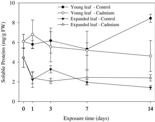
Soluble protein content 52
Antioxidant enzymes 53
Discussion 55
Plant growth 55
Metal accumulation and distribution 56
Chlorophyll content and PSII efficiency 56
Macro and micronutrient imbalances 57
Lipid peroxidation and membrane permeability 59
Soluble protein content 59
Antioxidant response to Cd 60
Conclusions 60
References 61
Chapter 3 Cadmium genotoxic effects in plants 3.1 – Evaluation of Cd genotoxicity in Lactuca sativa L. using nuclear microsatellites 69
Abstract 69 Introduction 70
Materials and Methods 72
Plant culture and growth conditions 72
Cadmium analysis 72
Microsatellite analysis 73
Results 74
Discussion 78
References 82
3.2 – Microsatellite instability in Lactuca sativa chronically exposed to cadmium 87 Abstract 87 Introduction 88
Materials and Methods 89
Plant culture and growth conditions 89
Cadmium analysis 90
Microsatellite analysis 90
Statistical analysis 91 Results 92
Cadmium accumulation and plant growth 92
3.3 – Flow cytometric assessment of Cd genotoxicity in three plants with different
metal accumulation and detoxification capacities 105
Abstract 105
Introduction 106
Materials and Methods 107
Plant culture and growth conditions 107
Cadmium accumulation assessment 108
Flow cytometric analysis 108
Statistical analysis 109
Results 109
Cadmium accumulation assessment 109
Toxicity symptoms and plant growth 111
Flow cytometric analysis 111
Discussion 113
Conclusions 116
References 117
Chapter 4 Trophic transfer of Cd from plants to isopods Does subcellular distribution in plants dictate the trophic bioavailability of Cd to Porcellio dilatatus (Crustacea, Isopoda)? 123
Abstract 123
Introduction 124
Materials and Methods 126
Plant culture and growth conditions 126
Subcellular Cd distribution in plant leaves 127
Isopod culture and feeding test conditions 127
Feeding experiment 1 128
Feeding experiment 2 129
Cadmium analysis 130
Statistical analysis 130
Results 130
Cadmium subcellular distribution in plant leaves 130
Feeding experiment 1 131
Isopod growth 131
Plant consumption and assimilation efficiency 133
Cadmium consumption, assimilation, and AE 133
Feeding experiment 2 133
Growth 133
Cadmium AE from purified subcellular fractions 135
Thlaspi caerulescens 137
Thlaspi arvense 138
Cadmium assimilation from plants 138
Confounding effects 139
Cadmium assimilation efficiencies 139
Conclusions 142
References 144
Chapter 5 General discussion and concluding remarks General discussion and concluding remarks 147
General Introduction
Preamble
Cadmium (Cd) is a naturally occurring element, and its presence has been detected in more than 1,000 species of aquatic and terrestrial flora and fauna (Eisler, 1985). With one known exception, there is no evidence that Cd is biologically essential or beneficial; on the contrary, it has been implicated in several human health diseases and various deleterious effects in wildlife (Eisler, 1985). The exception is a Cd-dependent carbonic anhydrase found in the marine diatom Thalassiosira weissflogii (Lane and Morel, 2000; Lane et al., 2005); a similar role has been postulated for the metal hyperaccumulating plant, Thlaspi caerulescens (Liu et al., 2008). In all life-forms, including microorganisms, higher plants and animals (in particular humans), Cd is toxic when present in sufficient concentrations (Eisler, 1985).
The identification of Cd as a distinct element is relatively recent. The German scientist Friedrich Stromeyer was at the origin of the discovery of Cd in 1817 (Robards and Worsfold, 1991). Its toxicity was soon recognized and early recorded cases of Cd poisoning were generally as a consequence of industrial exposure involving inhalation of Cd dusts (Robards and Worsfold, 1991).
Pollution of the biosphere with this toxic metal has accelerated dramatically since the beginning of the industrial revolution (Nriagu, 1996) and Cd accumulation in soil and water now poses a major environmental and human health problem. In 1955 in Japan, Cd toxicity was found to be the cause of Itai–itai disease. For the first time, Cd pollution was shown to have severe consequences on human health. Cadmium contaminations were attributed to the effluents from a zinc mine located in the upper reaches of Jinzu river and profoundly affected the health of the human population living in that area (Inaba et al., 2005).
Historically, the study of metal uptake by plants has focused on micronutrient metals important in agricultural production, whereas non-essential metals, such as Cd, Hg and Pb, have generally received less attention. However, over the last three decades Cd has been the subject of several investigations in plant research mainly because of its potential for bioaccumulation through soil-plant-animal food-chain. For example, the consequences of soil contamination by Cd, through application of treated sewage sludge (biosolids) (McLaughlin et al., 2006) and Cd-enriched phosphate fertilizers (e.g. He and Singh, 1994b, a) to soils have been extensively studied. However, the driving force of this research area has been the concern for the risk to human health, not for the state of the plant itself.
Most of the research on Cd pollution focused on the processes involved in Cd accumulation in crop plants and on the consequences of this accumulation on human health (Wagner, 1993). Cadmium phytotoxicity is, however, a relevant problem, especially in some highly metal polluted regions, where a decrease in agricultural crop productivity has been observed (Vassilev and Yordanov, 1997). More recently, metal hyperaccumulator plant species have been used to re-examine mechanisms of metal uptake by plants in light of the potential for phytoremediation of metal-contaminated soils (Chaney et al., 1997; Pilon-Smits, 2005; Padmavathiamma and Li, 2007). On the other hand, Cd toxicity to plants has great impact and relevance not only for plants but also to the ecosystem, in which the plants form an integral component. Therefore, understanding Cd uptake and physiological responses of plants is critical to the long-term safety and conservation of agricultural resources and ecosystems. In addition, plants as sedentary organisms offer unique advantages for in situ monitoring of soil contamination (Grant, 1999) and can potentially be used as biomonitors of environmental quality through the use of biomarkers. In plants it is well known that Cd interferes with photosynthesis, respiration and nitrogen metabolism, induces oxidative stress and genotoxicity, all of which can culminate in poor growth and low biomass production (Sanitá di Toppi and Gabbrielli, 1999; Fodor, 2002). The biochemical pathways involved in these processes offer a battery of biochemical biomarkers that not only provide mechanistic endpoints of toxicity, but also improve our understanding on the toxic modes of action and exposure assessment. Hence, the purpose of the first part of the present dissertation is to contribute to a better understanding of the overall process of Cd-induced senescence, describing the cascade of events and the enzymatic protection strategies that plants can adopt against Cd-induced oxidative stress (Chapter 2) in order to shed light on a selection of relevant plant biomarkers for further use as biomonitoring tools in the assessment of environmental Cd pollution. Special emphasis is given to genotoxicity of Cd; in Chapter 3 the clastogenic and mutagenic effects of Cd in exposed plants are examined.
Another issue concerning Cd accumulation in plants centres on the fact that Cd could pose a risk to animal health if they consume plants contaminated with Cd, even if plant tissue concentrations are not generally phytotoxic (McLaughlin, 2002). Indeed, the ability of some plant species to uptake and hyperaccumulate Cd in edible parts increases the risk of Cd assimilation by animal consumers through trophic transfer. Because there is very limited knowledge regarding the trophic transfer of metallic contaminants between plants and consumers of plants, the second part of this dissertation presents an examination of the subcellular distribution of Cd within plant leaves, and subsequent
significance of that distribution on the bioavailability of Cd to a consumer - a detritivore isopod (Crustacea). A subcellular fractionation procedure developed by Wallace and co-workers (Wallace et al., 2003; Wallace and Luoma, 2003) that has been largely applied in the dietary accumulation of metals, particularly in marine food chains, was adopted to try to explain the variability observed in metal assimilation by isopods fed plants with different patterns of Cd accumulation. Indeed, this method is considered a simple and pragmatic approach in the prediction of trophic transfer of metals and a first step towards a practical tool that could explain most of the variability observed in metals accumulation and toxicity in organisms (Vijver et al., 2004).
Cadmium – a priority pollutant
What is Cd
Cadmium is an element that occurs naturally in the earth’s crust as a result, for instance, of volcanic emissions. Pure Cd is a soft, silver-white metal, and it is not usually present in the environment as a pure metal, but as a mineral compound combined with other elements. Cadmium is most often present in nature as complex oxides, sulphides, and carbonates in zinc, lead and copper ores (ATSDR, 1999).
Cadmium is generally considered to be a so called “heavy metal” due to its high density (8.6 g.cm-3), high atomic weight (112.4 g.mol−1) or even for its toxic properties.
However, many different definitions have been proposed for this commonly used term; some based on density, on atomic number or weight, and others on chemical properties or toxicity (Nieboer and Richardson, 1980). Despite early suggestions for the use of other nomenclature (Nieboer and Richardson, 1980), the term “heavy metal” has been widely used by scientific community over the past three decades. More recently the term “heavy metal” has been considered meaningless and misleading in an IUPAC technical report due to the contradictory definitions and its lack of a coherent scientific basis (Duffus, 2002). Therefore, in the present dissertation the term “heavy metal” is used restrictively, specifically when citing other works.
Sources of Cd pollution
Cadmium is included in the list of 33 priority pollutants established by European Community (2455/2001/EC, 2001) and is one of 129 priority pollutants listed by EPA
(Environmental Protection Agency, USA). The release of Cd into the environment constitutes a significant pollution problem. Cadmium occurs naturally in the environment. It is estimated that about 25,000 to 30,000 tons of Cd are released to the environment each year; about half from the weathering of rocks into river water; and a further proportion from forest fires and volcanoes (ATSDR, 1999). Release of Cd from human activities is estimated to be about 4,000 to 13,000 tons per year, with major contributions from mining activities, and burning of fossil fuels (ATSDR, 1999). The Cd-yellow oil colours used by landscape painters, including Claude Monet (Figure 1.1) is just one of the many valuable uses of Cd. Other important applications of Cd are in metallurgical industry and in the manufacture of nickel–cadmium batteries, pigments, plastic stabilizers and anti-corrosive products, phosphors for television sets, scintillation counters and X-ray screens, semiconductors and ceramic glazes (Robards and Worsfold, 1991). As a consequence of this widespread and diverse usage, large quantities of Cd end up in sewage.
Treated sewage sludge (“biosolids”) and phosphate fertilizers (He and Singh, 1994b, a; Speir et al., 2003; McLaughlin et al., 2006; Singh and Agrawal, 2007) are important sources of Cd contamination in agricultural soils. The usage of Cd in developed countries has, however, begun to decline because of its toxicity. For instance, Cd is one of six substances banned by the European Union's Restriction on Hazardous Substances (RoHS) directive, which bans carcinogens in computers (2002/95/EC, 2002).
a b
a b
Figure 1.1 – Cadmium, a beautiful toxic colour. a) Poplars in the sun by Claude Monet, 1891 (www.monet-on-canvas.com/prod197.htm), showing the powerful use of Cd yellow pigments and b) a lettuce leaf reflecting the toxic effects of Cd exposure (arrow: Cd-induced necrosis).
Cadmium characteristics
The most important feature which distinguishes metals from other toxic pollutants is that they are not biodegradable and, once they become resident in the biosphere, they remain as a persistent pollutant. For instance, in estuarine coastal systems the residence time of Cd has been estimated as a relatively low 1-2 years (Robards and Worsfold, 1991). On the other hand, estimates of the residence time in ocean water range from 7,000 to 250,000 years (Robards and Worsfold, 1991). The bioavailability of metals and their subsequent toxicity to organisms within the biosphere is controlled largely by their physico-chemical form. Cadmium, among the metals, is likely to have high mobility in soils because it does not bind as strongly to organic matter as do metals such as Hg and Pb (Nelson and Campbell, 1991).
Besides of a long environmental persistence, Cd has a long biological half-life, which accounts for its bioaccumulation in individuals (John and Leventhal, 1996). For example, in man Cd accumulates in the liver and kidneys and has a long biological half-life ranging from 17 to 30 years (Goyer, 1997). Existing data on Cd bioaccumulation in a range of animals and plants (Robards and Worsfold, 1991; Greger, 1999) verify the ability of these species to amplify the concentration of Cd relative to their environment. Bioaccumulation occurs within an organism and is the increase in concentration of a substance in an individual’s tissue as a consequence of uptake from their environment, including diet (Connell et al., 1999). The extent of contaminant bioaccumulation, which is the net outcome of two competing processes, uptake and depuration, is related to the level of environmental contamination and depends upon a number of physico-chemical (e.g. chemical speciation, partitioning) and environmental factors (e.g. season, temperature) and biological variables (e.g. specie, feeding habitat, physiology) that may alter the distribution and bioavailability of individual contaminants (Robards and Worsfold, 1991; Connell et al., 1999). Among the factors that can affect Cd bioaccumulation are its physico-chemical form, the presence of other metals, pH, salinity, temperature, season, cation-exchange capacity of soils and the species taking up the Cd (Robards and Worsfold, 1991 and references therein).
Metal biomagnification is defined as the progressive accumulation of a metal with increasing trophic levels towards higher consumers (Connell et al., 1999). Among metals, this type of amplification at higher trophic levels was previously thought to occur only for mercury, but it has also been demonstrated in a few studies with Cd (Croteau et al., 2005). Croteau and co-workers (2005) have demonstrated that Cd was progressively enriched
among trophic levels in two discrete epiphyte-based food webs composed of macrophyte-dwelling invertebrates or fishes. Cadmium concentrations were biomagnified 15-fold within the span of two trophic links in both food webs.
Cadmium uptake: from soils to plants
Cadmium in soils
Among metals, Cd is of particular concern because of its mobility in the plant-soil system. Wagner (1993) estimated that non-polluted soil solutions contain Cd concentrations ranging from 0.04 to 0.32 µM. Cadmium concentrations in nonpolluted soils are however highly variable, depending on sources of minerals and organic material. For instance, Eisler (1985) reported Cd concentrations of 0.01-1.00 mg/kg in soils of nonvolcanic origin and up to 4.50 mg/kg in soils of volcanic origin. Soil solutions which have a Cd concentration varying from 0.32 to about 1 µM are considered as moderately polluted (Sanitá di Toppi and Gabbrielli, 1999). Topsoil concentrations are often more than twice as high as subsoil levels as the result of atmospheric fallout and contamination (Pierce et al., 1982). Cadmium levels up to 800 mg/kg have been reported for soils in polluted areas (IARC, 1993). Jung and Thornton (1996) have found Cd concentrations up to 40 mg/kg in surface soils taken from a mining area in Korea; and more recently, Cd contaminated river water (65-240 μg/l, 0.58-2.13 μM) downstream from a mining area in Bolivia has increased the soil concentration of Cd to 20 mg/kg and the concentration of Cd in soil solutions to 27 μg/l (0.24 μM)(Oporto et al., 2007).
Contamination of topsoil is likely the most important route for human exposure to Cd, mediated through uptake of soil Cd into edible plants (IARC, 1993). Cadmium concentrations of 0.5 mg/kg or more have been found in rice grown in Cd-polluted areas of Japan (Nogawa et al., 1989) and China (Cai et al., 1990). Furthermore, in a recent field study in Europe performed by Peris et al. (2007) the Cd content in edible parts of vegetables such as lettuce were found to be above the maximum levels established by the Commission Regulation no. 466/2001 for horticultural crops (466/2001/EC, 2001).
The Cd concentration of 100 μM to grow/contaminate plants hydroponically was chosen for use in all different approaches of the present dissertation. Previous studies have used similar approaches and similar concentrations (Azevedo et al., 2005a; Azevedo et al., 2005b, c). Also, this concentration is twice the maximum permitted concentration in irrigation water by Portuguese legislation (0.05 mg/l) (Decreto-Lei, n.º236/98), and therefore represents worst case scenario.
Uptake and transport of Cd by plants
Cadmium accumulation by higher plants can occur through foliar or root uptake. However, the primary point of entry for Cd into plants is through the roots. Cadmium uptake by plants grown in contaminated soils has been extensively studied, particularly in sludge-amended soils (e.g. Jackson and Alloway, 1991; Speir et al., 2003; McLaughlin et al., 2006; Singh and Agrawal, 2007) and in soils treated with Cd-enriched phosphate fertilizers (Crews and Davies, 1985; He and Singh, 1994b, a; Huang et al., 2003). In general, metals have to be in an available form to be taken up by plants. Alternatively plants must have mechanisms to make the metals available. The degree to which higher plants are able to take up Cd depend on its concentration in the soil and its bioavailability. Cadmium bioavailablity in soils is modulated by the presence of organic matter, pH, redox potential, temperature, light intensity, cation exchange capacity and concentrations of other elements (He and Singh, 1993; Greger, 1999; Sanitá di Toppi and Gabbrielli, 1999). In particular, Cd ions seem to compete with other micro and macro-nutrients such as calcium and zinc for the same transmembrane carriers (Sanitá di Toppi and Gabbrielli, 1999), which might lead to plant nutrient deficiencies (Krupa et al., 2002). As is the case for other metals, Cd uptake tends to be reduced at low pHs because of competition with H+ ions at root uptake sites; however, Cd bioavailability increases with decreasing pH in
soil (Greger, 1999). The presence of colloids from which there is a release of metals at low pH, increases the metal concentration in pore water and thus also in the roots (Greger, 1999). For instance, acid rain and the resulting acidification of soils and surface waters are known to increase the geochemical mobility of Cd (Campbell, 2006). Cadmium uptake also appears to be decreased in the presence of dissolved organic matter because ligands on the organic matter effectively bind Cd ions (He and Singh, 1993; Prasad, 1995). Chloride levels would also be expected to affect Cd availability as soil sodium chloride has an antagonistic effect on metal toxicity (Bhartia and Singh, 1994).
In the present dissertation hydroponic culture of plants was chosen as the most suitable culturing method because it avoids taking in account the above factors that can alter bioavailability of Cd in soils for plant uptake. Hence, hydroponics provides the most consistent and reproducible levels of contamination required for the present objectives. In addition, a previous study demonstrated that >90% of the Cd remained in solution in the Hoagland’s nutrient solution used in almost all experiments in this work, and was therefore available for uptake (Mann et al., 2005).
Cadmium is believed to enter the root through the cortical tissue till the stele either by apoplastic and/or a symplastic pathway (Sanitá di Toppi and Gabbrielli, 1999). The apoplast continuum of the root epidermis and cortex is readily permeable to solutes. The cell walls of the endodermal cell layer act as a barrier for apoplastic diffusion into the vascular system. In general, solutes have to be taken up into the root symplasm before they can enter the xylem (McLaughlin, 2002). The cell membrane plays a key role in metal homeostasis, preventing or reducing entry into the cell. However, examples of exclusion or reduced uptake mechanisms in higher plants are limited (Benavides et al., 2005). The mechanism for metal transport across the plasma membrane to the stele still not completely understood (McLaughlin, 2002). For all cationic metals, such as Cd, the main route for uptake across the plasma membrane is the large negative electrochemical potential produced as a result of the membrane H+ translocating adenosine triphospatase
(ATPases) (McLaughlin, 2002). Costa and Morel (1994) reported that in lettuce grown in hydroponic solution with Cd concentrations from 0.05 µM to 5 µM, high amounts of Cd in roots were correlated with high contributions from H+-ATPase in the active process of Cd uptake. Other authors contend however, that the main route for uptake of divalent metals is via ion channels, such as Cd2+ and Mg2+ channels (McLaughlin, 2002 and references
therein). Subsequent to metal uptake into the root symplasm, three processes govern the movement of metals from the root into the xylem: sequestration of metals inside root cells, symplastic transport into the stele and release into the xylem (Clemens et al., 2002).
During their transport through the plant, metals become bound to cell walls, which can explain why normally Cd2+ ions are mainly retained in the roots, and only small
amounts are translocated to the shoots (Cataldo et al., 1983; Greger, 1999). But once loaded in the xylem sap, Cd is translocated to the aerial parts of plants through the transpiration stream, where they might be present as a divalent ion (Greger, 1999) or complexed by several ligands, such as amino acids, organic acids and/or, perhaps, phytochelatins (Salt et al., 1995; Briat and Lebrun, 1999; Sanitá di Toppi and Gabbrielli, 1999; Gong et al., 2003).
Phytotoxicity effects of Cd as environmental markers of Cd stress
General effects
Plants can play a crucial role in the monitoring and assessment of environmental metal pollution. Plants respond to metal accumulation by expressing various
manifestations of toxicity that can be detected and analyzed at various levels of organization ranging from gross morphology to cellular, biochemical or molecular levels, and thus can be useful to monitor as well as assess environmental metal pollution. Moreover, the sedentary nature of plants is a major advantage of plant-based assays for monitoring toxic chemicals in the environment.
Cadmium is a toxic element without any known physiological function in plants that can affect plants on various organizational and functional levels. Several symptoms of Cd stress have been described in plants and they include chlorosis, necrotic lesions, wilting, reddish coloration and growth reduction (Prasad, 1995; Hagemeyer, 1999; Sanitá di Toppi and Gabbrielli, 1999). Disturbances in plant water relations are widely known as one of the first effects of Cd toxicity. Indeed, some authors have proposed that water stress caused by Cd is the beginning of the cascade of physiological and metabolic processes, including photosynthesis impairment (Barceló and Poschenrieder, 1990).
The photosynthetic apparatus is particularly susceptible to Cd toxicity. Photosynthesis can be inhibited at several levels: CO2-fixation, stomatal conductance,
chlorophyll synthesis, electron transport and enzymes of the Calvin cycle (Mysliwa-Kurdziel and Strzalka, 2002). One of the most usual symptoms of Cd stress is chlorosis of the leaves due to an impairment of photosynthetic pigment biosynthetic pathways (Mysliwa-Kurdziel and Strzalka, 2002), but also to a strong interaction between Cd and Fe that reduces uptake of Fe and causes Fe deficiency in leaves (Krupa et al., 2002). Cadmium can alter both chlorophyll biosynthesis by inhibiting protochlorophyllide reductase and the photosynthetic electron transport by inhibiting the water-splitting enzyme located at the oxidising site of photosystem II (Mysliwa-Kurdziel et al., 2002; Mysliwa-Kurdziel and Strzalka, 2002). Moreover, Cd2+, like other metals, can interfere with
photosynthetic pigments through the substitution of the Mg2+ ion in the chlorophyll
molecules by Cd2+ (Mysliwa-Kurdziel and Strzalka, 2002). These substituted chlorophylls have much lower fluorescence quantum yields when compared to Mg-chlorophylls (Krupa et al., 2002). Several authors have reported decreased levels of chlorophyll pigments in different plant species due to Cd stress (e.g. Krupa and Moniak, 1998; Lagriffoul et al., 1998; Láng et al., 1998; Chugh and Sawhney, 1999). Since the chlorophyll concentration may directly influence the functioning of the photosynthetic apparatus and thus affect overall plant metabolism, it is considered a key factor when assessing the impact of Cd stress (Fodor, 2002). The ratio Chl a/Chl b is another related endpoint relevant for Cd toxicity assessment. Although, there is no known direct influence of metal ions on the process of transformation of Chl a to Chl b, changes on Chl a/Chl b ratio are commonly
reported on metal stressed plants (Mysliwa-Kurdziel and Strzalka, 2002). Both increases and decreases in this ratio have been found in plants treated with Cd2+ (Mysliwa-Kurdziel and Strzalka, 2002 and references therein).
The reduction in photosynthetic rate is a common response in plants exposed to several metals (Mysliwa-Kurdziel and Strzalka, 2002). Profound anatomical changes in leaves and structural disorganization of chloroplasts are the basis of the inhibition of photosynthesis (Mysliwa-Kurdziel et al., 2002). The maximum photochemical efficiency of photosystem II (PSII) was found to be reduced in different plant species exposed to Cd (Chugh and Sawhney, 1999; Linger et al., 2005; He et al., 2008). Dark-adapted values of Fv/Fm (Fv, variable fluorescence; Fm, maximal fluorescence induction) reflect the potential
quantitative efficiency of PSII and are used as a sensitive indicator of plant photosynthetic performance (Maxwell and Johnson, 2000).
Another unfavourable effects of toxic metals on plants are the inhibition of the normal uptake and utilization of mineral nutrients (Fodor, 2002). One of the crucial factors of Cd2+ influence on plant metabolism and physiological processes is its relationship with other mineral nutrients. As mentioned above, Cd2+ transport across cell membranes is
most likely facilitated by metal transporters that normally act to mobilize essential metals. Thus, by substituting for essential divalent cations, Cd2+ limits their uptake. Alternatively,
Cd2+ may bind to specific groups of proteins and lipids or channel proteins of membranes,
thereby inhibiting transport and disturbing the uptake of many macro and micronutrients. Furthermore, destruction of the cell membranes can also alter the ratio of essential elements and cause the decrease in their content, thereby inducing nutrient deficiencies (Cseh, 2002).
One of the most important mechanisms for impairment of the uptake of nutrients by Cd is via the inhibition of Fe transport into the shoot, which has a pronounced effect on many aspects of the structure and function of the photosynthetic apparatus (Krupa et al., 2002). The induced iron shoot deficiency reduces the pool of Fe-containing electron carriers in the photosynthetic electron transport chain, causes disorganization of the chloroplast structure and even reduces RuBisCO (ribulose 1,5-bisphosphate carboxylase/oxygenase) content (Siedlecka and Krupa, 1996). Cadmium is also known to cause other important disturbances in nutrient levels that can severely affect normal plant metabolism. Specifically it can decrease the levels of Mg, K, P, Ca and Zn, and increase Mn content (Krupa et al., 2002).
Oxidative stress
A common consequence of most abiotic and biotic stresses is that they result, at some stage of exposure, in an increase in reactive oxygen species (ROS) (Mittler, 2002). The ROS intermediates are partially reduced forms of atmospheric oxygen (O2); they
typically result from the excitation of O2 to form a singlet (1O2), or from the transfer of one,
two or three electrons to O2 to form, respectively, a superoxide radical (O2-), hydrogen
peroxide (H2O2) or a hydroxyl radical (HO-) (Mittler, 2002). Metals have been
demonstrated to stimulate the formation of ROS, either by direct electron transfer involving metal cations, or as a consequence of metal-mediated inhibition of metabolic reactions (Dietz et al., 1999).
Although Cd is known to produce oxidative stress, in contrast with other metals, it does not seem to act directly on the production of ROS (via Fenton and/or Haber Weiss reactions) (Dietz et al., 1999). Metals without redox capacity, such as Cd can enhance the pro-oxidant status of a plant by reducing the antioxidant glutathione (GSH) pool, activating calcium-dependent systems and affecting Fe-mediated processes (Dietz et al., 1999). These metals can also disrupt the photosynthetic electron chain, leading to the production of ROS (O2- and 1O2) (Dietz et al., 1999).
The ROS are generated in plant cells during normal metabolic processes, such as respiration and photosynthesis (Mittler, 2002). Although some of them function as important signalling molecules that alter gene expression and modulate the activity of specific defence proteins, all ROS can be extremely harmful to organisms at high concentrations (Apel and Hirt, 2004). Reactive oxygen species may lead to the oxidation of proteins, lipids and nucleic acids (this particular aspect is discussed further in section 3.3), often leading to lipid peroxidation, membrane damage, mutagenesis and inactivation of enzymes, thus affecting cell viability (Apel and Hirt, 2004). As a consequence, tissues injured by oxidative stress generally contain increased concentrations of carbonylated proteins and malondialdehyde (MDA) (Apel and Hirt, 2004).
The balance between the steady-state levels of different ROS are determined by the interplay between different ROS-producing and ROS-scavenging mechanisms (Mittler, 2002; Apel and Hirt, 2004). One of the plant responses to ROS production is the increase in anti-oxidant enzyme activities providing protection from oxidative damage induced by several environmental stresses (Apel and Hirt, 2004). A variety of proteins function as scavengers of superoxide and hydrogen peroxide. Among the major ROS-scavenging enzymes in plants are catalase (CAT), peroxidase (POX) and superoxide dismutase
(SOD) (Mittler, 2002). The superoxide released by processes such as oxidative phosphorylation is first converted to hydrogen peroxide and then further reduced to give water. This detoxification pathway is the result of multiple enzymes, with superoxide dismutases catalysing the first step and then catalases and various peroxidases removing hydrogen peroxide (see 1.2). In addition, the anti-oxidative enzymes are supplemented with non-protein scavengers, including ascorbate and glutathione (Mittler, 2002).
O2 Oxygen .O 2 -Superoxide Superoxide dismutase H2O2 Peroxidases Catalase H2O Water Hydrogen peroxide
Figure 1.2 - Enzymatic pathway involving anti-oxidant enzymes for detoxification of ROS (adapted from Apel and Hirt (2004)).
As for other stresses, activation or inhibition of anti-oxidative enzymes due to metal stress depends not only on stress intensity and duration but also on the tissue type and the age of the plant (Dietz et al., 1999). Cadmium can inhibit and/or stimulate the activity of several anti-oxidative enzymes (Dietz et al., 1999). Several studies demonstrated that Cd stress induced antioxidant enzymes, whereas some others showed that exposure to high concentrations of Cd resulted in a decrease in antioxidant capacities (Schützendübel and Polle, 2002 and references therein). For instance, catalase activity has been shown to be suppressed in diverse plant species exposed to Cd (Chaoui et al., 1997; Chaoui and El Ferjani, 2005).
Finally, available data suggest that Cd, when not detoxified rapidly enough, may trigger, via the disturbance of the redox control of the cell, a sequence of reactions leading to growth inhibition, stimulation of secondary metabolism, lignification, and subsequent cell death (Schützendübel and Polle, 2002). Thus, in the present dissertation the antioxidant capacities of the plant were chosen as an appropriate endpoint for the assessment of Cd stress in plants.
.
Genotoxicity effects of Cd in plants
In addition to the various biological effects referred above, Cd exposure may induce genotoxicity in plants; i.e. like other metals, Cd can damage the genome or DNA of plants (Panda and Panda, 2002). There are different types of genotoxic effects:
mutagenesis, which is a permanent change in DNA sequence within a gene; clastogenesis that refers to a damage in chromosome structure, usually resulting in a gain, loss or rearrangement of chromosome pieces within the genome; aneugenesis, which refers to the gain or loss of one or more chromosomes (aneuploidy) or to a complete haploid set of chromosomes (euploidy) (Panda and Panda, 2002).
The genotoxic effects of Cd have been extensively studied in mammals and particularly in humans. Cadmium and its compounds were classified as Category 1 human carcinogens by the International Agency for Research on Cancer (IARC, 1993); exposure to this metal has been linked to several types of cancer, such as lung, prostate and renal cancer, and has been shown to induce tumours in experimental animals and exposed human cell lines (Waalkes, 2003) and to induce large deletion mutations in mammalian cells (Filipic et al., 2006). However, the molecular mechanisms underlying the genotoxic and carcinogenic potential in organisms are still not well understood. Two models are currently favoured (Figure 1.3).
Figure 1.3 - Proposed mechanisms of Cd induced mutagenesis (adapted from Filipic et al., 2006). Cadmium can initiate genotoxicity: (a) by induction of intracellular ROS formation, which would directly produce critical mutations, (b) by direct binding of Cd2+ to DNA,
possibly at guanine, adenine and thymine centres thereby damaging DNA (Hossain and Huq, 2002) or (c) by interference with DNA repair mechanisms, which would increase the level of spontaneous and/or exogenously produced mutations.
DNA
Cd
2+ ROS induction Inhibition of DNA repair DNA damage Exogenous / endogenousAccumulation of DNA damage (a)
(b)
(c)
According to one, Cd may interfere with DNA repair (Hartwig, 1994) acting as a mutagen by direct inhibition of an essential DNA mismatch repair, resulting in a high level of genetic instability (Hartwig, 1994; Jin et al., 2003; Slebos et al., 2006). Alternatively, genotoxicity may be induced indirectly by promoting the production of reactive oxygen species (ROS e.g. O2−, H
2O2 and OH−), which may then damage nucleic acids (Hartwig,
1994; Valverde et al., 2001; Apel and Hirt, 2004). Furthermore other authors have also shown that Cd2+ can directly damage DNA, binding to DNA, possibly at guanine, adenine
and thymine centres (Hossain and Huq, 2002) (see Figure 1.3).
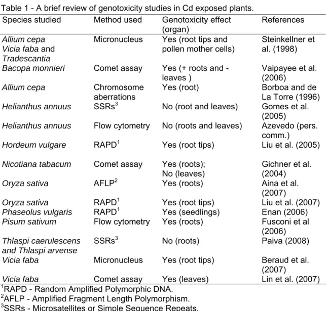
As in animals, continuous exposure to Cd might then significantly contribute to the inherited change of many phenotypic traits in the progeny of exposed plants. Thus, evaluation of the mutagenicity and/or induced genetic instability in plants by this metal is of the utmost importance in environmental studies. Furthermore, plants have been shown to provide ideal models for genotoxicity assays for screening as well as monitoring of environmental mutagens or genotoxins (Grant, 1994; Knasmuller et al., 1998; Grant, 1999). Several different techniques have been used in plant bioassays for the detection of environmental metal pollution, such as micronucleus (MCN) induction, chromosome aberration, comet assay, sister chromatid exchange (SCE), random-amplified polymorphic DNAs (RAPD), amplified fragment length polymorphism (AFLP) and simple sequence repeats or microsatellite markers (SSRs). A brief compilation of relevant works and their main achievements in the study of Cd genotoxicity in plants is presented in Table 1.
Some of these plant assays, such as the Allium cepa chromosome aberration, micronucleus tests and the Tradescantia tests have relatively low sensitivity and they cannot provide information on the effects of toxicity at the DNA level (Panda and Panda, 2002).
Chapters 3.1 and 3.2 present an evaluation of Cd genotoxic effects on plants using simple sequence repeats or microsatellite markers (SSRs) to assess genetic instability. Microsatellite markers are tandem repeats of DNA sequences of 1-6 base pair (bp) long units spread throughout the genome. These markers have a high abundance, random occurrence and are highly polymorphic, and thus extremely useful for fine-scale genetic analysis (Gupta et al., 1996; Tóth et al., 2000); they can be used in the detection of genomic DNA damage and/or mutational events (e.g. deletions, insertions, point mutations) (Tóth et al., 2000). Microsatellite markers are likely to be one of the most reproducible techniques, especially when compared to RAPDs, which has the main disadvantage of low reproducibility with a consequent inconsistency of results (Powell et al., 1996; Jones et al., 1997).
Table 1 - A brief review of genotoxicity studies in Cd exposed plants. Species studied Method used Genotoxicity effect
(organ)
References
Allium cepa Vicia faba and Tradescantia
Micronucleus Yes (root tips and pollen mother cells)
Steinkellner et al. (1998)
Bacopa monnieri Comet assay Yes (+ roots and -
leaves )
Vaipayee et al. (2006)
Allium cepa Chromosome
aberrations
Yes (root) Borboa and de
La Torre (1996)
Helianthus annuus SSRs3 No (root and leaves) Gomes et al.
(2005)
Helianthus annuus Flow cytometry No (roots and leaves) Azevedo (pers.
comm.)
Hordeum vulgare RAPD1 Yes (root tips) Liu et al. (2005) Nicotiana tabacum Comet assay Yes (roots);
No (leaves) Gichner et al. (2004)
Oryza sativa AFLP2 Yes (roots) Aina et al.
(2007)
Oryza sativa RAPD1 Yes (root tips) Liu et al. (2007) Phaseolus vulgaris RAPD1 Yes (seedlings) Enan (2006) Pisum sativum Flow cytometry Yes (roots) Fusconi et al
(2006)
Thlaspi caerulescens and Thlaspi arvense
SSRs3 No (roots) Paiva (2008)
Vicia faba Micronucleus Yes (root tips) Beraud et al.
(2007)
Vicia faba Comet assay Yes (leaves) Lin et al. (2007) 1RAPD - Random Amplified Polymorphic DNA.
2AFLP - Amplified Fragment Length Polymorphism. 3SSRs - Microsatellites or Simple Sequence Repeats.
Because of these advantages, SSRs have already been used to study genotoxic effects in several animal species (e.g. Zienolddiny et al., 2000; Jin et al., 2003; Ohshima, 2003; Slebos et al., 2006). For instance, some metals have been found to induce microsatellite instability (MSI). Nickel (Ni) has been reported to promote genetic instability in hamster (Ohshima, 2003) and human (Zienolddiny et al., 2000) cell lines, and exposure of human cell lines to environmentally relevant quantities of Cd led to statistically significant increases in MSI (Jin et al., 2003; Slebos et al., 2006). In plant research, SSRs are already a powerful tool in taxonomy (e.g. Prasad et al., 2000) genetic mapping (Gupta and Varshney, 2000; Ma et al., 2004) and environmental population genetics focusing on the relationships between environmental selective agents (stressors) and genotypic variability of plant natural populations (D'Surney et al., 2001; Mengoni et al., 2001; van Rossum et al., 2004; Berckmoes et al., 2005). Furthermore, SSRs have the potential to be used in the surveying of plant genomic DNA for evidence of genetic instability as in a
genotoxic bioassay for the detection of DNA damage induced by environmental contaminants. However, apart from a survey performed by Kovalchuck et al. (2000) using SSRs to monitor germline mutations in plants upon chronic exposure to ionizing radiation produced by the Chernobyl accident, the application of SSRs in higher plant bioassays remains unexplored. Therefore, in Chapters 3.1 and 3.2 these molecular markers were applied as a technique for the assessment of genetic instability at the level of DNA in plants exposed in vivo to Cd.
Conventional cytogenetic studies, such as chromosome aberration and micronucleus tests are very elaborate and time consuming. Flow cytometry (FCM) is largely used in health and biological research for many different purposes and appeared as a relatively rapid test applicable to any organism or tissue from which cellular or nuclear suspensions can be obtained. A FCM assay has been developed to detect the changes in nuclear DNA that result from the breakage of chromosomes providing a quantitative measurement of genetic damage at the cellular level (Otto and Oldiges, 1980). This technique has the potential to detect minute differences in nuclear DNA (nDNA) content and chromosomal damage produced by clastogenic agents through the quantification of the increase of the coefficient of variation (CV) of the G0/G1 peak (Otto
and Oldiges, 1980). Flow cytometry measurement of the dispersion in the nDNA content as induced by the interactions of DNA with environmental agents, emerged then as a powerful tool in cytogenetic investigations and in genotoxicity testing (Otto et al., 1981). This technique has subsequently been successfully employed in both laboratory and field studies with several animal species (e.g. Otto et al., 1981; Bickham et al., 1998; Matson et al., 2005; Oliveira et al., 2006; Barbee et al., 2008). However, the use of FCM as an assay for the assessment of genotoxicity in plants remains much less common; it has been used to detect genotoxic effects in maize plants exposed to coal fly ash (McMurphy and Rayburn, 1993) and to the fungicides captan (Rayburn et al., 1993) and triticonazole (Biradar et al., 1994).
More recently, FCM has been used to assess metal genotoxicity in plants; Rayburn and Wetzel (2002) found an increase in the CV values of the G0/G1 peak in
maize and wheat plants grown in soil with high levels of aluminium, and Citterio et al. (2002) reported that the exposure of Trifolium repens to Cd and Cr resulted in a decrease in the DNA index with increasing concentrations of Cr, and to an increase of debris background at the highest concentrations of Cd and Cr. Preliminary FCM assays performed in our laboratory revealed no genotoxic effects in lettuce plants exposed to Cd; no changes in nDNA content and in CV values were detected neither in five-week-old
lettuce plants exposed to 100 µM Cd for 14 days (Monteiro et al., 2004) nor in lettuce plants germinated and grown for 2 months in 10 µM Cd and analysed every 15 days (Monteiro et al., 2005). In Chapter 3.3 a FCM assay is used to detect putative genotoxic effects (e.g. clastogenic effects) in three plant species germinated and exposed to Cd for 28 days to increasing concentrations of Cd.
Mechanisms of tolerance
Metal-binding ligands
Plants, like all living organisms, have evolved a suite of mechanisms that control and respond to the uptake and accumulation of both essential and nonessential metals. These mechanisms include the chelation and sequestration of heavy metals by particular ligands and, in some cases, the subsequent compartmentalization of the ligand-metal complex in vacuoles.
The vacuole of plant cells plays an important role in the homeostasis of the cell (Barkla and Pantoja, 1996). In most plant cells the vacuole comprises more than 80-90% of the cell volume and acts as a central storage compartment for ions, amino acids, sugars and CO2 in the form of malate and also play a key role in the sequestration of toxic
ions and xenobiotics (Barkla and Pantoja, 1996; Briat and Lebrun, 1999). The vacuolar membrane, named tonoplast, functions as an effective and selective metal diffusion barrier (Briat and Lebrun, 1999). Vacuolar compartmentalization prevents the free circulation of Cd ions in the cytosol and forces them into a limited area (Sanitá di Toppi and Gabbrielli, 1999). Several studies have shown that the vacuole is the site of accumulation of a number of metals including Cd (Ma et al., 2005; Ueno et al., 2005). One example is the accumulation of Cd and phytochelatins (PCs) in the vacuole involving an ATP-binding cassette (ABC) transporter (Hall, 2002). Oat root tonoplast vesicles were found to accumulate Cd2+ by a 2H+/ion antiport mechanism (Salt and Wagner, 1993).
Several metal-binding ligands have now been recognized in plants and include organic acids, amino acids, peptides, and polypeptides (Rauser, 1999). Among the metal-binding ligands in plant cells the PCs and metallothioneins (MTs) are the best characterized. MTs are cysteine-rich polypeptides encoded by a family of genes whereas PCs are a family of enzymatically synthesized cysteine-rich peptides (Cobbett and Goldsbrough, 2002).
In plants, PC-Cd complexes are sequestered in the vacuole (Cobbett and Goldsbrough, 2002). In mesophyll protoplasts derived from tobacco plants exposed to Cd, almost all of both the Cd and PCs accumulated was confined to the vacuole (Vogeli-Lange and Wagner, 1990). Lactuca sativa and Thlaspi arvense plants also possess detoxification mechanisms in which PCs play an important role (Ebbs et al., 2002; Maier et al., 2003). Thlaspi caerulescens was found to mainly store Cd2+ in electron-dense
granules inside vacuoles by means of complexation with malate (Ma et al., 2005; Ueno et al., 2005).
Plant metal accumulation and hyperaccumulation
Plants respond to high concentrations of environmental metals in three main ways: metal excluders maintain low and constant metal concentration in their shoots up to a critical soil value; indicator species have internal metal concentrations that reflect the external metal levels, whereas metal accumulators have high accumulation of metal at very low external metal concentration (Greger, 1999). The term hyperaccumulator describes a plant with a highly abnormal capacity for metal accumulation (Reeves and Baker, 2000). Hyperaccumulator plants are found in metalliferous soils, such as calamine (with high levels of Zn, Pb and Cd) and serpentine soils (with high levels of Ni, Cr and Co) (Greger, 1999).
Although Cd is not an essential or beneficial element for plants, they generally exhibit measurable Cd concentrations, particularly in roots, but also in leaves, most probably as a result of inadvertent uptake and translocation (Assunção et al., 2003). A Cd foliar concentration above 100 µg/g DW (0.01%) is considered exceptional and it is used as a threshold value for Cd hyperaccumulation (100 mg/Kg DW) (Reeves and Baker, 2000). The metal hyperaccumulation characteristic is not common in higher terrestrial plants and less than 0.2% of all angiosperms have been identified as metal hyperaccumulators (Reeves and Baker, 2000). The Brassicaceae plant family is well represented among the reported hyperaccumulators. Thlaspi caerulescens is the best known hyperaccumulator plant with a capacity to hyperaccumulate Zn, Cd and Ni (Assunção et al., 2003). Thlaspi caerulescens plants have been found by Reeves and Baker (2000) to contain more than 100 mg/KgCd frequently, and more than 1000 mg/Kg Cd occasionally, with very large variations between sites and populations, and considerable intrasite variability. Several studies have shown that T. caerulescens ecotype from metalliferous soils of a Zn/Pb mine spoil in the southern France (Ganges
ecotype) is far superior in Cd accumulation to other ecotypes (e.g. Prayon from Belgium); in hydroponic conditions it was able to accumulate >10,000 mg/kg Cd in the shoots without showing any symptoms of phytotoxicity (Lombi et al., 2000).
Three plants with different patterns of Cd accumulation were the object of study in the present dissertation: lettuce (Lactuca sativa L.) is a Cd-accumulating plant and an important human food crop; the alpine pennycress (Thlaspi caerulescens J. & C. Presl, Ganges ecotype), which is a hyperaccumulator plant commonly used as a model in metal transport and accumulation studies with a view to their use in phytoremediation (Pence et al., 2000; Assunção et al., 2003; Zhao et al., 2003); and the related non-accumulator, field pennycress (Thlaspi arvense L).
Trophic transfer of Cd
A key pathway for metal exposure to animal species, including humans results from the uptake by plants of elements from the soil. However, the study of the trophic transfer of metals from plants to animals is a largely unexplored field. As indicated above, plants have developed mechanisms for sequestering metals in their systems in such a way that the metal is not phytotoxic, but these plants may still pose a threat to the animals that consume them, becoming a risk to ecological and human food chains (McLaughlin, 2002).
Factors affecting trophic transfer of metals
The bioaccumulation of metals is known to differ among species and metals because of differences in uptake and loss rates, exposure pathways and influences of environmental parameters (Fisher and Reinfelder, 1995; Wang and Fisher, 1999). However, less is known about the influence of these factors in the internal storage and detoxification of accumulated metal and subsequent impacts on trophic transfer. Since the ingestion of metal-contaminated food can serve as a source of metals to consumers and can result in sub-lethal toxicity (e.g. Fisher and Hook, 2002), understanding the mechanisms that influence metal trophic transfer is a critical step in the management of metal contaminated ecosystems. In general, to completely understand metal cycling through trophic levels, several factors which control the bioavailability of tissue-bound metals to predators must be considered and understood (e.g. tissue metal distributions and concentrations, duration of exposure, nutritional status and exposure history of
predator). Different species will accumulate and partition metals in varying ways depending on the detoxification mechanisms employed. The subsequent bioavailability of those partitioned metals to a consumer will be dictated by digestive and assimilative mechanisms of its digestive tract and gut passage time (Wang and Fisher, 1999). Added to this complexity is the varying ability of consumers to discriminate between different foods and contaminants, their nutritional status at the time of consumption, the degree of exposure, and the exposure history for the metal in question, all of which can influence the degree of metal assimilation (Wang and Fisher, 1999).
Metal assimilation and assimilation efficiency
One critical parameter in understanding the trophic transfer and accumulation of a metal is its assimilation efficiency (AE) in animals from the ingested food (Wang and Fisher, 1999). Assimilation efficiency has been defined as the fraction of ingested metal that is assimilated across the gut lining into the body tissue (Wang and Fisher, 1999). Assimilation efficiency measurements are difficult to make and often yield variable results (Fisher and Reinfelder, 1995); they can be determined by the mass balance method in which ingested and egested masses are compared to each other or to the mass retained in the animal after an appropriate gut clearance period (Fisher and Reinfelder, 1995).
Determination of AEs is an important endpoint when addressing contaminant bioavailability, it is considered a first-order physiological parameter that can be quantitatively compared among different chemicals, species, and food particles under various environmental conditions (Wang and Fisher, 1999). Furthermore, AE for metals has been shown to be directly proportional to metal bioaccumulation, which highlights the significance of AE in understanding and predicting metal bioaccumulation (Fisher et al., 1996).
The various factors that affect metal assimilation are reflected in the wide variety of Cd AEs that have been reported in organisms of different food chains fed biologically contaminated food. For instance AEs ranging from 1% have been reported in rats fed snail viscera (Hispard et al., 2008), to 4.7% in the lizard Podarcis carbonelli fed crickets (Mann et al., 2006), to 52% in the isopod P. dilatatus fed lettuce (Calhôa et al., 2006), and up to 76.2 to 94.2% for whelk Thais clavigera fed five different species of prey (Cheung and Wang, 2005).
Subcellular partition of metals
The internal distribution and detoxification of metals within an organism can be used to explain trophic transfer of metals but also to predict metal toxicity for the organism itself. The internal metal sequestration strategies of different species are complex and variable and the determination of the metal concentrations in different compartments can be used to understand the complex relationship between metal accumulation and toxicity.
Over the past decades, chemistry-orientated models have been developed to predict the bioavailability and toxicity of metals focusing on identifying which metal forms are present in the aquatic environment, and investigating their interaction with the biological site of action (Paquin et al., 2002). The free ion activity model (FIAM) relied on the free metal ion activity and assumed that uptake from solution was determined by the availability of free metal ions, whereas the biotic ligand model (BLM) which is an extension of the FIAM, assumes that the effect is proportional to the concentration of metal bound to the target site (biotic ligand) and that this site is in direct contact with the external environment. These models perform well in the prediction of metal bioavailability in water-borne exposures of aquatic organisms, but also for plants (Antunes et al., 2006) and soft-bodied organisms (Peijnenburg, 2002). When considering the contribution of the dietary route of metal exposure the gut/instestine can also act as a biotic ligand (Hogstrand et al., 2002) and metal speciation and/or dietary form is likely to be an important factor for metal assimilation.
Metals can be present in various chemical forms in an organism, including the following: (a) free ionic form or complexed ion species (e.g., CdCl2, CdCl+, CdCl3-); (b)
bound in the active center of functional proteins and enzymes; (c) bound to low molecular weight organic acids (e.g., citrate, malate); (d) bound to sequestration proteins (MTs and PCs); (e) bound in vesicles of the lysosomal system, as intracellular granules; (f) precipitated in extracellular granules, mineral deposits, residual bodies, and exoskeletons; (g) bound to cellular constituents potentially causing dysfunction (e.g. DNA) (Vijver et al., 2004).
The various internal metal fractions all have their own binding capacity for metals, which has implications for food-chain transfer to higher trophic levels. A study on the relationship between subcellular Cd distribution in an oligochaete and its trophic transfer to a predatory shrimp showed that only metal present in the soluble fraction (organelles and protein fraction)) of prey is available for the predator (Wallace et al., 1998). Factors influencing the subcellular distribution in the prey will directly alter trophic transfer to