Effects of Chromium
and Diclofenac
on tomato’s N and C
primary
metabolisms.
Glutamine
Synthetase
as a key metabolic
point to enhance
plant stress
tolerance
Maria João de Araújo Martins
Dissertação de Mestrado apresentada à
Faculdade de Ciências da Universidade do Porto em
Área Científica
2018
E ff ec ts of C hromi um and D iclof en ac on tomato’ s N and C pri mary met ab oli sm s . G lutami ne sy nthetase as a key met ab oli c po int to enhance plant str es s tolerance M ar ia João d e A raújo M ar tins FCUP 2018 2.º CICLOon
tomato’s N and C primary
metabolisms. Glutamine Synthetase
as a key metabolic point to enhance
plant stress tolerance
Maria João de Araújo Martins
Mestrado em Biologia Funcional e Biotecnologia de Plantas
Departamento de Biologia2018
Orientador
Jorge Teixeira, Professor Auxiliar, Faculdade de Ciências da Universidade do Porto
Coorientador
Fernanda Fidalgo, Professora Auxiliar, Faculdade de Ciências da Universidade do Porto
O Presidente do Júri,
“The only place success comes before work
is in the dictionary” - Vince Lombardi
Aknowledgements
Terminado este trabalho e este longo ano, resta-me agora agradecer a todas as pessoas que fizeram parte do meu percurso.
Antes de mais, quero primeiramente agradecer ao Professor Jorge! O professor desde cedo acompanhou o meu percurso e foi essencial para o sucesso deste trabalho. O professor foi a primeira pessoa que me levou a descobrir o maravilhoso mundo das plantas e agradeço-lhe por isso. Obrigada por toda a confiança que o professor me transmitiu ao longo do tempo, pela paciência quando eu desanimava, por me ter sempre tentado motivar a fazer mais e melhor nas horas e horas de conversas.
À Professora Fernanda, co-orientadora deste trabalho, muito obrigada pelo rigor, pela disponibilidade, por todos os conselhos e conhecimentos transmitidos e por toda a motivação que sempre me deu.
Aos meus “mini-bosses” Leonor e Cris: nem tenho palavras para vos agradecer o vosso apoio, ajuda, transmissão de conhecimentos, pelo carinho e pela amizade desenvolvida. Sem vocês, tenho a certeza que este trabalho não era o mesmo. Vocês são o meu exemplo de cientistas e fazem de mim uma cientista melhor!
Aos colegas de laboratório Jorge e Bruno, muito obrigada por todos estes anos (que já são alguns) de partilha, alegria, viagens de comboio e muita, muita luta diária no 2.62! Para além disto, muito obrigada pelas plantinhas, sem o vosso cuidado, este trabalho, garantidamente, não estaria feito. Aos meus “escravos” Miguel, David e João, muito obrigada pelo apoio e confiança nos meus conhecimentos.
À Ana Marta, muito obrigada pela ajuda, pela paciência a aturares os meus desesperos com o real-time (mas consegui!), pelas “regras da Marta” (prometo não me esquecer!), por toda a motivação e pelos conhecimentos.
Às “girls das plantas” Diana e Bruna, obrigada por estes 2 anos, foram muito mais fáceis com vocês do meu lado. Obrigada pelas conversas, pelos desabafos, pelo estudo e por toda a motivação! À Diana só tenho a dizer que sem ti, as horas de almoço a falar da vida não seriam a mesma coisa (:D).
Às duas meninas que na fase mais difícil estiveram lá, incondicionalmente, para me dar uma palavra de conforto e carinho: Maling e Catarina, muito obrigada! Que esta amizade se mantenha por muitos anos.
À Inês (ou Tina), que, embora longe, estará sempre por perto, obrigada por tudo! Foi um enorme gosto partilhar contigo todos estes anos e desejo que o futuro nos reserve o melhor!
Sara, obrigada por seres a minha pessoa, o meu juízo e a minha inspiração durante estes anos. Foste, és e serás das melhores amizades que eu levo destes 5 anos! Ao Pedro e à Joana, obrigada por não se limitarem a ser meros amigos, mas sim, realmente, a família que eu escolhi! Obrigada pela paciência, pelo orgulho que têm em mim e por todos os momentos que ao longo destes anos partilhamos juntos.
Ao Micael, que ao longo destes 3 anos me tem aturado mais do que qualquer um e faz de mim uma melhor pessoa e melhor cientista. Não há palavras para ti, mas sei que não preciso de dizer nada. Obrigada, por tudo!
Para terminar, como não poderia deixar de ser, um último (e talvez o maior) agradecimento à minha família por sempre me apoiarem, motivarem e educarem a querer fazer sempre mais e melhor. Obrigada por sempre me deixarem lutar pelos meus objetivos e por estarem lá quando eu os conquisto. Pai, avós, tios, irmãs e primos, e em especial mãe: esta é para vocês!
A todos vocês que, de alguma maneira, contribuíram para este trabalho: MUITO OBRIGADA!
Resumo
O crescimento mundial da população humana, bem como as atividades antropogénicas, têm levado a um aumento na utilização de metais pesados e produtos farmacêuticos, causando sérias perturbações no meio ambiente. O crómio (Cr) é um metal pesado muito perigoso, sendo libertado para o meio ambiente em largas quantidades e com efeitos nefastos conhecidos em vários organismos, incluindo nas plantas. O diclofenac (DCF) é um medicamento anti-inflamatório não esteroide extremamente utilizado, que não é totalmente removido nos processos de tratamento de águas residuais, atingindo assim todos os ecossistemas e apresentando-se como uma ameaça a todos os organismos. Através de uma abordagem integrada, combinando parâmetros bioquímicos com técnicas de biologia molecular, este trabalho teve como objetivo avaliar a fitotoxicidade de Cr e DCF em tomateiro, focando-se em dois processos primários das plantas: a assimilação de azoto e a fotossíntese. Além disso, pretendeu-se sobreexpressar o cDNA que codifica a GS2, como uma ferramenta para aumentar a tolerância das plantas. A exposição a concentrações crescentes de Cr (0, 5 e 10 µM) e DCF (0, 0,5 e 5 mg L-1) revelou que a glutamina sintetase (GS) foi diferencialmente afetada por ambos contaminantes ao nível da expressão génica, atividade e proteína, e que a atividade da enzima glutamato desidrogenase (GDH) aumentou, bem como os níveis de prolina. De um modo geral, após exposição ao Cr e DCF, foi observada uma diminuição nos transcritos dos genes relacionados com a fotossíntese, bem como uma redução na quantidade de amido. No entanto, nas plantas tratadas com Cr e DCF, os níveis de pigmentos fotossintéticos e o aparelho fotossintético não sofreram alterações. Adicionalmente, estes contaminantes induziram alterações no perfil do conteúdo polipeptídico solúvel. Os resultados obtidos sugerem que a GDH tem um papel importante e alternativo na assimilação do azoto, bem como na produção de prolina, em resposta ao stress. Além disso, o aparecimento de polipeptídeos de baixa massa molecular aponta para o papel de proteínas relacionadas com o stress na tolerância ao Cr e DCF. A clonagem do cDNA que codifica a GS2 em orientações opostas foi bem-sucedida, sendo este o primeiro passo para obtenção das primeiras plantas de tomateiro a sobreexpressar a GS2 com um aumento da tolerância ao stress.
Abstract
The growing of human population worldwide, as well as human activities, led to an increase in heavy metals (HMs) and pharmaceuticals utilization, causing serious disturbances in the environment. Cr is a dangerous HM, that is discharged to the environment in huge quantities and with known negative effects in various organisms, including plants. DCF is an extremely used non-steroidal anti-inflammatory drug (NSAID) that is not entirely removed by wastewater treatment processes, thus reaching all ecosystems and being a serious threat to all organisms. Through an integrated approach, where biochemical parameters and molecular biology techniques were combined, this work aimed to evaluate the phytotoxicity of Cr and DCF on tomato plants, focusing on two primary plant processes: nitrogen assimilation and photosynthesis. Moreover, the overexpression the GS2-encoding cDNA as a tool to increase plant tolerance was started. The exposure to increased concentrations of Cr (0, 5 and 10 µM) and DCF (0, 0.5 and 5 mg L-1) revealed that GS was differentially affected by both contaminants at the gene expression, activity and protein levels, and that GDH activity was enhanced, followed by an increase in proline levels. Upon exposure to Cr (VI) and DCF, an overall decrease in photosynthetic-related genes’ transcripts accumulation was observed paired with a reduction in the starch content. However, the pigments’ contents and the photosynthetic apparatus did not alter in Cr (VI)- and DCF-treated plants. Additionally, these contaminants induced some alterations in the soluble polypeptide content profile. The obtained results suggest that GDH plays an important and alternative role in N assimilation, as well as in proline production, in response to stress. Furthermore, the appearance of low molecular weight polypeptides indicates a role of some stress-related proteins in Cr (VI)- and DCF-induced stress tolerance. The cloning of SlGS2-encoding cDNA in opposite directions was successfully achieved, being the first step to obtain the first tomato plants overexpressing GS2 and with increased tolerance to stress.
Keywords
Nitrogen assimilation, glutamate dehydrogenase, heavy metals, pharmaceuticals, photosynthesis, overexpression (glutamine synthetase), abiotic stress; Solanum
Table of Contents
Aknowledgements ... II Resumo ... IV Abstract ... V Keywords ... V Figure Index ... XTable Index ... XIV
1. Introduction ... 1
1.1 Heavy metal contamination ... 1
1.1.1 Chromium VI [Cr (VI)] and its phytotoxicity ... 2
1.2 Pharmaceuticals contamination ... 4
1.2.1 Diclofenac (DCF) and its phytotoxicity ... 6
1.3 Nitrogen (N) metabolism ... 8
1.3.1 Glutamine synthetase and its role in NUE improvement ... 9
1.3.2 Glutamate Dehydrogenase ... 11
1.4 GS and GDH - a role in proline production under stress ... 12
1.5 Solanum lycopersicum L. cv Micro-Tom as a perfect model species for molecular biology studies ... 12
1.6 Main Objectives ... 14
2. Material and Methods ... 15
2.1 Plant material and growth conditions ... 15
2.2 Analytical determinations ... 15
2.2.1 Determination of Cr content in plant tissues ... 15
2.3 Biochemical determinations ... 16
2.3.1 Glutamine synthetase activity determination (GS; EC 6.3.1.2) ... 16
2.3.2 GDH activity determination (GDH, EC 1.4.1.2) ... 16
2.3.3 Sodium dodecyl sulphate polyacrylamide gel electrophoresis (SDS-PAGE) 17 2.3.4 Western Blotting analysis... 17
2.3.5 Evaluation of photosynthesis-related parameters ... 18
2.3.5.1 Photosynthetic pigments evaluation ... 18
2.3.5.2 Gas exchange analysis ... 19
2.3.5.3 Histochemical coloration of starch ... 19
2.3.6 Proline quantification ... 19
2.4 Bioinformatics characterisation of Solanum lycopersicum GS-encoding gene family (SlGSs) ... 20
2.5 Evaluation of expression of Solanum lycopersicum GS gene family ... 20
2.5.1 RNA extraction, quantification and assessment of its purity ... 20
2.5.2 Reverse Transcription (cDNA Synthesis) ... 21
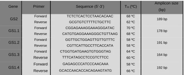
2.5.3 Primer design ... 21
2.5.4 Gene Sequencing ... 22
2.5.5 Expression of SlGS genes by Semi-quantitative RT-PCR ... 23
2.5.6 Evaluation of SlGS gene expression by Real-time PCR (qPCR) ... 23
2.6 Evaluation of photosynthetic-related genes expression by qPCR ... 24
2.7 Overexpression of GS2-encoding cDNA ... 24
2.7.1 Obtaining SlGS2-encoding cDNA sequence ... 24
2.7.1.1 Genomic DNA extraction ... 24
2.7.1.2 Obtaining the SlGS2 gene ... 25
2.7.1.3 Obtaining SlGS2-encoding cDNA ... 25
2.7.2 Bacterial strains and culture conditions ... 26
2.7.3 Induction of chemical competency in E. coli DH5α ... 26
2.7.4 Induction of electro competency in E. coli DH5α ... 26
2.7.5 Ligation to the pJET 1.2 plasmid ... 26
2.7.6 Transformation of chemical competent E. coli DH5α ... 27
2.7.7 Electro Transformation of E. coli DH5α ... 27
2.7.8 Plasmid Isolation ... 27
2.7.9 Plasmid DNA extraction using NZYMiniprep kit ... 28
2.8 Statistical Analysis ... 29
3. Results ... 30
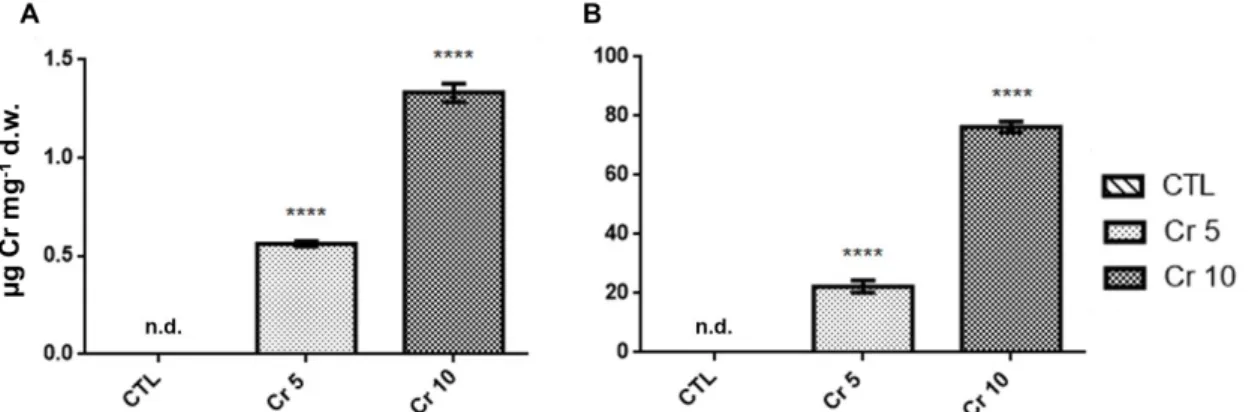
3.1 Cr accumulation on tomato plants’ shoots and roots ... 30
3.2 Effects of Cr (VI) and DCF on nitrogen assimilation ... 30
3.2.1 GS and GDH activity ... 30
3.2.2 Western Blotting Analysis ... 32
3.3 Soluble polypeptide accumulation analysis after Cr (VI) and DCF exposure . 33 3.4 Evaluation of Cr (VI) and DCF impact on physiological parameters associated with nitrogen metabolism ... 35
3.5 Assessment of the Cr (VI) and DCF effects on several photosynthetic endpoints 35 3.5.1 Photosynthetic pigments ... 36
3.5.2 Photosynthetic apparatus ... 36
3.5.3 Photosynthesis-related gene expression ... 37
3.5.4 Starch accumulation ... 38
3.6 Bioinformatics characterisation of Solanum lycopersicum L.’s GS-encoding gene family ... 39
3.6.1 Phylogenetic analysis of S. lycopersicum cDNAs coding for GS (SlGSs) 39 3.6.2 SlGSs relative expression analysis using the eFP browser ... 40
3.6.3 SlGS gene family relative expression analysis by RT-PCR ... 41
3.7 Study of Cr (VI) and DCF impact on SlGS gene family expression ... 43
3.7.1 Changes in transcript levels of tomato GS genes under Cr (VI)- and DCF-induced stress ... 43
3.8 Cloning of the SlGS2-encoding cDNA... 46
4. Discussion ... 49
4.1 The accumulation of Cr occurs predominately in roots ... 49
4.2 GS – from gene to protein ... 49
4.3 Cr (VI) and DCF differentially influenced N metabolism at gene expression, enzyme activity and polypeptide levels ... 50
4.4 Proline accumulation is positively affected by both contaminants ... 54
4.4.1 GDH - An alternative pathway that allow proline accumulation under stressful conditions ... 55
4.5 The exposure of tomato plants to Cr (VI) and DCF did not impair photosynthetic activity ... 56
4.6 Both Cr (VI) and DCF induced changes in soluble polypeptide content and profile 59 4.7 Development of new tools that will allow the overexpression of SlGS2-encoding cDNA 60 5. Concluding remarks ... 62
6. Future Perspectives ... 63
7. References ... 64
Figure Index
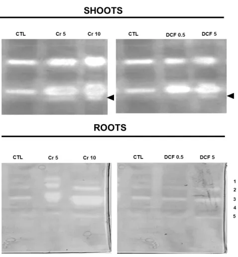
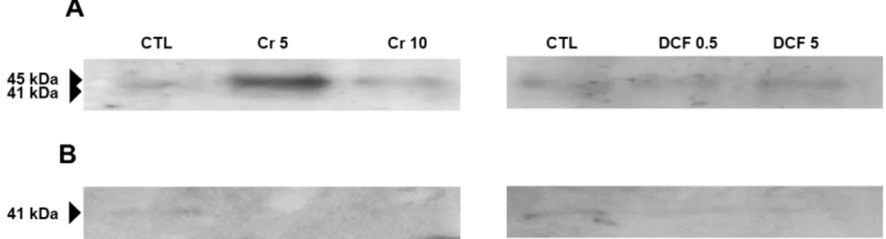
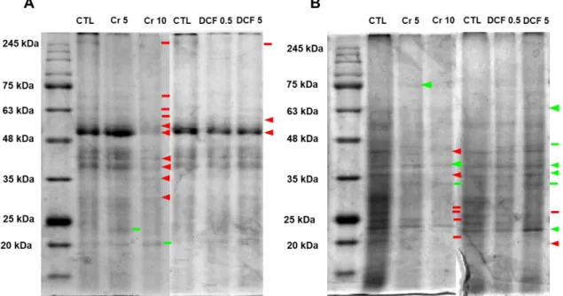
Figure 1. Sources of Cr (VI) in the environment and some effect of Cr (VI) in plants. ... 4 Figure 2. Different sources of pharmaceuticals and the pathways used to reach the aquatic environment (based on Lapworth et al. (2012)). ... 5 Figure 3. Diclofenac chemical structure (from: Vieno and Sillanpää (2014)). ... 7 Figure 4. Biochemical reactions catalysed by GS and GOGAT (Adapted from Hodges (2002)). ... 9 Figure 5. Solanum lycopersicum L. cv Micro-Tom. ... 13 Figure 6. Chromium accumulation levels in shoots (A) and roots (B) of tomato plants. CTL: Control; Cr 5: 5 µM Chromium (VI); Cr 10: 10 µM Chromium (VI); n.d.: non-detected. Values presented are mean plus SD. **** above bars indicate significant statistical differences from control at p ≤ 0.0001 ... 30 Figure 7. GS activity in shoots (A) and roots (B) of tomato plants exposed to Cr (VI) and DCF expressed as nkat mg-1 protein. CTL: Control; Cr 5: 5 µM Chromium (VI); Cr 10: 10 µM Chromium (VI); DCF 0.5: 0.5 mg L-1 Diclofenac; DCF 5: 5 mg L-1 Diclofenac. Values presented are mean ± SD. *** and **** above bars indicate significant statistical differences from control at p ≤ 0.001 and p ≤ 0.0001, respectively. ... 31 Figure 8. Typical GDH activity results for shoots and roots by native PAGE analysis. CTL: Control; Cr 5: 5 µM Chromium (VI); Cr 10: 10 µM Chromium (VI); DCF 0.5: 0.5 mg L-1 Diclofenac; DCF 5: 5 mg L-1 Diclofenac... 32 Figure 9. One-dimensional western blotting analysis of shoot (A) and root (B) tomato GS using GS antibodies raised to Pinus cytosolic GS. CTL: Control; Cr 5: 5 µM Chromium (VI); Cr 10: 10 µM Chromium (VI); DCF 0.5: 0.5 mg L-1 Diclofenac; DCF 5: 5 mg L-1 Diclofenac. ... 33 Figure 10. One-dimensional western blotting analysis of shoot (A) and root (B) tomato NAD–GDH using antibodies raised to Vitis vinifera NAD-GDH. CTL: Control; Cr 5: 5 µM Chromium (VI); Cr 10: 10 µM Chromium (VI); DCF 0.5: 0.5 mg L-1 Diclofenac; DCF 5: 5 mg L-1 Diclofenac. ... 33 Figure 11. Typical SDS-PAGE analysis of soluble proteins on shoots (A) and roots (B) of S. lycopersicum. CTL: Control; Cr 5: 5 µM Chromium (VI); Cr 10: 10 µM Chromium (VI); DCF 0.5: 0.5 mg L-1 Diclofenac; DCF 5: 5 mg L-1 Diclofenac. Arrows indicate common bands that suffered a decreased (red) or an increased accumulation; - indicates qualitative changes: red – disappearance; green – de novo polypeptide. The used ladder was BLUE Wide Range CSL-BBL Prestained Protein Ladder (Cleaver Scientific Ltd). 34 Figure 12. Proline levels in shoots (A) and roots (B) of tomato plants exposed to Cr (VI) and DCF. CTL: Control; Cr 5: 5 µM Chromium (VI); Cr 10: 10 µM Chromium (VI); DCF
0.5: 0.5 mg L-1 Diclofenac; DCF 5: 5 mg L-1 Diclofenac. Values presented are mean ± SD. *, ** and *** above bars indicate significant statistical differences from control at p ≤ 0.05, p ≤ 0.01 and p ≤ 0.001, respectively. ... 35 Figure 13. Total chlorophylls (A) and carotenoids (B) levels of tomato plants exposed to Cr (VI) and DCF. CTL: Control; Cr 5: 5 µM Chromium; Cr 10: 10 µM Chromium; DCF 0.5: 0.5 mg L-1 Diclofenac; DCF 5: 5 mg L-1 Diclofenac. Values presented are mean SD. **, *** and **** above bars indicate significant statistical differences from control at
p ≤ 0.01, p ≤ 0.001 and p ≤ 0.0001, respectively. ... 36
Figure 14. Expression profile of two genes coding for protein subunits of PSII, D1 protein (PSIIa) and CP47 (PSIIb), respectively, of tomato leaves exposed to Cr (VI) and DCF. CTL: Control; Cr 5: 5 µM Chromium; Cr 10: 10 µM Cr (VI); DCF 0.5: 0.5 mg L-1 Diclofenac; DCF 5: 5 mg L-1 Diclofenac. Values presented are mean SD. ** and **** above bars indicate significant statistical differences from control at p ≤ 0.01 and p ≤ 0.0001, respectively. ... 37 Figure 15. Expression profile of the small and large subunits of RuBisCO, rbcS and rbcL, respectively, of tomato leaves exposed to Cr (VI) and DCF. CTL: Control; Cr 5: 5 µM Chromium; Cr 10: 10 µM Cr (VI); DCF 0.5: 0.5 mg L-1 Diclofenac; DCF 5: 5 mg L-1 Diclofenac. Values presented are mean SD. * and **** above bars indicate significant statistical differences from control at p ≤ 0.05 and p ≤ 0.0001, respectively. ... 38 Figure 16. Lugol staining of starch in tomato leaves exposed to Cr (VI) and DCF. CTL: Control; Cr 5: 5 µM Chromium (VI); Cr 10: 10 µM Chromium (VI); DCF 0.5: 0.5 mg L-1 Diclofenac; DCF 5: 5 mg L-1 Diclofenac. ... 39 Figure 17. Phylogenetic tree constructed with all S. lycopersicum GS cDNA sequences recovered from the databases referred in the text. The bootstrap consensus tree was generated using the Neighbour-Joining method with MEGA7, with 1000 bootstrap replicates. The percentage of replicate trees in which the associated taxa clustered together in the bootstrap test are shown next to the branches. Bar represents the scale length. ... 40 Figure 18. Agarose gel (0.8% (w/v)) electrophoresis evidencing the PCR products of GS2, GS1.1, GS1.2, GS1.3 and GS1.4 of S. lycopersicum when gDNA was used as template. The used ladder was NZYDNA Ladder III (Nzytech®, Portugal). ... 42 Figure 19. Results for GS2, GS1.1, GS1.2, GS1.3 and GS1.4 RT-PCR analysis by 0.8 % (w/v) agarose gel electrophoresis in both shoots and roots; Expected sizes: 189 bp; 178 bp, 191 bp; 164 bp; 192 bp, respectively. GS1.1- corresponds to the negative control reaction for GS1.1. ... 43
Figure 20. Total RNA extracted from shoots and roots of tomato plants exposed to Cr (VI) and DCF. For quality assessment of total RNA, it was separated on agarose gel at 0.8 % (w/v). CTL: Control; Cr 5: 5 µM Chromium (VI); Cr 10: 10 µM Chromium; DCF 0.5: 0.5 mg L-1 Diclofenac; DCF 5: 5 mg L-1 Diclofenac. ... 43 Figure 21. Expression profile of GS2 gene in shoots (A) and roots (B), of tomato leaves exposed to Cr (VI) and DCF. CTL: Control; Cr 5: 5 µM Chromium (VI); Cr 10: 10 µM Chromium (VI); DCF 0.5: 0.5 mg L-1 Diclofenac; DCF 5: 5 mg L-1 Diclofenac. Values presented are mean SD. * and *** above bars indicate significant statistical differences from control at p ≤ 0.05 and p ≤ 0.001, respectively. ... 44 Figure 22. Expression profile of GS1.1 gene in shoots (A) and roots (B), of tomato leaves exposed to Cr (VI) and DCF. CTL: Control; Cr 5: 5 µM Chromium (VI); Cr 10: 10 µM Chromium (VI); DCF 0.5: 0.5 mg L-1 Diclofenac; DCF 5: 5 mg L-1 Diclofenac. Values presented are mean SD. * and **** above bars indicate significant statistical differences from control at p ≤ 0.05 and p ≤ 0.0001, respectively. ... 45 Figure 23. Typical results for GS1.2, GS1.3 and GS1.4 semi-quantitative RT-PCR analysis by 2 % (w/v) agarose gel electrophoresis in both shoots (A) and roots (B) of tomato plants exposed to increasing concentrations of Cr (VI); Expected sizes: 191 bp; 164 bp; 192 bp, respectively. CTL: Control; T1: 5 µM Cr (VI); T2: 10 µM Cr (VI). ... 45 Figure 24. Typical results for GS1.2, GS1.3 and GS1.4 semi quantitative RT-PCR analysis by 2 % (w/v) agarose gel electrophoresis in both shoots (A) and roots (B) of tomato plants exposed to increasing concentrations of DCF; Expected sizes: 191 bp; 164 bp; 192 bp, respectively. CTL: Control; T1: 0.5 mg L-1 DCF; T2: 5 mg L-1 DCF. ... 46 Figure 25. Agarose gel (0.8% (w/v)) electrophoresis evidencing the PCR products of GS2 of S. lycopersicum when gDNA was used as template, using 54.7, 53.4 and 51 ºC as annealing temperatures (lanes 1 to 3, respectively). The chosen temperature is highlighted. The ladder used was NZYDNA Ladder III (Nzytech®, Portugal). ... 46 Figure 26. Schematic representation of the GS2 cDNA insert depicting the sites for the restriction enzymes Sacl, Ncol and Xhol. The cDNA size is1684 bp. ... 47 Figure 27. Agarose gel (0.8 % (w/v)) electrophoresis evidencing three distinct minipreps digested with XhoI (A) and NcoI (B). The green and red arrows highlight the SlGS2-encoding cDNA in the sense and antisense orientations, respectively. 1, 2 and 3 represent the three distinct minipreps. The ladder used was GeneRuler 100 bp Plus DNA ladder, ready-to-use (ThermoFisher Scientific). ... 47 Figure 28. Agarose gel (0.8 % (w/v)) electrophoresis evidencing two distinct minipreps digested with SacI. 1 and 2 represent minipreps 1 and 2, respectively. The arrows
evidence two bands resulting from the restriction. The used ladder was GeneRuler 100 bp Plus DNA ladder, ready-to-use (ThermoFisher Scientific). ... 48 Figure 29. Two pathways that provide the glutamate for proline production and accumulation, under stressful conditions. The dashed arrow corresponds to the GDH pathway, which is an alternative route for amino acid’s production, while the GS/GOGAT cycle corresponds to the principal pathway of glutamate, and thus, proline production. ... 56
Table Index
Table 1. Concentrations of different pharmaceuticals in crops from agricultural fields and subsequent phytotoxicities of each compound (adapted from Bartrons and Peñuelas (2017)). ... 6 Table 2. Summary of properties of Diclofenac (Feito et al., 2012)... 7 Table 3. Gene-specific primers, their Tm and respective expected amplicon sizes for the performed RT-PCR reactions. ... 22 Table 4. Photosynthetic gene-specific primers and respective expected amplicon sizes. ... 24 Table 5. Gas exchange measurement of plants exposed to Cr (VI) and DCF, considering the following parameters: A, CO2 uptake (µmol m-2 s-1); E, transpiration rate (mmol m-2 s -1); Gs, stomatal conductance (mmol m-2 s-1); WUE, water use efficiency (nmol mol-1); Ci/Ca, internal and environmental CO2 ratio. CTL: Control; Cr 5: 5 µM Chromium (VI); Cr 10: 10 µM Cr (VI); DCF 0.5: 0.5 mg L-1 Diclofenac; DCF 5: 5 mg L-1 Diclofenac. Values presented are mean ± SD. ** above bars indicate significant statistical differences from control at p ≤ 0.01 ... 37 Table 6. Effects of Cr (VI) treatments on photosynthetic-related genes expression. It was considered up regulated or down regulated if expression increased, or decreased (respectively) with p < 0.05, and no change if p > 0.05; CTL: Control; Cr 5: 5 µM Chromium (VI); Cr 10: 10 µM Chromium (VI); DCF 0.5: 0.5 mg L-1 Diclofenac; DCF 5: 5 mg L-1 Diclofenac. ... 38 Table 7. Relative expression levels of the tomato GS-encoding genes on leaves and roots. ... 40 Table 8. Summary of chosen temperatures, expected sizes and intron presence of all SlGS-encoding genes. ... 42 Table 9. Summary of the effects of Cr (VI) and DCF exposure on the mRNA accumulation of the tomato GS-encoding genes. (=) means no changes, (-) means decreased accumulation and (+) means increased accumulation relative to the control. (x) means no mRNA accumulation. ... 52
Abbreviations and Acronyms
2-OG – 2- oxoglutarate;
ADP – adenosine diphosphate;
ALAD – δ-aminolevulinic acid dehydratase; AMP – ampicillin; Ca – calcium; Car – carotenoid; Chla – chlorophyll a; Chlb – chlorophyll b; Cr – chromium; DCF – 2-(2-(2,6-dichlorophenylamino)phenyl) acetic acid; diclofenac; DTT – 1,4-dithioreitol; EC – emerging contaminants; FW – fresh weight;
gDNA – genomic DNA;
GDH – glutamate dehydrogenase; GOGAT – glutamate synthase; GS – glutamine synthetase;
GS1 – cytosolic glutamine synthetase; GS2 – plastidic glutamine synthetase; HM – heavy metal; HS – Hoagland solution; LB – Luria Broth; MTT-3-(4,5-dimethylthiazol-2-yl)-2,5-diphenyltetrazolium bromide NR – nitrate reductase;
NUE – Nitrogen Use Efficiency; ON – Overnight;
PPCP – pharmaceutical and personal care product;
PSII – photosystem II;
PVPP – polyvinylpolypyrrolidone;
ROS – reactive oxygen species;
RT – room temperature;
RT-qPCR – reverse transcription coupled to real time PCR;
RuBisCO – ribulose-1,5-biphosphate carboxylase oxygenase;
SDS – sodium dodecyl sulphate;
SN – supernatant;
TEMED – N,N,N’N-Tetramethylethylenediamine;
Tm – melting temperature;
1. Introduction
The exponential growth of the world population, as well as anthropogenic activities, such as accelerated industrialization, intensive agriculture and extensive mining, is resulting in several disturbances in the environmental compartments (air, water, soil and biota). The contamination of the environment became an important public health problem and raised concern over the last years (Cristaldi et al., 2017; Gorito et al., 2017). By definition, a contaminant is a substance that is present in non-expected locations and in concentrations that overcome a set limit (Chapman, 2007). Well-known contaminants include toxic HMs, some pesticides, asbestos and petroleum and polyaromatic hydrocarbons. However, over 80,000 synthetic substances are released into the environment per year, either as industrial waste or as part of production processes. Some of these substances are considered emerging contaminants (ECs), either due to their recent development or the discovery of their presence in the environment, and they include pharmaceuticals and personal care products (PPCP’s) (Naidu et al., 2016; Bartrons and Peñuelas, 2017). Since plants are the basis of food chains and have the ability to uptake some contaminants into their tissues, the study of the impact of ECs on these organisms has become a matter of special interest, not only because of the effects of these substances on plant physiology, but also due to food quality and safety (Zhuang et al., 2009; Lajayer et al., 2017; Al-Farsi et al., 2018).
1.1 Heavy metal contamination
Various anthropogenic activities can cause a vast amount of perturbations in the biosphere. Consequently, these activities increase the accumulation of HMs, causing serious concerns of ecological, nutritional and environmental motives. HMs are a group of metals and metalloids that possess high atomic mass (over 20 g cm-3) and density (more than 5 g cm-3) (Emamverdian et al., 2015; Lajayer et al., 2017). HMs enter the environment mainly through two sources: natural and anthropogenic sources. Natural sources include volcanic activities, soil erosion and rock and mineral disaggregation, while anthropogenic ones comprise agricultural and industrial activities, fuel combustion, street run-off, mineral processing and landfills (Burakov et al., 2018). HMs have genotoxic, cytotoxic and mutagenic effects on humans, animals and plants. Despite these severe effects, some of these substances are essential elements for plants and animals due to their important biochemical and physiological roles in these living beings. Indeed, some HMs are an integral part of several enzymes and participate in redox reactions (Nagajyoti et al., 2010).
1.1.1 Chromium VI [Cr (VI)] and its phytotoxicity
Cr is a silver-coloured HM belonging to the group VI-B of the periodic table with atomic number 24, molecular weight 51.1 g mol-1 and density 7.19 g cm-3 and is the 21st most abundant metal of the Earth’s crust (Shanker et al., 2005; Shahid et al., 2017). This metal is one of the 18 main hazardous air pollutants (HAPs), 33 urban air toxicants, and the Agency for Toxic Substances and Disease Registry classified Cr as 7th among the top 20 hazardous substances (Oh et al., 2007). Furthermore, Cr has the 5th place within the HMs in the Comprehensive Environmental Response, Compensation, and Liability Act (Ma et al., 2007) and it is also considered as the 1st carcinogen, according to the International Agency for Research on Cancer (Cancer, 1987) and the National Toxicology Program (Shahid et al., 2017).
Despite having several valence states (from -2 to +6), the most common and stable forms of Cr are the trivalent - Cr (III) - and the hexavalent - Cr (VI) – forms (Ashraf et al., 2017; Shahid et al., 2017). These two states differ in bioavailability, mobility and toxicity (Panda and Choudhury, 2005). Cr (VI) is the most toxic form due to its high reactivity with other elements (Shahid et al., 2017), and it generally associates with oxygen, resulting in chromate (CrO42-) or dichromate (Cr2O72-) oxyanions. Cr (III) is less mobile and toxic, and forms complexes with organic matter in soil and aquatic environments. Cr (III) plays an important role on lipid and sugar metabolism of animals, including humans (Oliveira, 2012), however, it is not essential for plants (Shanker et al., 2005).
The release of high levels of Cr in soils and ground waters leads to environmental contamination. Consequently, crops growing in contaminated soils may accumulate this metal in their tissues, allowing Cr’s entrance in food chains, ultimately affecting human health (Broadway et al., 2010; Ahmed et al., 2016). This problem raised the attention of the scientific community worldwide over the years (Shanker et al., 2005; Shahid et al., 2017). Cr occurs naturally in rocks, soil, water, plants, animals, and volcanic dust and gases (Shanker et al., 2005). The main sources of Cr are industrial activities such as leather tanning, metallurgical, Cr plating, wood processing and preservation, anodizing aluminium, catalytic manufacture, cleaning agents, organic synthesis, textile dyeing and textile pigment production, and alloy preparation industries (Sinha et al., 2018). According to Santos and Rodriguez (2012), the most important sources of this element in the environment are Cr fugitive emissions from industrial cooling towers and road dust. Mohan and Pittman Jr (2006) found that 30, 896 and 146 metric tons/year of Cr were discharged worldwide in air, soil and water, respectively. In 2012, Cr concentration varied
from 0.1 to 0.5 mg L-1 in fresh water and from 0.0016 to 0.05 mg L-1 in sea waters (Kumar and Puri, 2012).
Plants’ contamination by Cr is dependent on the speciation of the metal, which defines its mobilization, uptake and phytotoxicity. Because Cr is not an essential element for plant metabolism, these organisms do not possess any specific mechanism for its uptake (Shanker et al., 2005; Oliveira, 2012). Even yet, plants can uptake Cr (III), through a passive mechanism that does not require energy consumption. Moreover, due to its structural similarity with phosphate and sulphate, the uptake of Cr (VI) can be achieved by an active process mediated by phosphate or sulphate carriers (de Oliveira et al., 2014; de Oliveira et al., 2016). Besides, Cr (VI) also affects the uptake of Ca, Fe, K, Mg, Mn and P (Gardea-Torresdey et al., 2004).
The majority of Cr is accumulated in roots, since this metal is poorly translocated to the aerial parts of the plant. In fact, the concentration of Cr in roots can be 100-fold higher than in shoots (Shanker et al., 2005; Oliveira, 2012). Cr is highly toxic to plants as it affects not only plant morphology but also several crucial processes. For example, root growth is strongly disturbed by Cr, due to the low translocation index of this metal to the aerial parts of plants. Consequently, this process disrupts some other processes such as water and nutrient absorption and their transportation to shoots, thus impairing shoot growth. On the other side, Cr (VI) directly influences foliar tissues by reducing leaf number and area, which is associated to the decrease in cell division and number of cells in leaves. In addition, at long-term exposure, this HM can cause chlorosis and tissue necrosis (Singh et al., 2013; Shahid et al., 2017).
Cr (VI)-induced stress culminates in several photosynthetic damages. It is well documented that Cr (VI) decreases chlorophyll a (chl a), chlorophyll b (chl b) and carotenoids contents (Singh et al., 2013). The decrease in chlorophyll content in response to Cr (VI) exposure is related to the degradation of key enzymes involved in chlorophyll biosynthesis, such as δ-aminolevulinic acid dehydratase (ALAD or porphobilinogen synthase, EC 4.2.1.24) and protochlorophyllide reductase (EC 1.3.1.33) (Ganesh et al., 2008). Moreover, Cr-induced reduction in absorption of Mg and N, which are fundamental components of the chlorophyll molecule, can also contribute to its negative effects on chlorophyll content (Sela et al., 1989). Since the production of photosynthetic pigments is affected, CO2 assimilation is also committed. Cr (VI) also inhibits the electron transport chain, therefore affecting the photosynthetic rate (Vernay et al., 2007; Liu et al., 2008; Subrahmanyam, 2008). Some authors report that this metal also prejudices Calvin cycle enzymes such as ribulose-1,5-biphosphate carboxylase
oxygenase (RuBisCO), stomatal conductance, transpiration rate and substomatal CO2 concentration (Rodriguez et al., 2011; Santos and Rodriguez, 2012).
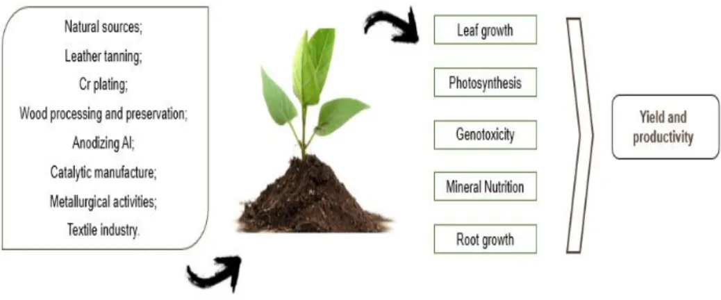
Cr (VI) can affect mineral nutrition of plants in a complex manner, due to its similar structure to other essential elements (Shanker et al., 2005; Oliveira, 2012; Shahid et al., 2017). The Cr (VI)’s competitive binding to common carriers, as above described, can reduce the uptake of many essential nutrients. Moreover, Cr-induced stress causes the inhibition of the activity of plasma membrane H+-ATPase (Shanker et al., 2005). High levels of Cr (VI) might displace some essential nutrients from their physiological binding sites which, consequently, decreases their uptake and translocation (Oliveira, 2012; Shahid et al., 2017). In fact, it is well documented that Cr (VI) can interfere with the uptake of some macronutrients such as N, P, K, and Mg. Given that N is an essential macronutrient for plants and being its uptake compromised by Cr (VI)-induced stress, N metabolism can also be negatively influenced by the presence of this HM. Indeed, some authors reported that Cr (VI) affects key enzymes involved in N metabolism such as nitrate reductase (NR, EC 1.6.6.1), nitrite reductase (NiR, EC 1.7.7.1) glutamine synthetase (GS, EC 6.3.1.2), glutamate synthase (GOGAT, EC 1.4.1.14; EC 1.2.7.1) and glutamate dehydrogenase (GDH, EC 1.4.1.2) (Dubey and Rai, 1987; Kumar and Joshi, 2008; Gangwar and Singh, 2011; Singh et al., 2013). As well as N, C and starch metabolism are also negatively affected by Cr (VI) (Singh et al., 2013). All the effects provoked by Cr (VI), indirectly affect plant yield and crop productivity (Figure 1) (Shanker et al., 2005; Oliveira, 2012).
Figure 1. Sources of Cr (VI) in the environment and some effect of Cr (VI) in plants.
1.2 Pharmaceuticals contamination
Over the last years, increasing urbanisation, growing per-capita income, ageing population and other factors have contributed to a major increase in the production and the consumption of pharmaceuticals worldwide, a problem that tends to continue in the
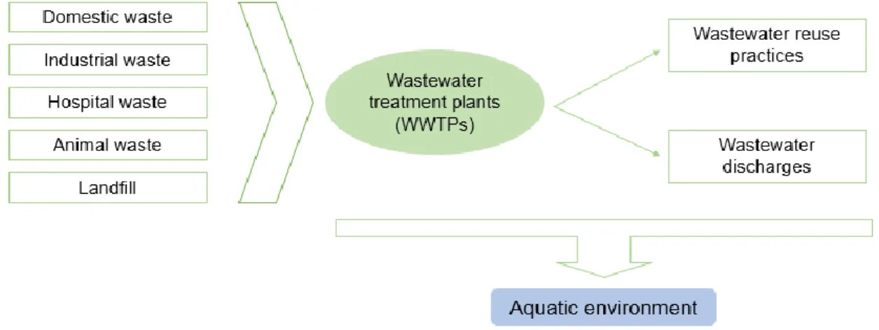
future. Pharmaceuticals, used in both human and animal medicine, agriculture and aquaculture facilities, are a wide range of chemical compounds frequently used in illnesses’ prevention, diagnosis, treatment or cure. The intensive usage of these substances enabled their presence in the environment, particularly in the aquatic compartment (Fatta-Kassinos et al., 2011; Bartrons and Peñuelas, 2017). Indeed, the presence of pharmaceuticals in aquatic environment was detected many decades ago by several authors (Tabak and Bunch, 1970; Norpoth et al., 1973). Different pathways allow the entrance of pharmaceuticals into the aquatic environment (Figure 2), but all of them result from human activities.
Figure 2. Different sources of pharmaceuticals and the pathways used to reach the aquatic environment (based on
Lapworth et al. (2012)).
Human bodies cannot completely metabolise some pharmaceuticals, being these compounds excreted as the parent drug or as its derived metabolites via urine and faeces, therefore reaching conventional wastewater treatments plants (WWTPs) (Ribeiro et al., 2016). Conventional WWTPs were not planned to completely remove organic substances, like pharmaceuticals, from wastewater effluents. Consequently, some of these substances can pass through the wastewater treatment process without being treated (Christou et al., 2017; Gorito et al., 2017) and reach several aquatic systems such as wastewater, ground water, surface water and drinking water (Odendaal et al., 2015; Balakrishna et al., 2017; Yang et al., 2017).
The re-use of wastewater effluents for crop irrigation, of manure for the fertilization of agricultural soils, as well as the application of biosolids, are considered serious threats to human health. In fact, these compounds are absorbed by plants, which are the basis of food chains, thus affecting humans by food ingestion (Bartrons and Peñuelas, 2017; Madikizela et al., 2018). Upon exposure to pharmaceuticals, plant growth and development can be directly or indirectly affected by disturbing plant’s microbiota
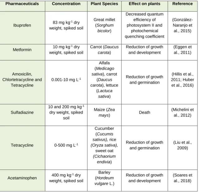
(Madikizela et al., 2018). As shown in Table 1, several studies reported phytotoxicity caused by pharmaceuticals in important crop plants (Table 1).
Table 1. Concentrations of different pharmaceuticals in crops from agricultural fields and subsequent phytotoxicities of
each compound (adapted fromBartrons and Peñuelas (2017)).
Pharmaceuticals Concentration Plant Species Effect on plants Reference
Ibuprofen 83 mg kg
-1 dry
weight, spiked soil
Great millet (Sorghum bicolor) Decreased quantum efficiency of photosystem II and photochemical quenching coefficient (González-Naranjo et al., 2015) Metformin 10 mg kg -1 dry
weight, spiked soil
Carrot (Daucus carota) Reduction of growth and development (Eggen et al., 2011) Amoxicilin, Chlortetracycline and Tetracycline 0.001-10 mg L-1 Alfafa (Medicago sativa), carrot (Daucus carota), lettuce (Lactuca sativa) Reduction of growth and germination (Hillis et al., 2011; Huber et al., 2016) Sulfadiazine 10 and 200 mg kg-1
dry weight, spiked soil Maize (Zea mays) Death (Michelini et al., 2012) Tetracycline 0-500 mg L-1 Cucumber (Cucumis sativus), rice (Oryza sativa), sweet oat (Cichaorium endivia) Reduction of growth and germination (Liu et al., 2009) Acetaminophen 400 mg kg -1 dry
weight, spiked soil
Barley (Hordeum vulgare L.) Reduction of growth and development (Soares et al., 2018)
As it is clear from the data in Table 1, plant growth and development can be differently affected by pharmaceuticals, depending on the compound and its concentration, and the plant species (Madikizela et al., 2018).
1.2.1 Diclofenac (DCF) and its phytotoxicity
Diclofenac (2-(2-(2,6-dichlorophenylamino)phenyl)acetic acid) (DCF) is a non-steroidal inflammatory drug (NSAID) (Figure 3), which is used as a pain killer or as an anti-inflammatory, and it is marketed under various trade names, being Voltaren the most known (Vieno and Sillanpää, 2014). Furthermore, it is consumed in large quantities worldwide (Zhang et al., 2008).
Figure 3. Diclofenac chemical structure (from: Vieno and Sillanpää (2014)).
This drug is administered as oral tablets or topical gel and, once in the human body, DCF is not completely metabolised, as 1 to 10% is excreted in its native ingested form (Davies and Anderson, 1997). Some properties of this substance are summarised in Table 2. Table 2. Summary of properties of Diclofenac (Feito et al., 2012).
Diclofenac
Cas NO. 15307-79-6
Molecular formula C14H10Cl2NNaO2
Molecular weight 318.13 Water solubility 50 mg L-1 Main metabolites 4’-hydroxydiclofenac 3’-hydroxydiclofenac 4’,5-hydroxydiclofenac
Excretion without metabolization 1-10%
Excretion 65-70% in urine, 20-30% in faeces
Biodisponibility 54%
Daily consumption per patient 200-300 mg
Annual world consumption 940 tons
Like other PPCP’s, upon being excreted via urine or faeces, DCF enters the environment through WWTP’s, being one of the most detected pharmaceuticals (aus der Beek et al., 2016; He et al., 2017), as well as one of the lowest removed (Vieno and Sillanpää, 2014). In WWTP’s, DCF reaches concentrations of nearly µg L-1, whereas in surface water bodies it is detected in ng L-1 levels (Lonappan et al., 2016). Within the aquatic
environment, DCF has been noticed in rivers, estuaries, lakes (Buser et al., 1998; Öllers et al., 2001; Metcalfe et al., 2003; Kim et al., 2007), in groundwater and even in drinking water (Rabiet et al., 2006; Benotti et al., 2008).
The ecological concern of DCF started in the beginning of the current century when, due to its usage for cattle treatments, this drug was associated with the massive decline of a vulture population in India (Oaks et al., 2004).
So far, numerous studies on the ecotoxicological effects of DCF have been conducted, mostly from animal and aquatic invertebrates and different biomarkers and endpoints have been used in these organisms to clarify and monitor the ecotoxicological effects of this drug (Huber et al., 2016). Comparing to animals, not much is known regarding the effects of DCF on plants.
In 2012, Huber et al., firstly reported the metabolism of DCF on plants, using Hordeum
vulgare L. and Armoracia rusticana L. cultures. Later, the same authors revealed the role
of peroxidases on the metabolism of DCF in cultures of Armoracia rusticana L. (Huber et al., 2016) and Fu et al. (2017) studied the uptake and metabolism of DCF on
Arabidopsis thaliana. Concerning the effects of DCF on plant physiology and
development, there is an even more accentuated gap in knowledge. Kummerová et al. (2016) reported the negative effects of this drug on biomass production, pigments content and oxidative stress in Lemna minorand, a recent work of Pierattini et al. (2018), besides studying the plant uptake of DCF, also evaluated some physiological responses such as growth parameters and stress enzymes activity. Yet, further investigation is required for adequately unveil the consequences of DCF in plant metabolism and development, especially regarding N and C assimilation processes, once they are part of the plants’ primary metabolism.
1.3 Nitrogen (N) metabolism
It is well-known that N is a vital macronutrient and an essential component of biomolecules like proteins, chlorophylls, nucleic acids, pyrimidines, purines, porphyrins, and co-enzymes. For this reason, N is indispensable for plant growth, development and productivity (Singh et al., 2013). N is obtained from the environment by three biological processes: nitrate (NO3-) reduction, ammonia uptake or N fixation (Hoffman et al., 2014). Most of plants acquire NO3- as the preferred source of N available forms to plants (Pathak et al., 2008). Then, NO3- is reduced to ammonium (NH4+) by NR and NiR, which is assimilated into the amino acids glutamine and glutamate by GS and GOGAT, respectively, through the GS/GOGAT cycle (Cren and Hirel, 1999). GDH also catalyses
the formation of glutamate, from NH4+ and 2-oxoglutarate (2-OG), when the concentration of NH4+ is high (Fontaine et al., 2012).
Knowing the importance of N to plant productivity and with the continuous increase of human population, the application of N fertilisers in crops became an important issue nowadays. The high costs of N fertiliser applications, along with the observed negative impact of this practise on natural ecosystems, is concerning the scientific community, which has been committed in improving the N use efficiency (NUE) (Miyashita and Good, 2008; Nguyen and Kant, 2018; Tiwari et al., 2018). This index, according to some authors, is defined as the biomass/grain yield per unit N accessible for uptake (Brauer and Shelp, 2010). So far, many strategies to improve NUE have been proposed, some of them being related to N metabolism (Tiwari et al., 2018).
1.3.1 Glutamine synthetase and its role in NUE improvement
Glutamine synthetase, also known as glutamate-ammonia ligase, is an enzyme that is involved in the first step of ammonium assimilation into glutamine (Thomsen et al., 2014). This enzyme catalyses the ATP-dependent fixation of NH4+ to the D-carboxyl group of glutamate to produce one molecule of glutamine, which, together with one molecule of 2-OG, originates two molecules of glutamate by the action of GOGAT. At the end of the GS-GOGAT cycle (Figure 4), through the catalytic action of GOGAT, two molecules of glutamate, at the expense of reducing power, as well as one molecule of glutamine are formed. One of them provides nitrogen groups for the biosynthesis of all nitrogenous compounds in the plant, while the other is recycled back to the GS-GOGAT cycle. Moreover, GS is responsible for the reassimilation of the NH4+ released during photorespiration in C3 plants (Lea and Miflin, 2003; Thomsen et al., 2014).
Figure 4. Biochemical reactions catalysed by GS and GOGAT (Adapted from Hodges (2002)).
GS-encoding genes, due to their old existence and function, act as a good molecular clock for phylogenetic analysis (Pesole et al., 1991; Kumada et al., 1993).In prokaryotes and eukaryotes, based on their gene sequence, molecular weight and quaternary structure, the GS superfamily is divided into three distinct types - GSI, GSII and GSIII
(Swarbreck et al., 2010; James et al., 2018). The GSI, GSII and GSIII proteins possess, on average, 360, 450 and 730 amino acids, respectively (van Rooyen et al., 2011). According to a research based on sequence similarity, the GSIII family does not exist in plants. Indeed, this GS isoenzyme is only described in various prokaryotes, whereas GSI and GSII are present in both eukaryotes and prokaryotes. Up to now, GSI function and role in plants is not well-established. However, recent studies proposed an association of this GS type with N and biotic stress signalling (Doskočilová et al., 2011; Silva et al., 2015).
The most predominant GS type in higher plants is the GSII (Bernard and Habash, 2009; Swarbreck et al., 2010). Within GSII, two types are identified in higher plants: a cytosolic (GS1) form and a plastidic (GS2) one (Cren and Hirel, 1999; Habash et al., 2001; Miflin and Habash, 2002). This GS type presents a decametric structure with two pentameric rings (Unno et al., 2006; Seabra et al., 2009; Torreira et al., 2014). Regarding the GSII gene family in plants, the majority of studies conducted to date revealed that most plant species possess only one nuclear gene encoding GS2, while GS1 seems to be encoded by two to five genes (Lam et al., 1996; Swarbreck et al., 2010). The GS2 polypeptide has an N-terminal transit peptide with almost 50 amino acids, which targets it to the chloroplast, and a 16-amino acid conserved region at the C-terminal part of the subunit, that is exclusive to this isoenzyme. GS1 is localised to the cytosol and possesses none of the above-mentioned conserved regions (Lightfoot et al., 1988; Cren and Hirel, 1999). Different localization of the GS genes’ expressions to specific cell and tissue types suggests distinct specific physiological functions (Swarbreck et al., 2010). GS1 (polypeptide molecular weight ~ 38-40 kDa) is a key factor in primary NH4+ assimilation in roots and re-assimilation of NH4+ released during leaf senescence and from protein breakdown during seed germination, while GS2 (polypeptide molecular weight ~ 42-45 kDa) is involved in the primary assimilation of NH4+ resulting from NO3- reduction in chloroplasts and in the reassimilation of NH4+ released during photorespiration. Previous studies suggested that GS2 also has a protective role against biotic and abiotic stresses (Lam et al., 1996; Miflin and Habash, 2002; Masclaux-Daubresse et al., 2010).
Taking into account the role of GS in N metabolism and the importance of this macronutrient in plant growth and development, it can be assumed that GS might be the rate-limiting enzyme during N incorporation into organic forms (Lam et al., 1996). In this way, GS can be considered as a good candidate to NUE improvement through genetic manipulations (James et al., 2018). Furthermore, due to the role of GS in the tolerance to abiotic and biotic stresses, the overexpression of GS-encoding genes appears to be
a good molecular approach to obtain genetically modified plants with an optimized NUE. Actually, up to date, numerous studies have already been carried out and the results are quite promising. For instance, in transgenic Lotus corniculatus plants overexpressing a soybean GS1 gene under the control of a cauliflower mosaic virus (CaMV) 35S promoter, it was observed an accelerated growth rate (Vincent et al., 1997). In addition, poplar trees (Gallardo et al., 1999; Fuentes et al., 2001; Pascual et al., 2008) and tobacco plants (Fuentes et al., 2001) with an overexpressed GS1 gene improved their vegetative growth and photosynthetic capacity. Also, it was observed an earlier flowering and seed development in transgenic wheat plants transformed with a Phaseolus vulgaris GS1 gene driven by the RuBisCO small subunit promoter (rbcS) (Habash et al., 2001). There are less studies regarding the overexpression of GS2, when compared to GS1. In 2000, (Migge et al.) reported a higher growth rate in transgenic tobacco seedlings overexpressing a GS2 cDNA. In rice leaves (Takabe et al., 2001) and protoplasts (Hoshida et al., 2000), the overexpression of a GS2 cDNA increased their photorespiration capacity and conferred salt and chilling tolerance. Moreover, Wang et al. (2013) reported an increase in leaf surface area, total protein and amino acid content, chlorophyll content, glucose and sucrose contents, and plant length in transgenic tobacco plants overexpressing the Arabidopsis Dof1 (a transcription factor that regulates GS gene expression) and GS1 and GS2 genes.
1.3.2 Glutamate Dehydrogenase
GDH is an enzyme that catalyses the deamination of glutamate to produce 2-OG and NH4+, with the production of reducing power, as well as the production of glutamate from NH4+ and 2-OG, using NADH or NADPH as a coenzyme (amination reaction). According to Ferraro et al. (2012), there are three to four genes in the GDH gene family, encoding two subunits (α and ), which can be randomly combined to form many NADH-GDH hexameric isoenzymes (Melo-Oliveira et al., 1996; Turano et al., 1997; Dubois et al., 2003; Purnell et al., 2005; Miyashita and Good, 2008). GDH is present in several plant organs, being located at several cell compartments, such as the cytosol, mitochondria and chloroplasts (Dubois et al., 2003).
The physiological role of GDH is not clearly understood yet, despite the efforts of the scientists to reveal it (Dubois et al., 2003; Purnell and Botella, 2007; Skopelitis et al., 2007; Lehmann and Ratajczak, 2008; Miyashita and Good, 2008; Masclaux-Daubresse et al., 2010). So far, it is known that under stressful conditions, the GS/GOGAT cycle is not completely enough neither to reduce the toxic levels of NH4+, nor to provide the necessary glutamate for the biosynthesis of some protective biomolecules. In this case, the dual role of GDH is crucial because the amination reaction leads to a reduction of
toxic amounts of NH4+ that are accumulated during stress, plus providing glutamate for the synthesis of other nitrogenous compounds (Forde and Lea, 2007), while the deamination activity is required to supply carbon skeletons for the tricarboxylic cycle (Chiraz et al., 2003; Jha and Dubey, 2004).
1.4 GS and GDH - a role in proline production under
stress
When subjected to various abiotic stresses, plants usually enhance the production of the amino acid proline, which has been proved to have multiple roles in response to abiotic stresses (Hare and Cress, 1997; Hellmann et al., 2000; Verbruggen and Hermans, 2008; Szabados and Savoure, 2010). Plants under stressful conditions can accumulate proline through two ways: either by a stimulation of its biosynthesis, or by inhibiting its oxidation/degradation. The major precursor for proline biosynthesis in stress-induced plants is glutamate, which results from the GS/GOGAT cycle (Díaz et al., 2010). Indeed, it was showed that the phloem-located GS is essential for controlling proline production and that a higher GS activity had a positive influence in proline synthesis in plants under water stress (Brugière et al., 1999). Also, in the previous year, Larher et al. reported that the conversion of amino acids into proline was compromised by the application of a GS inhibitor in rapeseed leaf discs (Larher et al., 1998). Moreover, a decrease in proline content was observed in gs2 mutants of Lotus japonicus under drought stress (Díaz et al., 2010). However, some studies showed that GS-GOGAT cycle was not the only source of glutamate for proline biosynthesis under stress-induced conditions (Lutts et al., 1999; Wang et al., 2007). These authors suggested that NAD-GDH is an alternative pathway to provide glutamate, since this enzyme is responsible for the conversion of 2-OG into glutamate by the amination reaction.
1.5 Solanum lycopersicum L. cv Micro-Tom as a perfect
model species for molecular biology studies
Tomato (Solanum lycopersicum L.), native from South America, belongs to the Solanaceae family which includes many economically important species like potato (Solanum tuberosum L.), tobacco (Nicotiana tabacum L.) and eggplant (Solanum
melongena L.). According to FAO, in 2014 tomato production reached ~160 millions of
tons year-1 and it is predicted that production of this crop continues to increase (Gerszberg and Hnatuszko-Konka, 2017). Nowadays, tomato not only has an agricultural and economic importance worldwide but is also considered a model system for genetic studies in plants (Bergougnoux, 2014).
Solanum lycopersicum cv. Micro-Tom (Figure 5) is a dwarf miniature tomato cultivar
which was originally created for gardening purposes. The phenotype of this cultivar derived from three mutations: self-pruning (producing a determinate phenotype), dwarf (reducing internode length and producing smaller, rugose, and dark-green leaves) and
miniature (likely to be associated with gibberellin signalling) (Meissner et al., 1997; Martí
et al., 2006).
Figure 5.Solanum lycopersicum L. cv Micro-Tom.
Due to its special characteristics such as small size, rapid life cycle (fruits mature in 70-90 days), a large number of plants per square meter (can reach 1357 plants m-2), easy to grow under laboratory conditions, a well-studied small diploid genome and high transformation frequencies, Micro-Tom became a good model system for agronomic and genetic studies. In fact, this cultivar is also named as “the laboratory tomato” (Meissner et al., 1997; Shibata, 2005). Up to now, there are many reports on Solanaceae and tomato genetics (Meissner et al., 1997; Shibata, 2005; Aoki et al., 2010), hormonal functions and interactions (Campos et al., 2010), microbial plant interaction (Park et al., 2007), carbohydrate and amino acids metabolisms (Obiadalla‐Ali et al., 2004; Scarpeci et al., 2007; Sorrequieta et al., 2010; Ferraro et al., 2012) and in molecular breeding of tomato fruit shelf-life (Okabe et al., 2012).
Thus, altogether, these characteristics, allied to the great economic importance, makes
1.6 Main Objectives
As previously stated, worldwide contamination by HMs and pharmaceuticals are fast-growing issues, posing important and determinant consequences to the dynamics of the agroecosystems, including the development of important crops, like tomato. Moreover, knowing that N is the major mineral nutrient influencing plant growth, this dissertation firstly aimed to uncover the effects of HM- and xenobiotic-exposure on the N metabolism and photosynthesis in S. lycopersicum plants. Given the recognized importance of GS, particular focus was given to the regulation and performance of this enzyme, at transcript, protein and activity levels.
In this way, to meet these goals, this work had several specific objectives:
i) To assess the impact of Cr (VI) and DCF in N metabolism, particularly on GS gene expression and protein accumulation and activity, as well as to investigate the role of GDH in N metabolism in tomato plants under these types of stresses, and check how these enzymes cooperate in proline biosynthesis;
ii) To evaluate the effects of these contaminants on photosynthesis and photosynthetic-related parameters;
iii)
To overexpress the SlGS2 cDNA as a tool to increase plant abiotic stress tolerance and NUE.By combining diverse and complementary approaches, through the application of several physiological, biochemical and molecular tools, this work will help to unravel the direct consequences of DCF and Cr (VI) in the growth and development of S.
lycopersicum, shedding some light on the phytotoxicity of DCF and strengthening the
scientific basis of Cr contamination risks for crops. Furthermore, this dissertation will be the first study reporting the overexpression of GS2 in tomato plants in an attempt to increase its tolerance to environmental contamination with EC and metals.
2. Material and Methods
2.1 Plant material and growth conditions
All experiments of this work were performed using certified Solanum lycopersicum L. cv. Micro-Tom (Tomato Genetics Resource Center (TGCR); germplasm LA3911) seeds. Prior to their use, the seeds were surface-sterilized with 70% ethanol for 10 min, followed by 20% commercial bleach containing 0.02% tween-20 for 5 min under constant agitation and then several washes with sterilized deionized water were done. After that, the seeds were distributed in Petri dishes (10 cm diameter) with Hoagland Solution (HS) solidified with 0.8% (w/v) agar (Taiz et al., 2015). To allow seed stratification, in order to synchronize the seed germination, the Petri dishes were placed at 4 ºC for two days, and then, transferred to a growth chamber (16 h light/ 8 h dark) at 25 ºC, with photosynthetically active radiation (PAR) of 60 µmol m-2 s-1 for 10 days. After this period, seedlings were transplanted to individual pots and cultivated with a mixture of expanded vermiculite:perlite (2:1). At this point, the seedlings were split in five growth conditions and grown in a growth chamber under the same conditions described above for 21 days. The five growth conditions consisted in: Control (CTL) – watered with HS; Cr 5 - watered with HS and 5 µM Cr (VI); Cr 10 - watered with HS and 10 µM Cr (VI); DCF 0.5 – watered with HS and 0.5 g L-1 Diclofenac; DCF 5 – watered with HS and 5 g L-1 Diclofenac. For each treatment, 12 biological replicates were prepared. After 21 days, plants were picked, and the shoots were separated from roots. Roots were washed carefully with tap water, followed by a wash with deionized water. Part of the plant material was directly handled for biochemical assays, another part was dried for posterior Cr and DCF quantifications in plant tissues, and the remaining material was frozen and grounded in liquid nitrogen (N2) and stored at -80 ºC for posterior biochemical and molecular assays.
2.2 Analytical determinations
2.2.1 Determination of Cr content in plant tissues
The levels of Cr on tomato plants tissues were quantified to verify the preferential organ for Cr accumulation. Both shoots and roots were collected (as above described) and dried at 60 ºC, until constant weight was registered. Then, the samples were ground to a fine powder and kept in a desiccator for further use. The samples were subjected to an acid digestion using a mixture of HCl:HNO3 to release the metals bound to proteins or other biological structures. The resultant digests were dissolved in a precise volume of water and the quantification of Cr content was performed using a Flame atomic
absorption spectrometer, based on a suitable external standard solution (Perkin Elmer, AAnalyst 200 model, Shelton CT) according to manufacturer’s instructions.
2.3 Biochemical determinations
2.3.1 Glutamine synthetase activity determination (GS; EC 6.3.1.2) The activity of GS was determined using samples from frozen leaf and root tissues. With a mortar on ice, aliquots of about 300 mg were homogenized in extraction buffer (750 µL for shoots and 500 µL for roots) composed of 25 mM Tris-HCl (pH 6.4), 10 mM Magnesium Chloride (MgCl2), 1 mM Dithiothreitol (DTT), 10% Glycerol, 0.05% Triton X-100 with quartz sand and 1% (w/v) Polyvinylpolypyrrolidone (PVPP). Posteriorly, homogenates were centrifuged at 15,000 g at 4 ºC for 20 min and the supernatant (SN) was collected. The enzymatic reaction started when 50 µL of SN were mixed with 50 µL of 6.4% (w/v) Sodium arsenate (pH 6.4) and 400 µL of reaction mixture composed of 100 mM Trizma-Base, 125 mM L-glutamine, 157 mM Hydroxylamine, 0.26 mM Manganese chloride tetrahydrate and 1.25 µM Adenosine 5’-diphosphate sodium salt (ADP), pH 6.4. For each SN, three replicates were prepared. Simultaneously, a blank was set by replacing the protein extract by extraction buffer. All the reactions were incubated at 30 ºC for 30 min and the reaction was stopped by adding 500 µL of Stop solution (0.16 M Iron Chloride (FeCl3), 0.25 M Trichloroacetic acid in 3.33% chloridric acid (HCl)). The absorbances were measured at 500 nm and the transferase activity of GS was determinate according to the formula (µmol min-1 mL-1):
GS activity = Abs (500 nm)
0.4 x protein extract x reaction time
The protein content of the extracts was determined by the Bradford method (Bradford, 1976) and GS activity was expressed as nkat mg-1 protein (1 kat = 60 mole min-1) (Cullimore and Sims, 1980). The extracts were mixed with glycerol to a final concentration of 40% and stored at -80 ºC until further use.
2.3.2 GDH activity determination (GDH, EC 1.4.1.2)
GDH activity was visualized in a non-denaturing 10% (w/v) resolving polyacrylamide gel (3.33 mL 30% Acrylamide stock, 1.25 mL 1.5 M Tris-HCl (pH 8.8), 1.15 mL 87% Glycerol, 75 µL 10% (w/v) Ammonium Persulfate (APS), 5 µL N,N,N’N-Tetramethylethylenediamine (TEMED) and distilled water to a final volume of 10 mL) and 4% stacking gel (660 µL 30% Acrylamide stock, 1.25 mL 0.5 M Tris-HCl (pH 6.8), 570
µL 87% Glycerol, 12,5 µL 10% (w/v) APS, 25 µL TEMED and distilled water to a final volume of 5 mL). The protein extracts used were those obtained from the GS activity assays and 40 µg protein were loaded with 1x sample buffer (10x sample buffer consisting of 50% (w/v) Sucrose, 0.1% (w/v) Bromophenol blue). The buffer system consisted in 25 mM Tris-HCl (pH 8.3) and 192 mM Glycine. Running was performed at non-limiting voltage, 15 mA per stacking gel, 20 mA per resolving gel and non-limiting time, under refrigeration. GDH activity bands were detected after an incubation overnight (ON) with gentle rocking in a staining solution (100 mM Tris-HCl pH 9.3, 55 mM L-glutamate, 450 µM NAD+, 480 µM 3-(4,5-dimethylthiazol-2-yl)-2,5-diphenyltetrazolium bromide (MTT), 130 µM Phenazine methosulphate and 500 µM CaCl2) (Loulakakis and Roubelakis-Angelakis, 1991). All images were captured with ChemiDoc™ XRS+ System (Bio-Rad®) and treated with Image Lab™ 5.2 (Bio-Rad®).
2.3.3 Sodium dodecyl sulphate polyacrylamide gel electrophoresis (SDS-PAGE)
SDS-PAGE (Laemmli, 1970) was carried out on 12.5% SDS-Polyacrilamide resolving gels (8.3 mL 30% Acrylamide stock, 5 mL 1.5 M Tris-HCl pH 8.8, 0.2 mL 10% SDS, 100 µL 10% (w/v) APS, 6.7 µL TEMED and distilled water to a final volume of 10 mL) and 5% SDS-Polyacrilamide stacking gels (670 µL 30% Acrylamide stock, 500 µL 1.0 M Tris-HCl pH 6.8, 40 µL 10% SDS, 40 µL 10% (w/v) APS, 4 µL TEMED and distilled water to a final volume of 4 mL). The extracts obtained from the GS activity assay were treated at 96 ºC for 5 min with 1x SDS sample buffer (10x sample buffer composed of 0.375 g Tris-HCl pH 6.8, 5 mL 99% Glycerol; 0.5 g SDS, 20 mg Bromophenol blue and water up to 10 mL) and 0.5% (v/v) -mercaptoethanol and 30 µg protein per lane were loaded. The running buffer consisted in 25 mM Tris, 192 mM Glycine and 1% (w/v) SDS and the electrophoresis occurred at non-limiting voltage, 15 mA per stacking gel, 20mA per resolving gel and non-limiting time. The gels were incubated ON in BlueSafe (Nzytech) with gentle rocking. Finally, the gels were rinsed with distilled water. The molecular weight of protein bands was estimated by comparison with a protein marker BLUE Wide Range CSL-BBL Prestained Protein Ladder (Cleaver Scientific Ltd). All images were captured with ChemiDoc™ XRS+ System (Bio-Rad®) and treated with Image Lab™ 5.2 (Bio-Rad®).
2.3.4 Western Blotting analysis
After separation by SDS-PAGE as described above, proteins were electroblotted onto a PVDF membrane (Protran, Whatman® Schleicher and Schuell), according to the