Suppresses Neuroinflammation
Wei Bi1,2., Xiuna Jing1., Lihong Zhu3, Yanran Liang1, Jun Liu1, Lianhong Yang1, Songhua Xiao1, Anding Xu2, Qiaoyun Shi4, Enxiang Tao1*
1Department of Neurology, Sun Yat-sen Memorial Hospital of Sun Yat-sen University, Guangzhou, People’s Republic of China,2Department of Neurology, First Affiliated Hospital of Jinan University, Guangzhou, People’s Republic of China,3Joint Laboratory for Brain Function and Health of Jinan University and the University of Hong Kong, School of Medicine, Jinan University, Guangzhou, People’s Republic of China,4Division of Cardiovascular Medicine, Center for Inherited Cardiovascular Disease, Stanford University School of Medicine, Stanford, California, United States of America
Abstract
Recently, researchers have focused on immunosuppression induced by rifampicin. Our previous investigation found that rifampicin was neuroprotective by inhibiting the production of pro-inflammatory mediators, thereby suppressing microglial activation. In this study, using 2-dimensional gel electrophoresis (2-DE) and mass spectrometry (MS), we discovered that 26S protease regulatory subunit 7 (MSS1) was decreased in rifampicin-treated microglia. Western blot analysis verified the downregulation of MSS1 expression by rifampicin. As it is indicated that the modulation of the ubiquitin-26S proteasome system (UPS) with proteasome inhibitors is efficacious for the treatment of neuro-inflammatory disorders, we next hypothesized that silencing MSS1 gene expression might inhibit microglial inflammation. Using RNA interference (RNAi), we showed significant reduction of IkBadegradation and NF-kB activation. The production of lipopolysaccharides-induced pro-inflammatory mediators such as inducible nitric oxide synthase (iNOS), nitric oxide, cyclooxygenase-2, and prostaglandin E2 were also reduced by MSS1 gene knockdown. Taken together, our findings suggested that rifampicin inhibited microglial inflammation by suppressing MSS1 protein production. Silencing MSS1 gene expression decreased neuroinflammation. We concluded that MSS1 inhibition, in addition to anti-inflammatory rifampicin, might represent a novel mechanism for the treatment of neuroinflammatory disorders.
Citation:Bi W, Jing X, Zhu L, Liang Y, Liu J, et al. (2012) Inhibition of 26S Protease Regulatory Subunit 7 (MSS1) Suppresses Neuroinflammation. PLoS ONE 7(5): e36142. doi:10.1371/journal.pone.0036142
Editor:Colin Combs, University of North Dakota, United States of America
ReceivedNovember 3, 2011;AcceptedMarch 26, 2012;PublishedMay 18, 2012
Copyright:ß2012 Bi et al. This is an open-access article distributed under the terms of the Creative Commons Attribution License, which permits unrestricted use, distribution, and reproduction in any medium, provided the original author and source are credited.
Funding:This work was funded by the Guangdong Provincial Department of Science and Technology (0400935505B33801003), the Ph.D. Programs Foundation of Ministry of Education of China (No. 20070558257), and the National Natural Science Foundation of China (81102449). The funders had no role in study design, data collection and analysis, decision to publish, or preparation of the manuscript.
Competing Interests:The authors have declared that no competing interests exist. * E-mail: tao_enxiang@yahoo.cn
.These authors contributed equally to this work.
Introduction
Microglial activation plays an important role in the pathophys-iology of neurodegenerative diseases, and the suppression of microglial activation has been shown to prevent the progression of Alzheimer’s disease (AD), Parkinson’s disease (PD), trauma, multiple sclerosis, and cerebral ischemia [1–5].
Rifampicin is a macrocyclic antibiotic that is used extensively against Mycobacterium Tuberculosis and other mycobacterial infections [6]. It has been reported that rifampicin is immuno-suppressive [7–10]. We previously found that rifampicin improved survival of catecholamine anda-synuclein-containing cells, which degenerate in PD, thus might be therapeutic in this disease [11]. Rifampicin suppressed the release of pro-inflammatory mediators including nitric oxide (NO), prostaglandin E2 (PGE2), tumor necrosis factor-a (TNF-a), and interleukin-1b (IL-1b) from BV2 microglial cells that were pre-treated with lipopolysaccharides (LPS). It acted as a neuroprotector to increase neuronal survival against microglia-induced neuron death [12]. Our results strongly supported rifampicin as a potential therapeutic for the treatment of neurodegenerative diseases. Despite the above findings, the
mechanism through which rifampicin inhibits neuroinflammation is not completely understood.
[18]. Based on the above evidence, we further hypothesized that rifampicin inhibited the expression of MSS1, thus suppressed IkBa degradation and the production of inflammatory media-tors.
In this study, we examined the effect of rifampicin on the expression of MSS1 in LPS-stimulated BV2 microglia by western blot to confirm the results of proteomics experiments. We demonstrated that after silencing the expression of MSS1 gene via RNA interference (RNAi), IkBaprotein degradation and NF-kB activity were both downregulated in LPS-stimulated BV2 microglia. We also showed that the production of inducible NO synthase (iNOS), NO, cyclooxygenase-2 (COX-2), and PGE2were significantly decreased after MSS1 gene knockdown in LPS-activated BV2 microglia. Our results implied that rifampicin inhibited IkBa degradation by suppressing the expression of MSS1, therefore regulated the production of inflammatory mediators.
Materials and Methods
Chemicals and Reagents
Rifampicin (purity .98%), LPS and dimethyl sulfoxide (DMSO) were purchased from Sigma (St. Louis, MO). Rifampicin was dissolved in less than 0.1% of DMSO solution. Antibodies against iNOS, COX-2, and IkBa were obtained from Cell Signaling Tech (Beverly, MA). Mouse beta-actin antibody was purchased from Sigma. Dulbecco’s-modified Eagle’s medium (DMEM) containing L-arginine (200 mg/L), fetal bovine serum (FBS), and other tissue culture reagents were purchased from Gibco (Grand Island, NY).
Cell Culture
BV2-immortalized murine microglial cells were provided by the Cell Culture Center of the Chinese Academy of Medical Sciences (China). Cells were cultured in DMEM supplemented with 10% FBS, 100 units/ml penicillin, and 100mg/ml streptomycin in a humidified atmosphere of 5% CO2at 37uC [12]. To examine the effect of rifampicin on the expression of MSS1 in LPS-stimulated BV2 microglia, 36105cells per well were seeded in 6-well plates and pretreated with 150mmol/L rifampicin for 2 hours (h) before the addition of LPS (1000 ng/ml).
2-dimensional Gel Electrophoresis and Image Analysis
LPS-treated cells were washed three times with ice-cold washing buffer (10mM Tris-HCl, 250mM sucrose, pH 7.0), collected in clean 1.5 ml eppendorf tubes. Lysis buffer [7 M urea, 2 M thiourea, 4% CHAPS (w/v), 1% dithiothreitol (DTT), 2% immobilized pH gradients (IPG) (v/v), pH 3– 10 NL] was added, and samples were centrifuged at 13,200g
for 30 min at 4uC. The supernatant was subjected to 2-DE using an Amersham Biosciences IPGphor IEF System and Hoefer SE 600 (GE healthcare, Uppsala, Sweden) electropho-resis units (13 cm), according to manufacturer’s instructions and a previously described protocol [19]. Protein lysates and 2-DE gels were processed in parallel. Protein concentrations were determined using the Bradford assay. After 2-DE, the gels underwent silver nitrate staining according to a previously described protocol [20], then were scanned using an Image Scanner (GE Healthcare). The images were analyzed using the ImageMaster 2D Platinum (GE Healthcare).
Matrix-assisted Laser Desorption/ionization Time-of-flight Mass Spectrometry (MALDI-TOF-MS) and Database Search
Only protein spots that were consistently different in at least three independent experiments were considered to be significant for analysis by MALDI-TOF-MS. Protein spots were excised from the silver-stained gels and transferred into siliconized 1.5 ml eppendorf tubes. Tryptic in-gel digestion was performed as previously reported with slight modifications [19]. Molecular mass analysis of the tryptic peptides was performed using ABI 4800 plus a MALDI-TOF-MS mass spectrometer (Applied Biosystems, Foster City, CA). Spectra were interpreted and processed using the Global Protein Server Workstation (V3.6, Applied Biosystems) via the internal MASCOT search engine (V2.1, Matrix Science, London, UK) to analyze MALDI-TOF-MS and MS/MS data. Based on combined MALDI-TOF-MS and MS/MS spectra, MASCOT protein scores of greater than 65 were considered statistically significant (p,0.05). The individual MS/MS spectrum with the best ion score (based on MS/MS spectra) that was statistically significant (p,0.05) was also accepted. Searches were performed against the IPI mouse database (V3.36) with param-eters as the following: the enzyme trypsin with one missed cleavage was allowed; variable modifications included acetamidation of cysteine and oxidation of methionine; peptide mass tolerance was set to 50 ppm and fragment ion mass tolerance was set to 0.2 Da; and only monoisotopic masses were included in the search.
MSS1 Gene silencing
Gene silencing was performed using small interference RNA (siRNA) targeting MSS1 mRNA for degradation. MSS1-specific siRNAs had the sense sequence of 59 -GUCGAACGCACAU-CUUUAATT-39, corresponding to a region that was 443–461 bases downstream of the first nucleotide of the start codon of
mouse MSS1 cDNA (GenBank Accession Number:
NC_000007.13). The sense sequence of scrambled siRNAs was 59-UUCUCCGAACGUGUCACGUTT-39. RNA duplexes were synthesized, purified and annealed by Dharmacon (Lafayette, CO). BV2 cells were transfected with targeting and scrambled RNA duplexes at a final concentration of 100 nM using Lipofectamine 2000 (Invitrogen, Grand Island, NY) in either 96-well or 6-96-well culture plates. The cells were assayed at 24 h post-transfection via western blotting.
Nitrite (Griess) Assay
The NO levels in the culture supernatants were determined by measuring nitrite levels using a Griess reaction [21]. Six wells of cells were treated with rifampicin per experiment. After the BV2 microglial cells were stimulated in 24-well plates for 24 h, 100ml of the cell culture medium was taken out and mixed with the same volume of the Griess reagent [1% sulfanilamide, 0.1% N-(1-naphthyl)-ethylenediamine dihydrochloride, 2.5% H3PO4]. The nitrite concentration was determined by evaluating the absorbance at 540 nm using a 96-well microplate spectrophotometer and calculated by referring to a standard curve.
Enzyme-linked Immunosorbent Assay (ELISA)
Western Blot Analysis
The BV2 microglial cells were harvested from each group and followed by western blot analysis that was conducted as previously described [22]. Cell pellets were briefly lysed in RIPA buffer [1 mM ethylenediaminetetraacetic acid (EDTA), 150 mM NaCl, 1% igepal, 0.1% sodium dodecyl sulfate (SDS), 0.5% sodium deoxycholate, and 50 mM Tris-HCl, pH 8.0]. Equal amounts of cellular proteins were separated by 8–12% sodium dodecyl sulfate polyacrylamide gel electrophoresis (SDS-PAGE), transferred to polyvinylidene fluoride (PVDF) membranes, blocked with 5% nonfat milk for 2 h, and incubated with antibodies against iNOS (1:1000), COX-2 (1:1000), IkBa(1:1000), andb-actin (1:5000) at 4uC overnight. The next day, the membrane was washed by Tris-Buffered Saline Tween-20 (TBST) three times, 10 minutes each, and incubated with the corresponding secondary antibodies that were horseradish peroxidase-conjugated for 1 h at room temper-ature. Antibody interactions were detected using enhanced chemiluminescence (ECL) followed by exposure to film.
NF-kB Reporter Gene Assay
A total of 16106BV2 microglial cells were transfected with 2mg NF-kB-Luciferase reporter plasmid and pCMV-gal control vector (Clontech, Mountain View, CA) using Lipofectamine reagents according to the manufacturer’s protocol (Invitrogen). BV2 microglial cells were plated in 96-well plates at a density of 16104cells per well and cultivated at 37uC overnight. Three wells
of cells were treated per experiment. After incubation with the appropriate DNA-Lipofectamine mixtures, cells were pre-incubat-ed with or without rifampicin for 2 h before the addition of LPS for 6 h. Cells were then washed, lysed, and centrifuged according to the manufacturer’s instructions (Promega, Madison, WI). 20ml of cell extract was mixed with 100ml of luciferase assay reagent at room temperature followed by the luciferase activity detection using a luminometer (Safire2, Tecan Instruments, Switzerland). Luciferase activity was normalized through dividing the mean luciferase relative light units (RLU) by the mean value of b -galactosidase RLU.
Statistical Analysis
Data were presented as the mean6standard error of the mean (SEM) derived from three or more independent experiments. Comparisons between two groups were analyzed using Student’s t-test. A value ofp,0.05 was deemed to be statistically significant.
Results
2-DE Maps and Protein Identification by MALDI-TOF-MS
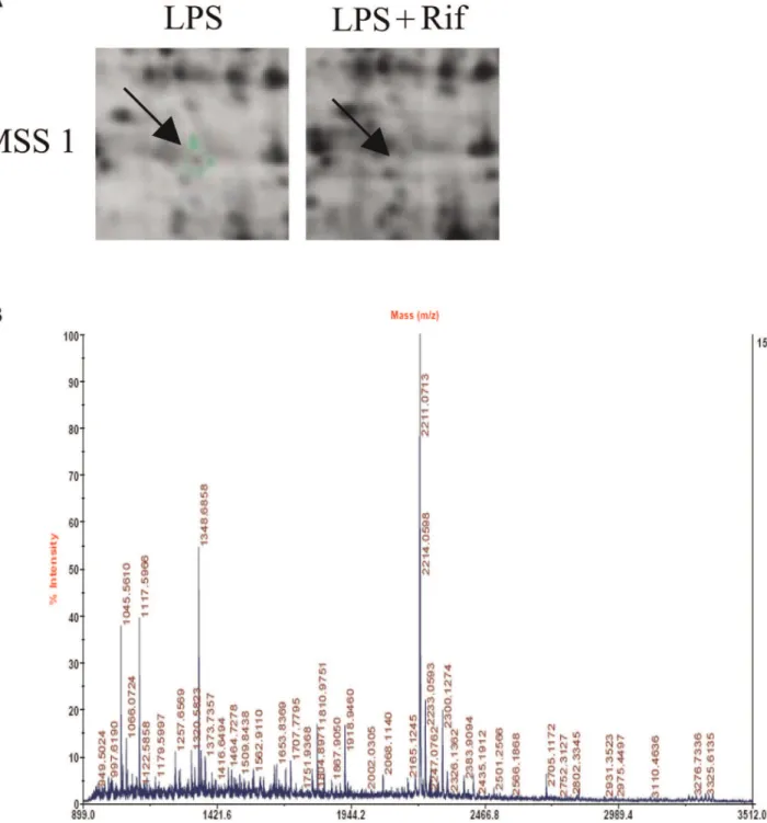
After matching, fifteen protein spots were extracted, digested, and submitted for identification by MALDI-TOF-MS. MSS1 protein was successfully identified. Its expression level was downregulated compared to the control (Figure 1). Detailed information about MSS1 proteins is listed in Table 1, including International Protein Index (IPI) accession number, molecular weight, pH indicated, and rifampicin treated-to-vehicle fluores-cence ratios.
Rifampicin Significantly Suppressed the Expression of MSS1 in LPS-activated BV2 Microglia
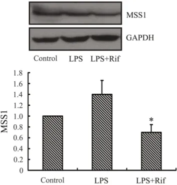
To examine the effect of rifampicin on the expression of MSS1 in LPS stimulated BV2 microglia, we measured the protein levels of MSS1 in LPS-stimulated BV2 microglia. Rifampicin treatment greatly inhibited the LPS-induced MSS1 protein expression
(Figure 2). The result suggested that rifampicin significantly reduced the expression of MSS1 in LPS-stimulated BV2 microglia.
Verification of MSS1 Gene Silencing
We used western blot analysis to confirm the gene knockdown of MSS1 by its targeting siRNAs. After transfection with siRNAs, the expression of MSS1 protein was decreased to 40% compared with the negative control cells. The difference was statistically significant (p,0.01, Figure 3).
IkBa Protein Degradation was Significantly Reduced by MSS1 Gene Knockdown in LPS-stimulated BV2 Microglia
To examine the regulation IkBadegradation by MSS1 in LPS-activated microglia, the BV2 cells were transfected with either MSS1-specfic or control siRNAs for 24 h followed by LPS stimulation at 1000 ng/mL for 30 min. Cell lysates were analyzed for the protein expression of IkBausing western blot. Our results demonstrated that IkBa protein degradation was significantly reduced after MSS1 gene knockdown in LPS-stimulated BV2 microglia (Figure 4).
Downregulation of Microglial NF-kB Activation by MSS1 Gene Silencing in Response to LPS Stimulation
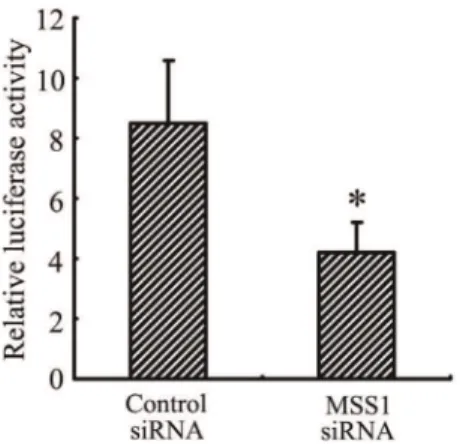
After MSS1 gene knockdown via RNAi, we assessed NF-kB activation using the NF-kB reporter gene assay. The BV2 cells were transfected with either MSS1-specfic or control siRNAs for 24 h. Cells were then incubated with LPS at 1000 ng/mL for 8 h. NF-kB activity was determined by measuring the relative luciferase activity. As shown in Figure 5, LPS markedly enhanced NF-kB activity, while transfection with MSS1-targeted siRNA significant-ly inhibited the enhancement.
Decrease of iNOS Expression and NO Production by MSS1 Gene Knockdown in LPS-induced BV2 Microglia
To investigate the effect of MSS1 gene silencing on iNOS expression and NO production, we measured their protein levels as well as the accumulation of nitrite, a stable metabolite of NO, in LPS-stimulated BV2 microglia. Transfection using MSS1-specific siRNA greatly inhibited the LPS-induced iNOS protein expression (Figure 6). We next evaluated the NO production in culture supernatants by detecting nitrite levels using a Griess reaction. Consistent with the downregulation of iNOS, transfection with MSS1-targeted siRNAs reduced the LPS-mediated NO pro-duction in BV2 microglia (Fig. 6). Our results indicated that MSS1 gene silencing suppressed the production of pro-inflammatory NO by inhibiting the expression of iNOS in LPS-stimulated BV2 microglia.
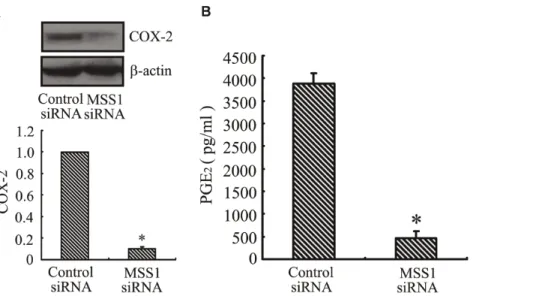
Inhibition of COX-2 Expression and PGE2Production by MSS1 Gene Knockdown in LPS-activated BV2 Microglia
Figure 1. 2D-DIGE gel images of proteins isolated from LPS-stimulated BV2 microglia with or without rifampicin pretreatment.
Arrows indicate proteins that were differentially expressed in rifampicin-treated cells compared with non-treated controls. Peptide mass fingerprint spectra produced by MALDI-TOF-MS. Representative spectra from three independent experiments are shown. The x-axis represents mass-to-charge ratio (m/z), and the y-axis represents relative abundance. The peptide masses are labeled and annotated with corresponding m/z.
doi:10.1371/journal.pone.0036142.g001
Table 1.Differential MSS1 protein expression identified by MALDI-TOF-MS.
Accession Number Name Molecular Weight (Dalton) pH Indicated
Ratio of Spot Density (Rifampicin/Vehicle)
IPI 00270326 26S proteasome regulatory subunit 7
52,867 5.97 21,000,000
Discussion
The immunosuppressive properties of rifampicin have been discussed in the literature for 30 years [23–27]. Calleja et al. discovered that rifampicin activated the human glucocorticoid receptor (hGR), which regulated the expression of various genes including those that encoded interleukins [28]. Further investiga-tions uncovered that rifampicin inhibited Toll-like receptor 2 (TLR2) via the suppression of the DNA binding of NF-kB, providing a novel mechanism contributing to the immunosup-pression of rifampicin [29]. Our results showed that the anti-inflammatory, neuroprotective properties of rifampicin were mediated through the inhibition of signaling molecules, such as NF-kB and mitogen activated protein kinases (MAPKs) in LPS-activated BV2 microglial cells [12]. However, the mechanism by which rifampicin reduces microglial inflammation is not com-pletely understood.
In this investigation, we used 2-DE and MALDI-TOF-MS to identify proteins affected by rifampicin in LPS-pretreated micro-glia. We successfully identified MSS1 protein and showed that its expression was downregulated (Figure 1A and 1B). The proteomic results were verified by western blot analysis, which also demonstrated the inhibition of the LPS-induced MSS1 expression by rifampicin (Figure 2). Our findings suggested that MSS1 was involved in microglial inflammation and its gene knockdown by RNAi alleviated neuroinflammation (Figure 3) in LPS-activated BV2 cells. Our results open the door to further study MSS1 as a potential therapeutic target for neuroinflammation.
UPS represent an ATP-dependent protein degradation mech-anism in eukaryotic cells. The 26S proteasome is a multi-catalytic
proteinase complex. It has a highly ordered structure composed of two complexes, a 20S core and a 19S regulator. The 19S regulator is composed of a base, which contains 6 ATPase subunits and 2 ATPase subunits, and a lid, which contains up to 10 non-ATPase subunits [30]. Proteasomes are distributed throughout eukaryotic cells at a high concentration and cleave peptides in an ATP/ubiquitin-dependent process in a non-lysosomal pathway [31]. Alterations in UPS are correlated with a variety of human pathologies, such as cancer, immunological disorders, inflamma-tion, and neurodegeneration [31–33].
Figure 2. Rifampicin significantly suppressed the expression of MSS1 in LPS-stimulated BV2 microglia.Cells were treated with the indicated doses of rifampicin for 2 h prior to the addition of LPS (1000 ng/ml). At 24 h post-LPS incubation, cell lysates were analyzed for the protein production of MSS1 using western blot. Rifampicin significantly inhibited the LPS-induced MSS1 expression at protein levels. Data were obtained from three independent experiments with four to six replicates each. *p,0.05 compared with untreated cells and
cells treated with LPS in the absence of rifampicin. doi:10.1371/journal.pone.0036142.g002
Figure 3. MSS1 gene knockdown reduced the expression of MSS1 at protein levels. In order to assess the efficacy of gene silencing, western blot analysis was performed after transfection with siRNAs targeting MSS1. The specificity of MSS1 gene silencing was determined by comparing with cells transfected with the scrambled RNA duplex. The BV2 cells were transfected with either MSS1-specfic or control siRNAs. At 24 h post-incubation, cell lysates were analyzed for the protein expression of MSS1 using western blot. Compared with the negative control group, the expression of MSS1 was significantly reduced by incubation with MSS1-targeted siRNAs. Data were obtained from three independent experiments with four to six replicates each. *p,0.05 compared with the negative control group.
doi:10.1371/journal.pone.0036142.g003
Figure 4. MSS1 gene silencing decreased IkBaprotein degra-dation in LPS-activated microglia.The BV2 cells were transfected with either MSS1-specfic or control siRNAs for 24 h, then cells were stimulated with LPS (1000 ng/mL) for 30 min before cell lysates were analyzed for IkBa expression using western blot. IkBa protein degradation was significantly reduced after the addition of siRNAs targeting MSS1 in LPS-induced BV2 microglia. Data were obtained from three independent experiments with four to six replicates each. *p,0.05 compared with the negative control group.
MSS1, also known as S7 or PSMC2, is a 433-amino-acid member of the AAA ATPase family. As a chaperone-like subunit of the 19S regulatory complex, MSS1 localizes in both the nucleus and the cytoplasm where it participates in proteasome events throughout the cell [18]. Additionally, MSS1 is thought to interact with several basal transcription factors and, via this interaction, play a role in transcriptional regulation [34]. Our data provided strong evidence for a novel mechanism of MSS1-mediated neuroinflammation. Additional investigations are needed to
further elucidate this pathway and address the potential of MSS1 gene silencing as a therapy for neurodegenerative disorders. NF-kB is an important transcription factor for the secretion of pro-inflammatory mediators [35]. LPS is shown to increase NF-kB activation, through IkB phosphorylation and the subsequent IkB degradation in macrophages [36]. In the canonical pathway of NF-kB induction, IkBs are phosphorylated at two amino-terminal serines, thus targeting them for polyubiquitination and the subsequent proteasomal degradation. IkB degradation enables NF-kB to translocate to the nucleus and bind to its target genes, including IkB. In addition, proteasomal degradation of transcrip-tionally active p65/RelA promotes the prompt termination of NF-kB responses [37]. The ubiquitin–proteasome pathway is consid-ered pivotal to signal-induced IkB degradation [38–39]. In this study, we investigated the regulation of IkB and NF-kB signaling pathways by MSS1 gene silencing using a reporter gene assay and western blot analysis. Our results demonstrated that LPS caused rapid degradation of IkBa, while MSS1 gene knockdown significantly reduced IkBa degradation in LPS-stimulated BV2 microglia (Figure 4). We also found that LPS markedly enhanced NF-kB activity, whereas treatment with MSS1-specific siRNA significantly inhibited the enhancement (Figure 5). These results suggested that MSS1 gene knockdown suppressed NF-kB activa-tion, likely through the blockage of IkBa degradation. The downregulation of IkBaprovided a novel mechanism for MSS1’s immunomodulation in LPS-activated microglia.
Intranuclear blockage of NF-kB has been reported to suppress the expression of iNOS and COX-2 [40]. Our results indicated that transfection with MSS1-targeted siRNAs decreased the production of pro-inflammatory NO by inhibiting the expression of iNOS in LPS-stimulated BV2 microglia (Figure 6A and 6B). We also found that gene knockdown of MSS1 reduced the production of pro-inflammatory PGE2 through suppressing COX-2 gene expression in LPS-induced BV2 microglia (Figure 7A and 7B).
In conclusion, we demonstrated that rifampicin inhibited the expression of MSS1, which subsequently decreased IkBa
Figure 5. MSS1 gene silencing inhibited microglial NF-kB activation in response to LPS stimulation. The BV2 cells were transfected with either MSS1-specfic or control siRNA for 24 h, then cells were incubated with LPS at 1000 ng/mL for 8 h. After that, cells were transfected with NF-kB-luciferase reporter plasmid and pCMV-gal control vectors using Lipofectamine reagents. NF-kB activation was detected and expressed as relative luciferase activity. Compared with the negative control group, treatment with MSS1-targetd siRNA significantly suppressed the enhancement of NF-kB activity by LPS. Data were obtained from three independent experiments with four to six replicates each. *p,0.05 compared with the negative control group.
doi:10.1371/journal.pone.0036142.g005
Figure 6. Decreased iNOS expression and NO production by MSS1 gene silencing in LPS-activated microglia.The BV2 cells were transfected with either MSS1-specfic or control siRNA for 24 h, then cells were stimulated for 24 h with LPS (1000 ng/mL). At 24 h post-LPS incubation, cell lysates were analyzed for the protein production of iNOS using western blot. The Griess assay was performed to measure the production of the NO metabolite, nitrite. Transfection with MSS1-specific siRNA suppressed the LPS-induced iNOS expression at protein levels, along with the production of nitrites Data were obtained from three independent experiments with four to six replicates each. *p,0.05 compared with the
negative control group.
degradation and the production of inflammatory mediators. Our results supported the potential application of MSS1 suppression, together with anti-inflammatory rifampicin, for the treatment of neuroinflammation and neurodegeneration.
Author Contributions
Conceived and designed the experiments: ET WB. Performed the experiments: WB XJ YL LZ SX AX ET. Analyzed the data: ET WB QS XJ LY. Contributed reagents/materials/analysis tools: ET QS JL AX. Wrote the paper: ET WB QS.
References
1. Liu B, Hong JS (2003) Role of microglia in inflammation-mediated neurodegenerative diseases: mechamisms and strategies for therapeutic in-tervention. J Pharmacol Exp Ther 304: 1–7.
2. Eikelenboom P, van Gool WA (2004) Neuroinflammatory perspectives on the two faces of Alzheimer’s disease. J Neural Transm 111: 281–294.
3. Koning N, Bo¨ L, Hoek RM, Huitinga I (2007) Downregulation of macrophage inhibitory molecules in multiple sclerosis lesions. Ann Neurol 62: 504–514. 4. Krause DL, Mu¨ller N (2010) Neuroinflammation, microglia and implications for
anti-inflammatory treatment in Alzheimer’s disease. Int J Alzheimers Dis 2010. pii, 732806.
5. Qian L, Flood PM, Hong JS (2010) Neuroinflammation is a key player in Parkinson’s disease and a prime target for therapy. J Neural Transm 117: 971–979.
6. Blanchard JS (1998) The ying and yang of rifampicin. Nat Med 4: 92–96. 7. Bellahse`ne A, Forsgren A (1980) Effect of rifampin on the immune response in
mice. Infect Immun 27: 15–20.
8. Tsankov N, Grozdev I, Kkzandjieva J (2006) Old drug–new indication. Rifampicin in psoriasis. J Dermatolog Treat 17: 18–23.
9. Tsiskarishvili NV, Tsiskarishvili NI (2009) The anti-tubercular drugs in the treatment of psoriasis.Georgian Med News 174: 25–28.
10. Namazi HJ (2008) Practice pearl: a novel use of rifampicin for treatment of carpal tunnel syndrome. Pain 9: 380–381.
11. Xu J, Wei C, Xu C, Bennett MC, Zhang G, et al. (2007) Rifampicin protects PC12 cells against MPP+-induced apoptosis and inhibits the expression of an alpha-Synuclein multimer. Brain Res 1139: 220–225.
12. Bi W, Zhu L, Wang C, Liang Y, Liu J, et al. (2011) Rifampicin inhibits microglial inflammation and improves neuron survival against inflammation. Brain Res 1395: 12–20.
13. Dai JN, Zong Y, Zhong LM, Li YM, Zhang W (2011) Gastrodin inhibits expression of inducible NO synthase, cyclooxygenase-2 and pro-flammatory cytokines in cultured LPS-stimulated microglia via MAPK pathways. PLoS One 6: e21891.
14. S Vallabhapurapu, M Karin (2009) Regulation and function of NF-kappaB transcription factors in the immune system, Annu. Rev. Immunol 27: 693–733. 15. Zhang W, Wei Q (2011) Calcineurin stimulates the expression of inflammatory factors in RAW 264.7 cells by interacting with proteasome subunit alpha type 6. Biochem Biophys Res Commun 407: 668–673.
16. De Mot R, Nagy I, Walz J, Baumeister W (1999) Proteasomes and other self-compartmentalizing proteases in prokaryotes. Trends Microbiol 7: 88–92.
17. Williams AJ, Dave JR, Tortella FC (2006) Neuroprotection with the proteasome inhibitor MLN519 in focal ischemic brain injury: relation to nuclear factor kappaB (NF-kappaB), inflammatory gene expression, and leukocyte infiltration. Neurochem Int 49: 106–12.
18. Yanagi S, Shimbara N, Tamura T (2000) Tissue and cell distribution of a mammalian proteasomal ATPase, MSS1, and its complex formation with the basal transcription factors. Biochem Biophys Res Commun 279: 568–573. 19. Wang Y, Cheung YH, Yang Z, Chiu JF, Che CM, et al. (2006) Proteomic
approach to study the cytotoxicity of dioscin (saponin). Proteomics 6: 2422–2432.
20. Jessie K, Pang WW, Haji Z, Rahim A, Hashim OH (2010) Proteomic analysis of whole human saliva detects enhanced expression of interleukin-1 receptor antagonist,thioredoxin and lipocalin-1 in cigarette smokers compared to non-smokers. International Journal of Molecular Sciences 11: 4488–4505. 21. Tsikas D (2007) Analysis of nitrite and nitrate in biological fluids by assays based
on the Griess reaction: appraisal of the Griess reaction in the L-arginine/nitric oxide area of research. J Chromatogr B Analyt Technol Biomed Life Sci 15; 851(1–2): 51–70.
22. Woo MS, Jang PG, Park JS, Kim, WK, Jon TH, et al. (2003) Selective modulation of lipopolysaccharide-stimulated cytokine expression and mitogen-activated protein kinase pathways by dibutyryl-cAMP in BV2 microglial cells. Molecular Brain Res 113: 86–96.
23. Dajani BM, Canadi MS, Thompson JS, Kasik JE (1972) Rifampicin: an immunosuppressant? Lancet 2: 1904.
24. Tsankov N, Angelova I (2003) Rifampin in dermatology. Clin Dermatol 21: 50–55.
25. Mlambo G, Sigola LB (2003) Rifampicin and dexamethasone have similar effects on macrophage phagicytosis of zymosan, but differ in their effects on nitrite and TNF-alpha production. Int Immunopharmacol 3: 513–522. 26. An N, Song Y, Zhang X, Ci X, Li H, et al. (2008) Pretreatment of mice with
rifampicin prolongs survival of endotoxic shock by modulating the levels of inflammatory cytokines. Immunopharmacol Immunotoxicol 30: 437–446. 27. An N, Song Y, Zhang X, Ci X, Li H, et al. (2005) Group B streptococci exposed
to rifampin or clindamycin (versus ampicillin or cefotaxime) stimulate reduced production of inflammatory mediators by murine macrophages. Pediatr Res 57: 419–423.
28. Calleja C, Pascussi JM, Mani JC, Maurel P, Vilarem MJ (1998) The antibiotic rifampicin is a nonsteroidal ligand and activator of the human glucocorticoid receptor. Nat Med 4: 92–96.
Figure 7. Inhibition of COX-2 expression and PGE2production by MSS1 gene knockdown in LPS-induced microglia.The BV2 cells
were transfected with either MSS1-specfic or control siRNA for 24 h, then cells were stimulated for 24 h with LPS (1000 ng/mL). At 24 h post-LPS incubation, cell lysates were analyzed for the protein production of COX-2 using western blot. We collected the supernatant and further analyzed the production of PGE2. MSS1 gene silencing suppressed the LPS-induced COX-2 expression at protein levels, as well as PGE2production. Data were
obtained from three independent experiments with four to six replicates each. *p,0.05 compared with the negative control group.
29. Kim SK, Kim YM, Yeum CE, Jin SH, Chae GT, et al. (2009) Rifampicin Inhibits the LPS-induced Expression of Toll-like Receptor 2 via the Suppression of NF-kappaB DNA-binding Activity in RAW 264.7 Cells. Korean J Physiol Pharmacol 13: 475–482.
30. Hershko A, Ciechanover A (1998) The ubiquitin system. Annu Rev Biochem 67: 425–479.
31. Finley D (2009) Recognition and processing of ubiquitin-protein conjugates by the proteasome. Annu Rev Biochem 78: 477–513.
32. Goldberg AL (2003) Protein degradation and protection against misfolded or damaged proteins. Nature 426: 895–899.
33. Sujashvili R (2008) Ubiquitin-26s proteasome system. New therapeutic implications. Georgian Med News 161: 48–51.
34. Yanagi S, Shimbara N, Tamur T (2000) Tissue and cell distribution of a mammalian proteasomal ATPase, MSS1, and its complex formation with the basal transcription factors. Biochem Biophys Res Commun 279: 568–573.
35. Ozato K, Tsujimura H, Tamura T (2002) Toll-like receptor signal in gand regulation of cytokine gene expression in the immune system. Biotechniques 66– 72.
36. Kim WG, Mohney RP, Wilson B, Jeohn GH, Liu B, et al. (2000) Regional difference in susceptibility to lipopolysaccharide-induced neurotoxicity in the rat brain: role of microglia. J Neurosci 20: 6309–6316.
37. Saccani S, Marazzi I, Beg A, Natoli G (2004) Degradation of promoter-bound p65/RelA is essential for the prompt termination of the nuclear factor kappaB response. J Exp Med 200: 107–113.
38. Chen F, Lu Y, Kuhn DC, Maki M, Shi X, et al. (1997) Calpain Contributes to Silica-Induced IkBa Degradation and Nuclear Factor-kB Activation. AR-CHIVES OF BIOCHEMISTRY AND BIOPHYSICS 342: 383–388. 39. YmamotoY, Gaynor RB (2004) IkappaB kinases: key regulators of the
NF-kappaB Pathway. Trends Biochem Sci 29: 72–79.