Biomarkers, Genomics, Proteomics, and Gene Regulation
Gla-Rich Protein Is a Novel Vitamin K-Dependent
Protein Present in Serum That Accumulates at Sites
of Pathological Calcifications
Carla S.B. Viegas,*
†Sofia Cavaco,*
Pedro L. Neves,
‡Ana Ferreira,
§Alexandre Joa˜o,
§Matthew K. Williamson,
¶Paul A. Price,
¶M. Leonor Cancela,*
†and Dina C. Simes*
†§ From the Centre of Marine Sciences * and GenoGla Diagnostics,† University of Algarve, Faro, Portugal; the Department of Nephrology,‡Faro Hospital, Faro, Portugal; the Department of Dermatology,§Lisbon Central Hospital, Lisbon, Portugal; and the Division of Biology,¶University of California San Diego, La Jolla, CaliforniaMineralization of soft tissues is an abnormal process that occurs in any body tissue and can greatly in-crease morbidity and mortality. Vitamin K-depen-dent (VKD) proteins play a crucial role in these processes; matrix Gla protein is considered one of the most relevant physiological inhibitors of soft tissue calcification know to date. Several studies have suggested that other , still unknown , VKD pro-teins might also be involved in soft tissue calcifica-tion pathologies. We have recently identified in sturgeon a new VKD protein , Gla-rich protein (GRP) , which contains the highest ratio between number of Gla residues and size of the mature pro-tein so far identified. Although mainly expressed in cartilaginous tissues of sturgeon , in rat GRP is present in both cartilage and bone. We now show that GRP is a circulating protein that is also ex-pressed and accumulated in soft tissues of rats and humans , including the skin and vascular system in which, when affected by pathological calcifica-tions , GRP accumulates at high levels at sites of mineral deposition , indicating an association with calcification processes. The high number of Gla res-idues and consequent mineral binding affinity properties strongly suggest that GRP may directly influence meral formation , thereby playing a role in processes in-volving connective tissue mineralization. (Am J Pathol 2009, 175:2288 –2298; DOI: 10.2353/ajpath.2009.090474)
Extracellular matrix (ECM) calcification can be either a physiological or a pathological process depending on site and time of occurrence. Physiological ECM calcifica-tion is restricted to bone and to the hypertrophic zones of growth plate cartilage, whereas pathological or ectopic ECM calcification, defined as inappropriate biomineral-ization occurring in soft tissues and consisting of calcium phosphate salts that include hydroxyapatite, is an abnor-mal process that can occur virtually in any tissue of the body.1However, skin, kidney, tendons, and the
cardio-vascular system appear particularly prone to develop this pathology.2
First considered to be a passive process occurring as a nonspecific response to tissue injury or necrosis, recent evidence now indicates that ECM calcification is a natu-rally occurring process that must be actively inhibited and starts to appear as soon as inhibitors are removed from the matrix.1,3,4In a healthy organism, cells appear to
synthesize natural inhibitors of mineralization that prevent ectopic calcification, which initiates when disequilibrium occurs between expression of calcification inhibitors and enhancers, emphasizing the need for a tight regulation to prevent ectopic calcifications.
Key genes known to be involved in the regulation of this complex process are those acting as calcification inhibitors such as matrix Gla protein (MGP), osteocalcin (BGP), bone sialoprotein (BSP), osteoprotegerin (Opg), and fetuin.1,3Among those, MGP, a vitamin K-dependent
Supported in part by Centre of Marine Sciences (M.L.C. and D.C.S.), project POCTI MAR 57921 2004 (D.C.S., M.L.C., C.S.B.V.) and NIH grant HL58090 (to P.A.P.). C.S.B.V. was the recipient of Ph.D. fellowship SFRH/ BD/9077/2002 from the Portuguese Science and Technology Foundation. Methods and tools described and obtained during the course of this work are included in United States priority application 61/136.315, August 27, 2008, and PCT patent application PCT/PT2009000046, August 27, 2009.
Accepted for publication August 21, 2009.
D.C.S., M.L.C., and C.S.B.V. are cofounders of GenoGla Diagnostics. Supplemental material for this article can be found on http://ajp. amjpathol.org.
Address reprint requests to Dina C. Simes, Ph.D., Centre of Marine Sciences (CCMAR), University of Algarve, Campus de Gambelas, 8005-139 Faro, Portugal, E-mail: [email protected].
protein (VKD), is widely accepted as playing a pivotal role in preventing soft tissue calcification, local mineral-ization of the vascular wall,5 and more recently, skin
elastic fiber mineralization in pseudoxanthoma elasticum (PXE)6 – 8 and in scleroderma with and without
calcino-sis.9It is also known that several factors, such as
insuf-ficient intake of vitamin K, mutations in the␥-carboxylase enzyme, and warfarin treatment, which can all induce arterial10 –12 and skin calcifications,7,13–15 may act by
reducing or abolishing␥-carboxylation of VKD proteins. Those pathologies have also been associated with a loss of MGP function, until now considered to be the central Gla protein for prevention of connective tissue mineral-ization, both in the vascular system and skin. Although many efforts have been made to understand the mech-anisms controlling these abnormal calcifications, the ex-istence of other potential, still unknown, calcification in-hibitors has been suggested to explain some reported phenotypes and occurrences that are not completely justified by the presence or absence of MGP.1,16,17
We have recently identified in sturgeon a new VKD protein, Gla-rich protein (GRP), with an unprecedented high content of Gla residues and uncommonly high ca-pacity to bind calcium, with orthologs in all taxonomic groups of vertebrates and highly conserved throughout evolution (78% identity between sturgeon and human GRP).18GRP mRNA was found to be highly expressed in
sturgeon cartilaginous tissues, and in rat skeletal tissues, both cartilage and bone, which invalidated the concept that this protein could be solely a specific marker for distal chondrocytes, as previously proposed by others.18
In this study we show, for the first time, that GRP is a circulating protein also expressed and accumulated in soft tissues like skin and vascular system of rats and humans and that it is clearly associated with calcification pathologies in these tissues, being highly accumulated at sites of ectopic mineral deposits. Furthermore, the exten-sive number of Gla residues (16 Gla residues in sturgeon and, by comparison, 15 in all mammals) and the absence of other identifiable functional domains, together with our
in vivoand in vitro evidence for a high mineral binding affinity, strongly suggest that GRP might be a potent physiological modulator of soft tissue calcification, acting by directly influence mineral formation and or recruit-ment, and an important new player in the complexity of phenotypes involving connective tissue mineralization, whose mechanisms and regulatory pathways remain to be fully understood.
Materials and Methods
Biological Material
This study was approved by the Faro Hospital and Lisbon Central Hospital ethics committee. We included in our study patients with stage 5 chronic kidney disease who underwent surgery for arteriovenous fistula creation. A sample of the radial artery wall was collected at the time of surgery from each patient. Calcified and noncalcified carotid samples were also collected at autopsy. Skin
biopsies were taken under local anesthesia from the af-fected skin of PXE, dermatomyositis with calcinosis, and scleroderma with calcinosis (lateral neck or axilla) pa-tients. All patients exhibited clinical signs and were diag-nosed for the corresponding pathology. Control skin bi-opsies were obtained from forearm regions of volunteer healthy subjects. Human blood was collected from vol-unteer healthy subjects by venipuncture at Faro Hospital. Informed consent was obtained from all participants.
Pig ears were obtained from the local slaughterhouse, immediately frozen for transport, and kept at⫺80° until further processing. Rat skin and blood samples were obtained from Mus musculus specimens maintained at the University of Algarve animal facilities.
Gene Expression by Quantitative Real-Time
Polymerase Chain Reaction
Total RNA was extracted from rat adult tissues (including bony, cartilaginous, and major soft tissues) as de-scribed.19One microgram of total RNA was treated with
RQ1 RNase-free DNase (Promega, Madison, WI) and reverse-transcribed at 37°C with MMLV-RT (Invitrogen, San Diego, CA) using specific reverse primers RnGRP1R (5⬘-CACTCAAAAACAAGACAAAGCAAACATCCG-3⬘), RnGAPDH_RT1R (5 ⬘-GAAGACGCCAGTAGACTCCACGA-CAT-3⬘) and RnHPRTI_RT1R (5⬘-CACAAGGGAAGTGA-CAATCTACCTGACG-3⬘). Quantitative real-time polymer-ase chain reaction (qPCR) was performed with an iCycler iQ apparatus (Bio-Rad, Amadora, Portugal), using primer sets RnGRP1F (5 ⬘-TCCTTCCTACCTCTACAACCGCCAA-AA-3⬘)/RnGRP1R to amplify rat GRP, RnGAPDH_RT1F (5⬘-CGGCAAGTTCAACGGCACAGTCAAG-3⬘)/RnGAPDH_RT1R to amplify rat glyceraldehyde-3-phosphate dehydroge-nase (GAPDH), and RnHPRTI_RT1F (5 ⬘-AAATGTCTGTT-GCTGCGTCCCTTTTGAT-3⬘)/RnHPRTI_RT1R to amplify rat HPRTI. PCR reactions, set up in duplicates, were performed as previously described18 using Absolute
QPCR SYBR green fluorescein mix (ABgene, Epsom, UK). Fluorescence was measured at the end of each extension cycle in the FAM-490 channel and melting profiles of each reaction were performed to check for unspecific product amplification. Levels of gene expres-sion were calculated using the comparative method (⌬⌬Ct) and normalized using gene expression levels of GAPDH or HPRTI housekeeping genes. Gene expression in lung was set to 1 and used as reference for relative expression in other tissues. qPCR was performed in qua-druplicates and a normalized SD was calculated.
Histological Sample Preparation
Samples were collected as previously described18 and
included either in paraffin or in Historesin Plus (Leica Microsystems, Lisbon, Portugal), according to the man-ufacturer’s instructions. Mineral deposits were detected with silver nitrate (Sigma-Aldrich, Taufkirchen, Germany) by the von Kossa method, and physiological structures were identified by counterstaining with hematoxylin and eosin or toluidine blue.20
In Situ Hybridization
A 417-bp fragment of rat GRP cDNA (spanning from nucleotide 417 to the 3⬘ end) cloned in pCRII-TOPO was
either linearized with ApaI and transcribed with SP6 RNA polymerase to generate an antisense riboprobe or linear-ized with KpnI and transcribed with T7 RNA polymerase to generate a sense riboprobe. A 364-bp fragment of the human GRP cDNA (spanning from nucleotide 459 to 822 according to EST sequences retrieved from GenBank sequence data base and previously identified as GRP),18
amplified by PCR with HsGRPis2F (5 ⬘-CATCCTATCTC-TACAACCGCCACC-3⬘) and HsGRPis1R (5⬘-TTCAG-CGTTTTTATTTGTAAGCCATA-3⬘) primers and genomic DNA, and cloned in pCRII-TOPO, was either linearized
with KpnI and transcribed with T7 RNA polymerase to generate an antisense riboprobe or linearized with ApaI and transcribed with SP6 RNA polymerase to generate a sense riboprobe. Probes were then labeled with digoxi-genin using RNA labeling kit (Roche, Mannheim, Ger-many) according to the manufacturer’s instructions. RNA
in situhybridization was performed on paraffin sections of rat and human tissues with digoxigenin-labeled antisense riboprobes, as previously described.18 Briefly, sections
were digested with 40g/ml proteinase K (Sigma) in 1X phosphate-buffered saline containing 0.1% Tween 20 (Sigma) for 30 minutes and then hybridized at 68°C over-night in a humidified chamber. After hybridization, sec-tions were washed and the signal revealed with the alka-line phosphatase-coupled antidigoxigenin-AP antibody (Roche) and nitro blue tetrazolium/5-bromo-4-chloro-3-indolyl phosphate substrate solution (Sigma). Negative controls for GRP mRNA detection were performed with sense probes.
Production of Polyclonal Rabbit Antibody
(CTerm-GRP) against GRP
Affinity-purified rabbit polyclonal antibody against GRP was obtained from SDI- Strategic Diagnostics (Newark, DE), using for immunization a synthetic amino acid pep-tide corresponding to the C terminus of rat GRP (RQWHYDGLYPSYLYNRQNI), synthesized by NeoMPS, Inc. (San Diego, CA), purified to 95% purity, and conju-gated to keyhole limpet hemocyanin. A cysteine residue was introduced at the N terminus of the peptide for bind-ing to keyhole limpet hemocyanin. The affinity purified antiserum was termed CTerm-GRP.
Protein Extraction and Detection from
Noncalcified Mammalian Tissues
Pig and rat skin (dermis and epidermis) and human blood vessels were cleaned from fat tissue, lyophilized, and reduced to fine powder. From the calculated weight a 10-fold excess (w/v) of 4 mol/L guanidine HCl (Sigma-Aldrich) extraction solution was added with vigorous stir-ring at 4°C for 24 hours, and the extracted material was separated by centrifugation for 10 minutes at 10000⫻ g. A portion of these crude guanidine extract was dialyzed
against 50 mmol/HCI, using 3500 molecular weight tub-ing (Spectra-Por 3, Spectrum, Gardena, CA) with four changes of medium over 2 days and analyzed for the presence of GRP by Western blotting using the purified CTerm-GRP antibody. Rat GRP was further isolated from the crude guanidine HCl extract by reverse-phase high performance liquid chromatography as previously de-scribed.18Resulting fractions were analyzed for the
pres-ence of GRP and Gla-containing proteins by dot blot using the CTerm-GRP and M3B (American Diagnostica, Stamford, CT) antibodies, respectively.
For further isolation, pig guanidine HCl extract was incubated with 0.1% hydroxyapatite (Calbiochem, San Diego, CA) for 24 hours at 4°C, with constant rotation. After incubation, hydroxyapatite was separated from the crude extract by centrifugation for 10 minutes at 10000⫻
g.Hydroxyapatite was further cleaned by washing twice with 6 mol/L guanidine HCl for 1 hour and twice with distillated water for 30 minutes at room temperature with constant stirring. The resulting hydroxyapatite powder was demineralized using a 10-fold excess of 10% formic acid for 4 hours at 4°C with vigorous stirring, as de-scribed for bone demineralization18,21and further dialyzed
against 50 mmol/L HCl as described above. Aliquots of the formic acid dialyzed extract were analyzed by Western blotting using the purified CTerm-GRP antibody.
Electrophoresis and Western Blotting
Aliquots of total protein were fractionated into a 4 to 12% gradient polyacrylamide precast gel containing 0.1% so-dium dodecyl sulfate (NuPage, Invitrogen), and protein profile revealed by staining the gel as described.18,21
Transfer onto nitrocellulose membranes (Amersham Bio-sciences, Carnaxide, Portugal) and protein immunode-tection was performed as described previously.21,22GRP
protein was detected by incubating blots overnight with 5 g/ml anti-CTerm-GRP antibody in 5% (w/v) nonfat dried milk powder in Tris-buffered saline/Tween 20 (15 mmol/L NaCl, 10 mmol/L Tris-HCl buffer, pH 8, 0.05% Tween 20) as primary antibody and alkaline phosphatase-labeled goat anti-rabbit IgG antibody (Sigma-Aldrich) diluted 1:30,000 in Tris-buffered saline/Tween 20, as secondary antibody. Visualization of immunoreactive bands was achieved using nitro blue tetrazolium/5-bromo-4-chloro-3-indolyl phosphate substrate solution (Calbiochem) as described.21,22
Dot Blotting
Samples of total protein were applied onto a nitrocellu-lose membrane (Invitrogen) as previously described.22
GRP detection was performed using the purified CTerm-GRP antibody as primary antibody and alkaline phos-phatase-labeled goat anti-rabbit IgG (Sigma-Aldrich), as secondary antibody. Gla-containing proteins were de-tected using 5 g/ml of the purified monoclonal M3B antibody (American Diagnostica) as primary antibody and alkaline phosphatase-labeled goat anti-mouse IgG as secondary antibody.
Immunolocalization
Immunohistochemical staining experiments were per-formed on paraffin and Historesin Plus-embedded tissue sections as described.20,21Briefly, the endogenous
per-oxidase activity was blocked with 3% H2O2 in Coons buffer (CBT: 0.1 mol/L Veronal, 0.15 mol/L NaCl, 0.1% Triton X-100) for 15 minutes. Nonspecific antibody bind-ing was blocked with 0.5% (w/v) bovine serum albumin directly after peroxidase activity blocking, or after treat-ment with chondroitinase ABC (Sigma-Aldrich) (0.1 U/ml) for 1 hour at 37°C. Incubation with the purified polyclonal CTerm-GRP or the M3B antibodies (5g/ml and 5 g/ml, respectively, diluted in CBT), as primary antibodies, was performed overnight in a humidified chamber at room temperature. Peroxidase activity was detected using as secondary antibodies the peroxidase-conjugated goat anti-rabbit and anti-mouse IgG, respectively Aldrich), and 0.025% 3,3-diaminobenzidine (Sigma-Aldrich) as described.20,21Negative controls consisted in
the substitution of the primary antibody with both normal rabbit serum and CBT. Counterstaining was performed with hematoxylin and eosin or toluidine blue.
Depletion of High Abundant Serum Proteins and
GRP Detection
Human and rat sera were prepared as described,23,24
quick-frozen in 1-ml aliquots on dry ice, and stored at ⫺80°C until use. Depletion of the high abundant proteins was performed for each 1 ml of human and rat serum aliquots using the ProteoMiner kit (Bio-Rad), according to the manufacturer’s instructions. Five microliters of each of the eluted fractions was applied onto a nitrocellulose membrane (Invitrogen). GRP detection was performed with the purified CTerm-GRP antibody as described in dot blotting.
Results
The GRP Gene Is Expressed Both in Skeletal
and in Soft Tissues
We have recently shown that GRP in sturgeon is ex-pressed exclusively by cartilage-associated cells, while in rat it was also expressed by bone cells. However, our
in silico analysis further suggested that GRP could be present in other nonskeletal tissues.18To continue these
studies and establish the pattern of GRP tissue distribu-tion, we have determined its spatial levels of expression in adult rat skeletal and soft tissues. Expression of GRP was detected by real-time PCR in all 16 tissues analyzed, the most significant levels being observed in cartilage-and bone-containing tissues such as skull, inner ear, tail, outer ear, and nose (see Supplemental Figure S1 at
http://ajp.amjpathol.org). However, the presence of higher levels of expression in outer ear and nose, a result con-firmed when using GAPDH to normalize GRP gene pression (results not shown), could not be easily
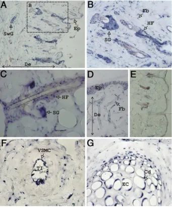
ex-plained. To further elucidate this question and clearly establish the identity of GRP-expressing cells, we per-formed in situ hybridization in sections from all rat tissues expressing GRP. Interestingly, in sections of outer ear (Figure 1) and nose (results not shown) GRP was found to be highly expressed in skin and its appendages, at the levels of epidermis and dermis. In rat outer ear skin, GRP mRNA was detected in epidermis (Ep) (Figure 1, A and D) and in all main structures housed in the dermis (De); in the fibroblasts (Fb) that compose the reticular layer of the dermis (Figure 1, B and D); in the hair follicles (HF) (Figure 1, B and C); and in sweat (SwG) (Figure 1A) and sebaceous (SG) (Figure 1, B and C) glands. Negative controls were performed through hybridization of consec-utive sections with GRP sense probe (Figure 1E), con-firming the specificity of the signal observed in the above described structures. Figure 1F shows GRP positive ex-pression in an artery from the dermis of rat outer ear, with higher expression found in vascular smooth muscle cells (VSMC) of the tunica intima (TI), and lower expression in the tunica media. In rat outer ear, GRP mRNA is also expressed by elastic cartilage (EC) chondrocyte cells (Cd) from all stages of differentiation (Figure 1G).
Figure 1. Rat GRP gene is highly expressed in nonskeletal tissues. Sites of
gene expression were determined by in situ hybridization with digoxigenin-labeled antisense probes in outer ear paraffin sections, and signal was revealed with alkaline phosphatase-coupled antidigoxigenin-AP antibody and nitro blue tetrazolium /5-bromo-4-chloro-3-indolyl phosphate substrate solution, yielding a characteristic blue color. GRP is highly expressed in the skin and its appendages in both epidermis (Ep, A and D) and dermis (De, A and D). Within the dermis GRP is detected in the fibroblasts (Fb, B and D), hair follicles (HF, C), and sebaceous (SG, B and C) and sweat glands (SwG,
A). In blood vessels, GRP is detected mainly in vascular smooth muscle cells
(VSMC) of the tunica intima (TI, F). In the elastic cartilage (EC) composing the outer ear, GRP is detected in chondrocyte (Cd) cells (G). Negative control performed through in situ hybridization with GRP sense probe is presented in E. Magnifications: A, D, E,⫻10; B, C, F, G, ⫻20.
These high levels of GRP expression found in both skin and its appendages, as well as in the elastic cartilage, which together constitute the main structures present in rat nose and in outer ear, can explain the highest GRP levels obtained in these tissues by qPCR (see Supple-mental Figure S1 at http://ajp.amjpathol.org). The results presented here, together with our previous findings, clearly indicate that GRP is present in all types of carti-lage. Also the presence of GRP mRNA in blood vessels could explain the levels of GRP obtained by qPCR in all irrigated organs.
In Soft Tissues, GRP Accumulates in Skin and in
the Vascular System
Specific GRP rabbit polyclonal antibodies (CTerm-GRP antibody) were produced using a 19-amino-acid syn-thetic peptide homologous to the C-terminal of rat GRP, and cross-reactivity against GRP from other species was checked by Western blot. The antibody was validated by Western blot using purified sturgeon GRP18; a crude rat
skin extract, prepared using 4 mol/L guanidine HCl; a crude pig skin extract, obtained as described above, followed by a direct incubation with hydroxyapatite from which proteins with affinity for this mineral phase were eluted; and a crude guanidine extract of medium-size human blood vessels with no apparent calcifications and collected at autopsy. In all cases, a single positive immu-noreactive band, with a migration profile similar to that of sturgeon GRP, was obtained (see Supplemental Figure S2A at http://ajp.amjpathol.org),18indicating that GRP is
not only expressed but also accumulated in skin and/or in vascular system in sufficient amounts to be detected from a crude extract. Furthermore, our data provided evidence that mammalian GRP shows a clear binding affinity for hydroxyapatite.
To further confirm the ␥-carboxylation status of the protein, we further purified rat GRP from the skin guani-dine crude extract using reverse-phase high perfor-mance liquid chromatography as previously described.18
Individual peak fractions were analyzed for the presence of GRP by dot blot using the CTerm-GRP antibody (re-sults not shown) and the GRP peak containing fraction was then further analyzed for the presence of Gla resi-dues using the anti-Gla M3B antibody. Positive M3B an-tibody immunoreaction was obtained by dot blot for rat skin GRP (see Supplemental Figure S2B (rGRP) at http://
ajp.amjpathol.org),18using as positive control, the purified
sturgeon GRP protein (see Supplemental Figure S2B (StGRP) at http://ajp.amjpathol.org)18whose
␥-carboxyla-tion status was previously confirmed by amino acid anal-ysis.18These results provided additional evidence for the
presence of␥-carboxylated residues in rat GRP isolated from skin.
Spatial Profile of GRP Accumulation in Rat
Tissues
Sites of GRP accumulation were determined by immuno-histochemistry in paraffin sections of several adult rat
tissues, using the CTerm-GRP antibody. In skeletal tis-sues, GRP antigen was mainly detected inside cartilagi-nous (Figure 2, A and B) and bony cells (Figure 2C), following a pattern similar to that previously observed for GRP mRNA.18,25 In the rat rib, GRP was localized in
mature (MC), columnar (CC), hypertrophic (HC) (Figure 2A), and immature (IC) (Figure 2B) chondrocytes, as well as in compact bone osteocytes (Oc) (Figure 2C). In ad-dition, when we performed a chondroitinase pretreatment of the sections, we were able to detect GRP in the extra-cellular matrix of calcified hypertrophic cartilage and in the matrix surrounding the rib cartilage (results not shown), in concordance with previously reported re-sults.25In skin, GRP accumulation also followed the
pat-tern of its mRNA distribution. It accumulated in epidermis (Ep) (Figure 2, D and E), within those fibroblasts (Fb) responsible for synthesis of dermis collagen elastic fibers (Figure 2E), and in skin appendages, eg, hair follicles (HF) (Figure 2D), sebaceous (SG) (Figure 2D), and sweat (results not shown) glands. In the elastic cartilage (EC) of
Figure 2. Rat GRP is highly accumulated in cartilage (A, B, F), bone (C), skin
and its appendages (D and E), and in the vascular system (G–J) as deter-mined by immunohistochemistry using the CTerm-GRP primary antibody and peroxidase-conjugated goat anti-rabbit IgG as secondary antibody. Per-oxidase activity was visualized using 3,3-diaminobenzidine substrate yielding a brown color. A and B: Sites of GRP accumulation in sections of rib cartilage showing protein detection inside chondrocytes in all stages of maturation: columnar chondrocytes (CC), mature chondrocytes (MC), hypertrophic chondrocytes (HC), and immature chondrocytes (IC). C: GRP detection in bone sections showing protein inside osteocytes (Oc). D–F: GRP detection in outer ear sections showing protein accumulation in skin epidermis (Ep) and dermis (De, D and E) and its appendages: hair follicles (HF, D), sebaceous glands (SG, D), and dermal fibroblast (Fb, E). In addition, GRP was detected in chondrocytes (Cd) of the elastic cartilage (EC, F). G–J: GRP accumulation in the vascular system in sections of outer ear (G and H), heart (I), and kidney (J), showing GRP highly accumulated in the walls of blood vessels (Bv) and capillaries (Cp). TM, tunica media; TI, tunica intima; EL, elastic lamina. Magnifications: A–E and J,⫻10; F–H and I, ⫻20.
the outer ear, GRP was mainly detected inside the chon-drocytes (Cd) (Figure 2F). The absence of signal in the extracellular matrix was probably due to lack of chon-droitinase pretreatment of these tissue sections. In addi-tion to cartilage, bone and skin, the vascular system also appears to be one of the primary sites of GRP accumu-lation. Small and medium blood vessels from several irrigated tissues showed high levels of GRP accumulated in their walls, as can be observed in sections of rat outer ear (Figure 2, G and H), heart (Figure 2I), and kidney (Figure 2J).
GRP Accumulated in Skin and in Vascular
Tissues Is
␥-Carboxylated
The pattern of distribution of Gla-containing proteins was compared, in selected tissues, to that of GRP accumula-tion through immunohistochemistry analysis. Studies were performed on consecutive sections of rat outer ear (Figure 3, A–C) and rib (Figure 3D) tissues using a mono-clonal antibody that specifically recognizes Gla residues, (M3B antibody). Until now, MGP was the only known Gla protein that was associated with skin elastic fibers, al-though mainly in pathological situations involving ectopic calcifications.6 MGP was also the only known Gla
con-taining protein accumulated in cartilage, although with a restricted pattern of distribution. We have previously identified distinct patterns of GRP and MGP expression in the cartilaginous cells of rat rib, clearly showing that MGP is not expressed by hypertrophic chondrocytes of the central zone of hyaline cartilage.18
The presence of Gla proteins within skin and cartilage tissue sections was confirmed using the M3B antibody. In rat skin, Gla proteins were detected in epidermis (Ep, Figure 3, A and B), dermis (De), fibroblasts (Fb) of elastic
fibers (Figure 3, A–C), hair follicles (HF, Figure 3B), and sebaceous (SG, Figure 3C) and sweat (results not shown) glands. In cartilage, Gla proteins were detected in all stages of chondrocyte differentiation, which in-cluded the hypertrophic chondrocytes (CHC) of the cen-tral zone (black star) where GRP is the only known Gla protein expressed (Figure 3D). Although we cannot ex-clude the possibility that, at these sites, other Gla con-taining proteins in addition to GRP are being recognized by the M3B antibody, the perfect co-localization ob-served between M3B and CTerm-GRP antibody immuno-localizations, together with our positive dot blot result showing the presence of Gla residues in GRP from rat skin obtained following reverse-phase high performance liquid chromatography (see Supplemental Figure S2B at
http://ajp.amjpathol.org),18strongly suggests that GRP is
in fact␥-carboxylated in these tissues.
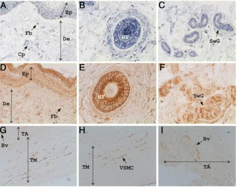
GRP Is Also Expressed in Normal Human Skin
and in Vascular Tissues
The presence of high levels of GRP in rat skin and vas-cular system raised the question of whether GRP had a similar pattern of accumulation and expression in human tissues. Human GRP mRNA localization and protein ac-cumulation were detected in paraffin embedded sections of healthy human skin by in situ hybridization (Figure 4, A–C) and immunohistochemistry using the previously val-idated CTerm-GRP antibody (Figure 4, D–F). The pattern of human GRP accumulation in skin (Figure 4, D–F) fol-lows the pattern of mRNA localization (Figure 4, A–C), which is similar to the results we obtained for rat GRP (Figure 1, A–C). In human skin, GRP is detected at the epidermis and dermis (Figure 4, A and D) levels, being highly expressed and accumulated in fibroblasts (Fb) of both papillary and reticular dermis (Figure 4, A and D), in hair follicles (HF, Figure 4, B and E), in sweat (SwG) (Figure 4, C and F) and sebaceous (results not shown) glands, and in small blood vessels and capillaries (Cp) that irrigate the skin (Figure 4A).
After showing by Western blot that GRP was present in human vascular system (see Supplemental Figure S2A at
http://ajp.amjpathol.org),18 the detailed pattern of GRP
accumulation was evaluated in carotid samples obtained at autopsy. Samples were embedded in paraffin and analyzed by immunohistochemistry using the CTerm-GRP antibody (Figure 4, G–I). Histomorphological evalu-ations were performed by staining consecutive sections with hematoxylin and eosin, and the presence of mineral deposits was monitored by von Kossa staining. At sites showing normal histomorphological features and ab-sence of calcification (results not shown), GRP was found mainly associated with vascular smooth muscle cells (VSMC) of the tunica media (TM, Figure 4, G and H), and in small blood vessels (Bv) irrigating the tunica adventitia (TA, Figure 4, G and I). The apparent lack of GRP in the extracellular matrix could be due to the absence of pro-tein accumulation in normal, non-pathological conditions, as also described for MGP in normal blood vessels.5
Figure 3. Gla proteins detection co-localize in rat skin and cartilage with
GRP accumulation. Immunolocalization of Gla proteins was performed using the Gla-specific monoclonal antibody M3B, and peroxidase-conjugated goat anti-mouse IgG as secondary antibody, in sections of rat outer ear (A–C) and rib (D), showing a positive co-localization (brown) with GRP, presented in Figure 2. De, dermis; Ep, epidermis; Fb, fibroblasts (arrowheads); SG, sebaceous gland; and HF, hair follicle. CHC, cartilage of the hypertrophic zone. Black star is located within the calcified cartilage. Magnification,⫻10.
GRP Is Highly Accumulated at Sites of Ectopic
Calcification in Human Skin and in the Vascular
System
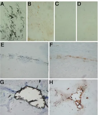
The in vivo calcium mineral binding properties of GRP were originally detected by our results in sturgeon, since the protein was found to accumulate in the mineralized branchial arches, being extracted using a selective acid demineralization procedure.18We now show, using an in
vitroassay, that pig GRP has the ability to bind to hy-droxyapatite crystals, suggesting the possibility of GRP being associated with ectopic mineralization of connec-tive tissue, either in skin or in vascular system or both. To address this question, the CTerm-GRP antibody was used for GRP immunolocalization in human samples de-rived from patients diagnosed with skin and vascular system-associated calcification pathologies (Figures 5
and 6, respectively). The presence of mineral deposits in samples from patients diagnosed with dermatomyositis with calcinosis (Figure 5, A–C) and PXE (Figure 5, D–F) were identified by von Kossa staining (Figure 5, C and F), and presence of GRP accumulated at the same sites was detected by immunohistochemistry (Figure 5, A, B, D, and E). Consecutive sections incubated in the same con-ditions but without the CTerm-GRP antibody (NC insets in Figure 5, A and D) were used as negative controls. GRP was found to be highly accumulated at sites of calcifica-tion in both pathological situacalcifica-tions, either when massive calcified material was deposited in the reticular dermis (Figure 5, A and B) or when small calcified spots were diffused along the elastic fibers (Figure 5, D and E). Results clearly show that GRP is associated with the mineralized material since mineral staining and GRP ac-cumulation are clearly co-localized.
Figure 4. GRP is present in human skin (A–F)
and vascular system (G–I). A–C: Sites of GRP expression determined by in situ hybridization with digoxigenin-labeled antisense probes (blue) in healthy human skin. D–F: Sites of GRP accumulation determined by immunohisto-chemistry with the CTerm-GRP primary anti-body and peroxidase-conjugated goat anti-rab-bit IgG as secondary antibody (brown), in healthy human skin. D–F show consecutive sec-tions of A–C. Human GRP is detected in skin at the levels of epidermis (Ep) and dermis (De, A and D), small capillaries (Cp, A), and skin ap-pendages (B, C, E, F), similarly to what is ob-served for rat GRP (Figures 1 and 2). Fb, fibro-blast; HF, hair follicle; SwG, sweat gland. G–I: GRP accumulation in noncalcified human ca-rotids showing GRP in vascular smooth muscle cells (VSMC) located in the tunica media (TM, G and H), and in small blood vessels (Bv) of the tunica adventitia (TA, G and I). Magnifications:
A–G,⫻10; H and I, ⫻20.
Figure 5. GRP is highly accumulated at sites of
pathological calcification in human skin diag-nosed with dermatomyositis with calcinosis (DC) (A–C) and PXE (D–F). Sites of GRP accu-mulation were determined by immunohisto-chemistry using the CTerm-GRP primary body and peroxidase-conjugated goat anti-rabbit IgG as secondary antibody (brown pointed by arrows, A and B, D and E). Mineral detection was achieved by staining consecutive sections of DC and serial sections of PXE with silver nitrate by the von Kossa staining method
(open arrows, C and F, respectively). In both
pathological situations, GRP is co-localized with sites of mineral deposition, either in DC samples when massive mineral deposits are present (A and B) or in PXE samples where disperse small mineral spots are detected (D and E). A–C were counterstained with toluidine blue and F with hematoxylin and eosin. Negative controls were performed by omitting the CTerm-GRP antibody in consecutive sections of DC (NC, inset in A) and PXE (NC, inset in D) samples. Magnifica-tion,⫻10.
Arterial calcification is a very common process that normally progresses with age, and in fact, up to 95% of men and women at autopsy show coronary artery calci-fication regardless of death cause.3 Also, among the
normal population, patients with chronic kidney disease (CDK) are a high risk group for development of vascular calcifications.26 The pattern of GRP accumulation was
thus studied in a group of patients with CDK (Figure 6, B and D) and in a group of samples collected at autopsy from vascular tissue showing ectopic calcifications (Fig-ure 6, F and H), as detected by von Kossa staining (Figure 6, A, C, E, and G). In both groups, the calcifica-tion observed by von Kossa staining was at the media level, characterized by the absence of macrophages and lipids.
Our results provided clear evidence that GRP was highly accumulated at sites of medial calcification, either in mildly calcified arteries, showing disperse mineral de-posits (Figure 6, B and F), or in extensive and advanced lesions (Figure 6H). In cases where calcification was homogenously dispersed along the fibers of the tunica media (Figure 6A), GRP was detected both inside the vascular smooth muscle cells and in the extracellular matrix (Figure 6B), in contrast with its almost absence in noncalcified areas (Figure 6, C and D). When calcifica-tion was localized, either as disperse (Figure 6E) or
mas-sive (Figure 6G) mineral deposits, GRP was easily iden-tified as co-localizing with the mineral (Figure 6, F and H). This high accumulation of the protein at sites of mineral deposits, in contrast with its absence in the extracellular matrix detected in normal situations, reveals that GRP is definitively associated with the processes of abnormal calcification in the vascular system.
Discussion
Our recent discovery of GRP, a new VKD protein origi-nally purified from sturgeon mineralized cartilage and presenting highly conserved orthologs in all vertebrate groups, an unprecedented high density of Gla residues, and a strong association with cartilaginous tissues, lead us to suggest a function for GRP in the regulation of calcium in the extracellular matrix environment.18
Al-though in sturgeon GRP was mainly associated with car-tilaginous tissues, in rat we have previously shown that GRP was also expressed in bone cells.18In the present
report we show that GRP in rat and human is also ex-pressed and accumulated in the skin and in the vascular system. Our results of immunohistochemistry and West-ern blot were performed using a newly developed anti-body that was validated, in addition to sturgeon GRP, for all mammalian species used, which included rat, pig, and human. Moreover, we were able to confirm that rat GRP is ␥-carboxylated using a Gla-specific antibody, reinforcing the previously proposed theory that GRP should be ␥-carboxylated in all species.18
The high levels of GRP found in the skin, although surprising, cannot be considered as unexpected. Since the late 1970s, a relation between Gla proteins and skin has been suggested. The presence of ␥-carboxylase activity in both epidermal and dermal tissue and the accumulation of non-carboxylated precursor proteins in both dermal and epidermal microsomes after warfarin treatment,17 together with the reported occurrence of
Gla-containing proteins in pathological depositions of calcified material in skin of patients suffering from sclero-derma and sclero-dermatomyositis,27,28pointed to the presence
of a still unknown Gla protein localized in skin, with a role in skin calcium metabolism and involved in the regulation of the calcification process.17In fact, the strongest
re-ported evidences for the presence of Gla-containing pro-teins in skin emerged from studies involving skin patho-logical situations with ectopic mineralization.
Human skin is an organ containing the complete bio-chemical machinery to develop calcification, as shown by the high abundance of reported pathologies ranging from mild to severe skin calcifications (dermatomyositis, scleroderma, PXE, Keutel syndrome, among others), al-though the molecular mechanisms behind this ectopic calcifications have only started to be elucidated. The reported effect of warfarin in these skin pathologies is still controversial. While sometimes a profound and rapid decrease of the calcified lesions with complete disap-pearance has been reported,29,30 more often other
au-thors have described a pathological effect of warfarin in the skin30,31and related it to the inhibition of
␥-carboxy-Figure 6. GRP is highly accumulated in the human vascular system at sites of
pathological calcification of human arteries from both CDK patients (A–D) and postmortem samples (pM) (E–H). Sites of GRP accumulation were determined by immunohistochemistry using the CTerm-GRP primary anti-body and peroxidase-conjugated goat anti-rabbit as secondary antianti-body (brown) (B, D, F, H), and mineral detection was achieved by staining consecutive sections of both CDK and pM with silver nitrate by the von Kossa staining method (A and C and E and G, respectively). In CDK samples GRP is highly accumulated at sites showing mineral deposition (A and B), when compared with non-calcified areas (C and D). In pM samples GRP is co-localized with sites of disperse (E and F) and massive (G and H) calcification.
lation of local Gla-containing proteins. Until now, the iden-tity of this skin Gla protein has not been clearly eluci-dated, but the recent and strong association of MGP with several skin pathologies,6 –9,32and in particularly to the
presence of different processed forms of this pro-tein,7,14,32 supports and points to an essential role of
MGP in the prevention of these skin calcifications. PXE, an autosomal recessive disease characterized by a progressive mineralization of connective tissue result-ing in skin, arterial, and eye disease, can be divided into the classical PXE, caused by mutations in the ABCC6 gene, which encodes an ABC transporter protein,13and
the so called PXE-like, identified more recently, as being caused by missense mutations in the␥-glutamyl carbox-ylase (GGCX) gene.15,33 Either by direct reduction of
carboxylation, caused by reduced GGCX activity,14,15or
by a deficiency in the reduced form of vitamin K, an obligatory cofactor for carboxylation, postulated to be transported from the liver into circulation and peripheral tissues by the ABCC6 transporter,34it is now assumed
that ectopic mineralization, the pathological hallmark of PXE clinical manifestations, is caused by a local tissue deficiency in vitamin K-dependent protein carboxylation. Again, functional MGP has being assumed as having an essential role in preventing aberrant mineralization.6,8,13,14
The discovery of GRP in skin and, moreover, its strong association with abnormal calcifications in both PXE and dermatomyositis patients, shown by the high accumula-tion of protein co-localized with the mineral deposits, strongly suggest that GRP may be the unknown skin Gla protein that remained elusive until now, and a new impor-tant player in modulating skin ectopic mineralization. Al-though MGP is widely accepted as one of the strongest physiological inhibitors of soft tissue calcification known to date, and its association with skin mineralization has been proved, it was never clearly established whether MGP is produced/accumulated in normal skin, as it is in cartilage, bone matrix, and arterial walls.5On the
con-trary, GRP is systemically synthesized and accumulated in skin, as shown by the high levels of mRNA and protein found both in rat and human, which suggests that GRP is a naturally occurring agent affecting ectopic skin calcifi-cation. Moreover, MGP-deficient knockout mice manifest extensive calcification of the aorta and articular cartilage, dying by artery rupture within the first 2 months of age.35
However, there is no reported phenotype relating it to skin calcifications. Furthermore, Keutel syndrome, which is characterized by loss-of-function mutations in the MGP gene, result in abnormal calcification of the soft tissues with diffuse cartilage calcification and short terminal pha-langes. Other findings include peripheral pulmonary ste-nosis, hearing loss, dysmorphic facies and mental retar-dation,36but abnormal skin changes are not reported in
patients with Keutel syndrome. However, a new variant of Keutel syndrome characterized by a new MGP mutation recently identified in a consanguineous family, showed overlapping features of cutis laxa.37Whether loss of MGP
function can explain all of the clinical manifestations ob-served within these patients, in particular the skin alter-ations, was further discussed and questioned, and other
possibilities such as additional unidentified mutations in other genes were suggested.16
Besides skin, we found GRP highly accumulated in small and medium size blood vessels in rat, and we were able to confirm its presence in the human vascular sys-tem, both by immunohistochemistry and Western blot. Furthermore, we show high accumulation of the protein co-localized with mineral deposits in situations of mas-sive and disperse mineralization, associated with media calcification. Although the presence of Gla proteins in the vascular system is known, as is the case of MGP and Gas6, our discovery of a new Gla protein also present in the same tissues raises many questions about previously reported work describing phenotypes based solely on general interference in␥-carboxylation mechanisms.
Our work underlines the need for a strict re-evaluation of the role of each target Gla protein involved in vascular calcification and the elucidation of their corresponding specific functions. It has been shown that inhibiting ex-trahepatic␥-carboxylation by warfarin induces vascular media calcification in rats,10,38consistent with the finding
that in human, high vitamin K intake is associated with low aorta calcification39and beneficial effects on the elastic
properties of the vessel wall.40Moreover a high vitamin K
intake can reverse pre-existing arterial calcification in rats.11As vitamin K is an essential cofactor in the
␥-car-boxylation reaction catalyzed by the␥-glutamyl carbox-ylase, and warfarin prevents␥-carboxylation, interfering with the VKOR enzyme, responsible for the vitamin K recycling, it is conceivable that some of the effects ob-served with warfarin and/or vitamin K administration are due to impairment and/or improvement of GRP function. Although emerging in vitro and whole animal data sug-gest that warfarin may induce vascular calcification,4,41
always through a proposed relation with the under-car-boxylation of MGP, this has not yet been well studied in humans.
In rats, MGP has been extensively shown to be a vascular calcification inhibitor, with widespread and ex-tensive vascular calcification in the MGP knockout mice35
and phenotype rescue after restoration of MGP expres-sion.42However, in humans, MGP loss of function, as in
Keutel syndrome, show less severe vascular calcifica-tions, suggesting additional agents or more complex mechanisms preventing arterial calcification. The exis-tence of other inhibitors, yet to be found, that function to prevent ECM calcification, in particular in tissues as skin, has been previously suggested.1The structural property
of GRP, with an uncommonly extensive Gla domain show-ing high mineral-bindshow-ing affinity, its presence in normal soft tissues like skin and vascular system, and its asso-ciation with ectopic mineral deposits, is consistent with a putative role for GRP as being a naturally occurring mod-ulator of calcium availability in the ECM and thus a po-tential inhibitor of cardiovascular and skin calcifications, constitutively expressed, acting by directly influencing mineral formation.
The complex chemical nature of GRP, as all other VKD proteins, which are strictly dependent on their Gla resi-dues for proper function,5,12indicates the need of
␥-car-boxylation status accumulated at the mineral deposits, both in skin and blood vessels. Antibodies specific to both forms,␥-carboxylated (Gla-GRP) and non-carboxy-lated (Glu-GRP), are currently under development (Geno-Gla Diagnostics, Faro, Portugal) and will be a valuable tool for further studies on GRP function. Similar to what was suggested for MGP,12,14 Gla and Glu-GRP forms
may have different patterns of accumulation, with differ-ent roles in the mineralization process, and this can be dependent on the vitamin K bioavailability in these pe-ripheral tissues.
To access whether GRP was present in blood serum, we used the ProteoMiner kit (Bio-Rad, Amadora, Portu-gal) to remove the most abundant serum proteins from rat and human serum. Using this approach together with the validated CTerm-GRP antibody, we were able to detect GRP, as one of the less abundant circulating proteins present in rat and human serum (results not shown). The evidence for the presence of GRP in blood opens new perspectives for future studies aiming to establish a re-lationship between levels of circulating Gla and Glu-GRP and the degree of vascular and skin calcifications. We are currently developing a GRP enzyme-linked immu-nosorbent assay (GenoGla Diagnostics, Faro, Portugal) that will be able to differentiate the possible forms of GRP circulating in serum. This assay could be used to monitor serum GRP levels in relation with a given pathology and establish its potential use as tool suitable for diagnostic and prognostic evaluation of skin and vascular calcifica-tion risk associacalcifica-tion. Further understanding of the mech-anisms and active players involved in the occurrence of pathological extracellular matrix calcification should offer a potential hope for the development of new therapeutic strategies to control these processes.
Acknowledgments
We thank Dr. Idale´cio Bernardo, Department of Nephrol-ogy, Faro Hospital, for collecting CDK samples during surgery for arteriovenous fistula creation and to Dr. Vera Maria for blood collection at Faro Hospital.
References
1. Schinke T, McKee MD, Karsenty G: Extracellular matrix calcification: where is the action? Nat Genet 1999, 21:225–229
2. Giachelli CM: Ectopic calcification: gathering hard facts about soft tissue mineralization. Am J Pathol 1999, 154:671– 675
3. Wallin R, Wajih N, Greenwood GT, Sane DC: Arterial calcification: a review of mechanisms, animal models, and the prospects for therapy. Med Res Rev 2001, 21:274 –301
4. Danziger J: Vitamin K-dependent proteins, warfarin, and vascular calcification. Clin J Am Soc Nephrol 2008, 3:1504 –1510
5. Cranenburg EC, Schurgers LJ, and Vermeer C: Vitamin K: the coag-ulation vitamin that became omnipotent. Thromb Haemost 2007, 98:120 –125
6. Gheduzzi D, Boraldi F, Annovi G, DeVincenzi CP, Schurgers LJ, Vermeer C, Quaglino D, Ronchetti IP: Matrix Gla protein is involved in elastic fiber calcification in the dermis of pseudoxanthoma elasticum patients. Lab Invest 2007, 87:998 –1008
7. Li Q, Jiang Q, Schurgers LJ, Uitto J: Pseudoxanthoma elasticum: reduced gammaglutamyl carboxylation of matrix gla protein in a
mouse model (Abcc6-/-). Biochem Biophys Res Commun 2007, 364:208 –213
8. Hendig D, Zarbock R, Szliska C, Kleesiek K, Go¨tting C: The local calcification inhibitor matrix Gla protein in pseudoxanthoma elasti-cum. Clin Biochem 2008, 41:407– 412
9. Davies CA, Jeziorska1 M, Freemont AJ, Herrick AL: Expression of osteonectin and matrix Gla protein in scleroderma patients with and without calcinosis. Rheumatology 2006, 45:1349 –1355
10. Price PA, Faus SA, Williamson MK: Warfarin causes rapid calcifica-tion of the elastic lamellae in rat arteries and heart valves. Arterioscler Thromb Vasc Biol 1998, 18:1400 –1407
11. Schurgers LJ, Spronk HM, Soute BA, Schiffers PM, DeMey JG, Ver-meer C: Regression of warfarin-induced medial elastocalcinosis by high intake of vitamin K in rats. Blood 2007, 109:2823–2831 12. Schurgers LJ, Spronk HM, Skepper JN, Hackeng TM, Shanahan CM,
Vermeer C, Weissberg PL, Proudfoot D: Post-translational modifica-tions regulate matrix Gla protein function: importance for inhibition of vascular smooth muscle cell calcification. J Thromb Haemost 2007, 5:2503–2511
13. Li Q, Jiang Q, Pfendner E, Va´radi A, Uitto J: Pseudoxanthoma elasticum: clinical phenotypes, molecular genetics and putative pathomechanisms. Exp Dermatol 2009, 18:1–11
14. Li Q, Schurgers LJ, Smith AC, Tsokos M, Uitto J, Cowen EW: Co-existent pseudoxanthoma elasticum and vitamin K-dependent coag-ulation factor deficiency: compound heterozygosity for mutations in the GGCX gene. Am J Pathol 2009, 174:534 –540
15. Vanakker OM, Martin L, Gheduzzi D, Leroy BP, Loeys BL, Guerci VI, Matthys D, Terry SF, Coucke PJ, Pasquali-Ronchetti I, De Paepe A: Pseudoxanthoma elasticum-like phenotype with cutis laxa and multi-ple coagulation factor deficiency represents a separate genetic en-tity. J Invest Dermatol 2007, 127:581–587
16. Nanda A, Anim JT, Al-Gareeb M, Alsaleh QA: Keutel syndrome with overlapping features of cutis laxa: a new variant. Am J Med Genet A 2006, 140:1487–1489
17. Boer-van den Berg MA, Verstijnen CP, Vermeer C: Vitamin K-depen-dent carboxylase in skin. J Invest Dermatol 1986, 87:377–380 18. Viegas CS, Simes DC, Laize´ V, Williamson MK, Price PA, Cancela ML:
Gla-rich Protein (GRP), a new vitamin K-dependent protein identified from sturgeon cartilage and highly conserved in vertebrates. J Biol Chem 2008, 283:36655–36664
19. Chomczynski P, Sacchi N: Single-step method of RNA isolation by acid guanidinium thiocyanate-phenol-chloroform extraction. Anal Bio-chem 1987, 162:156 –159
20. Ortiz-Delgado JB, Simes DC, Viegas CS, Schaff BJ, Sarasquete C, Cancela ML: Cloning of matrix Gla protein in a marine cartilaginous fish. Prionace glauca: preferential protein accumulation in skeletal and vascular systems. Histochem Cell Biol 2006, 126:89 –101 21. Simes DC, Williamson MK, Delgado JBO, Viegas CSB, Price PA,
Cancela ML: Purification of Matrix Gla protein (MGP) from a marine teleost fish. Argyrosomus regius: calcified cartilage and not bone as the primary site of MGP accumulation in fish. J Bone Min Res 2003, 18:244 –259
22. Simes DC, Williamson MK, Schaff BJ, Gavaia PJ, Ingleton PM, Price PA, Cancela ML: Characterization of osteocalcin (BGP) and matrix Gla protein (MGP) fish specific antibodies: validation for immuno-detection studies in lower vertebrates. Calcif Tissue Int 2004, 74:170 –180
23. Cranenburg EC, Vermeer C, Koos R, Boumans ML, Hackeng TM, Bouwman FG, Kwaijtaal M, Brandenburg VM, Ketteler M, Schurgers LJ: The circulating inactive form of matrix Gla Protein (ucMGP) as a biomarker for cardiovascular calcification. J Vasc Res 2008, 45:427– 436
24. Price PA, Nguyen TM, Williamson MK: Biochemical characterization of the serum fetuin-mineral complex. J Biol Chem 2003, 278:22153–22160
25. Surmann-Schmitt C, Dietz U, Kireva T, Adam N, Park J, Tagariello A, Onnerfjord P, Heinegard D, Schlo¨tzer-Schrehardt U, Deutzmann R, von der Mark K, and Stock M: Ucma, a novel secreted cartilage-specific protein with implications in osteogenesis. J Biol Chem 2008, 283:7082–7093
26. Moe SM, Chen NX: Pathophysiology of vascular calcification in chronic kidney disease. Circ Res 2004, 95:560 –567
gamma-carboxyglutamic acid in the proteins associated with ectopic calcifi-cation. Biochem Biophys Res Commun 1976, 73:349 –355 28. Lian JB, Boivin G, Patterson-Allen P, Grynpas M, Walzer C: Calcergy
and calciphylaxis: timed appearance of gamma-carboxyglutamic acid and osteocalcin in mineral deposits. Calcif Tissue Int 1983, 35:555–561
29. Berger RG, Featherstone GL, Raasch RH, McCartney WH, Hadler NM: Treatment of calcinosis universalis with low-dose warfarin. Am J Med 1987, 83:72–76
30. Cukierman T, Elinav E, Korem M, Chajek-Shaul T: Low dose warfarin treatment for calcinosis in patients with systemic sclerosis. Ann Rheum Dis 2004, 63:1341–1343
31. Lassoued K, Saiag P, Anglade MC, Roujeau JC, Touraine RL: Failure of warfarin in treatment of calcinosis universalis. Am J Med 1988, 84:795–796
32. van Summeren MJ, Spliet WG, van Royen-Kerkhof A, Vermeer C, Lilien M, Kuis W, Schurgers LJ: Calcinosis in juvenile dermatomyositis: a possible role for vitamin K-dependent protein matrix Gla protein. Rheumatology (Oxford) 2008, 47:267–271 33. Uitto J, Jiang Q: Pseudoxanthoma elasticum-like phenotypes: more
diseases than one. J Invest Dermatol 2007, 127:507–510
34. Borst P, van de Wetering K, Schlingemann R: Does the absence of ABCC6 (multidrug resistance protein 6) in patients with Pseudoxan-thoma elasticum prevent the liver from providing sufficient vitamin K to the periphery? Cell Cycle 2008, 7:1575–1579
35. Luo G, Ducy P, McKee MD, Pinero GJ, Loyer E, Behringer RR,
Karsenty G: Spontaneous calcification of arteries and cartilage in mice lacking matrix GLA protein. Nature 1997, 386:78 – 81 36. Parmar H, Blaser S, Unger S, Yoo SJ, Papsin B: Petrified ears in a
patient with Keutel syndrome: temporal bone CT findings. Pediatr Radiol 2006, 36:241–243
37. Hur DJ, Raymond GV, Kahler SG, Riegert-Johnson DL, Cohen BA, Boyadjiev SA: A novel MGP mutation in a consanguineous family: review of the clinical and molecular characteristics of Keutel syn-drome. Am J Med Genet A 2005, 135:36 – 40
38. Spronk HM, Soute BA, Schurgers LJ, Thijssen HH, De Mey JG, Vermeer C: Tissue-specific utilization of menaquinone-4 results in the prevention of arterial calcification in warfarin-treated rats. J Vasc Res 2003, 40:531–537
39. Geleijnse JM, Vermeer C, Grobbee DE, Schurgers LJ, Knapen MH, van der Meer IM, Hofman A, Witteman JC: Dietary intake of mena-quinone is associated with a reduced risk of coronary heart disease: the Rotterdam Study. J Nutr 2004, 134:3100 –3105
40. Braam LA, Hoeks AP, Brouns F, Hamulya´k K, Gerichhausen MJ, Vermeer C: Beneficial effects of vitamins D and K on the elastic properties of the vessel wall in postmenopausal women: a follow-up study. Thromb Haemost 2004, 91:373–380
41. Becker RC: Warfarin-induced vasculopathy. J Thromb Thrombolysis 2007, 23:79 – 81
42. Murshed M, Schinke T, McKee MD, Karsenty G: Extracellular matrix mineralization is regulated locally; different roles of two gla-containing proteins. J Cell Biol 2004, 165:625– 630