Genetic and epigenetic mechanisms involved in
regulation of STEAP1 gene expression in LNCaP
prostate cancer cells
Inês Margarida Marques de Sousa
Dissertação para obtenção do Grau de Mestre em
Bioquímica
(2º ciclo de estudos)
Orientador: Professor Doutor Cláudio Jorge Maia Batista
Co-orientador: Professor Doutor Manuel Carlos Loureiro de Lemos
iii
Acknowledgments
The realization of this master thesis would not have been possible without the collaboration and unconditional support of several people who already I would like to thank.
First of all, I would like to thank my supervisor, Professor Cláudio Maia for the opportunity to develop this project and for the endless support and availability. I would also like to thank my co-supervisor, Professor Manuel Lemos for his availability and helpful recommendations.
I would like to thank the University of Beira Interior and the Health Sciences Research Centre for having made possible the realization of this project.
I would also like to express my gratitude to all my colleagues of laboratory and research center, especially to Margarida, Cátia, Marília and Henrique for all the advice, teaching and support that they always showed.
Moreover, I would like to thank Eduarda Coutinho for the patience, willingness and Knowledge shared about sequencing.
I am also grateful to my friends Margarida, Adriana, Vera and Mariana for the friendship and all good times shared. To Margarida and Adriana, a special thank you for the support and encouraging words that helps me to overcome the difficulties and bad moments.
Finally, I would like to thank my family in especially to my dad, mom, and brother, the most important people in my life who always supported me, unconditionally.
v
Resumo
O cancro da próstata é o segundo tipo de cancro mais frequentemente diagnosticado e a quinta principal causa de morte por cancro nos homens em todo o mundo. O desenvolvimento do cancro da próstata é caracterizado por alterações progressivas nos mecanismos genéticos e epigenéticos o que conduz a uma desregulação da expressão genética. O gene Six transmembrane epithelial antigen of the prostate 1 (STEAP1) codifica uma proteína com seis domínios transmembranares. Nos tecidos normais, a expressão do STEAP1 é muito baixa, no entanto é sobre-expresso em vários tipos de cancro nomeadamente no cancro da próstata. Vários estudos indicaram que a sobre-expressão do STEAP1 parece promover o crescimento celular, sugerindo que este pode actuar como um oncogene. Estudos anteriores demonstraram também que o mRNA e a proteína STEAP1 apresentam uma maior estabilidade em linhas celulares de cancro da próstata LNCaP quando comparado com as linhas celulares da próstata não-neoplásicas PNT1A. Esta diferença pode ser devida a modificações pós-transcricionais e/ou pós-translacionais. No entanto, estas alterações não justificam a sobre-expressão do STEAP1 em células tumorais, sugerindo assim o envolvimento de outros mecanismos de regulação. Portanto, o objectivo do presente trabalho foi explorar a hipótese de que alterações genéticas e/ou epigenéticas poderão estar envolvidas na sobre-expressão do STEAP1. A fim de avaliar a possível presença de alterações genéticas na sequência do gene STEAP1, foi sequenciada a região promotora do STEAP1 em células LNCaP e PNT1A. Para estudar o envolvimento de mecanismos epigenéticos, foram comparados os padrões de metilação do STEAP1 entre as linhas celulares PNT1A e LNCaP. Para além disso, foi ainda avaliado o efeito de um tratamento com inibidores das DNA metiltransferases (DNMT) e histonas desacetilases (HDAC) na expressão do gene STEAP1 em células PNT1A. A análise da sequência da região promotora do STEAP1 revelou algumas variantes tanto nas células LNCaP como PNT1A quando comparada com a sequência genómica disponível. A análise in silico das variantes mostrou diferenças nos fatores de transcrição que se podem ligar a cada variante alelica incluindo a ligação de activadores transcripcionais ao alelo alterado das variantes. A análise do padrão de metilação do STEAP1 entre células PNT1A e LNCaP mostrou diferenças na região promotora próxima do local de início da transcrição. O tratamento com 5-Aza-2’-deoxicitidina (inibidor das DNMT) induziu um ligeiro aumento na expressão do STEAP1 (três vezes em comparação com o grupo de controlo, p<0.01), enquanto que o tratamento com ambos os inibidores 5-Aza-2’-deoxicitidina e TSA (inibidor das HDAC) induziu um aumento acentuado na expressão do STEAP1 (quinze vezes relativamente ao grupo de controlo, p<0.001). A diferença no padrão de metilação do STEAP1 entre as células LNCaP e PNT1A, juntamente com o aumento da expressão do STEAP1 em resposta ao tratamento com os inibidores de HDACs e DNMTs, indica que a expressão génica do STEAP1 parece ser regulada por mecanismos epigenéticos.
vi
viii
Resumo Alargado
O crescimento e envelhecimento da população associado a um aumento da adopção de factores de risco tornaram o cancro um dos maiores problemas de saúde a nível mundial. O cancro da próstata é o segundo tipo de cancro mais frequentemente diagnosticado e a quinta principal causa de morte por cancro nos homens de todo o mundo. O processo de desenvolvimento do cancro da próstata é caracterizado por alterações progressivas nos mecanismos genéticos e epigenéticos que regulam a expressão genética. Uma das alterações genéticas mais frequente no cancro da próstata é a fusão do gene TMPRSS2, cuja expressão é regulada pelos androgénios com os genes da família de factores de transcrição ETS. Quanto às alterações epigenéticas, a alteração que é mais frequentemente encontrada em casos de cancro da próstata e lesões pré-neoplásicas é a hipermetilação da região promotora do gene que codifica a enzima Glutationa S-transfrease π.
O gene Six transmembrane epitelial antigen of the protate 1 (STEAP1) foi o primeiro elemento da família de proteínas STEAP a ser identificado. O STEAP1 codifica uma proteína com seis domínios transmembranares que se encontra localizada nas junções celulares das células epiteliais. Pensa-se que esta proteína possa actuar como um canal iónico ou proteína transportadora de pequenas moléculas tendo assim um papel na comunicação intercelular. Enquanto que nos tecidos normais a expressão do STEAP1 é muito baixa ou mesmo nula, nos tecidos tumorais é sobre-expresso em vários tipos nomeadamente no cancro da próstata. Vários estudos indicaram que a sobre-expressão do STEAP1 parece promover o crescimento celular, sugerindo que este pode actuar como um oncogene. Estudos anteriores demonstraram também que o mRNA e a proteína STEAP1 apresentam uma maior estabilidade em linhas celulares de cancro da próstata LNCaP quando comparado com as linhas celulares da próstata não-neoplásicas PNT1A. Esta diferença pode ser devida a modificações pós-transcricionais e/ou pós-translacionais. No entanto, estas alterações não justificam a sobre-expressão do STEAP1 em células tumorais, sugerindo assim o envolvimento de outros mecanismos de regulação.
Portanto, o objectivo do presente trabalho foi explorar a hipótese de que alterações genéticas e/ou epigenéticas poderão estar envolvidas na sobre-expressão do STEAP1 no cancro da próstata. Para testar esta hipótese foi delineado um conjunto de tarefas. A fim de avaliar a possível presença de alterações genéticas na sequência do gene STEAP1, nomeadamente mutações, foi sequenciada a região promotora do STEAP1 em duas linhas celulares da próstata, uma neoplásica (LNCaP) e uma não-neoplásica (PNT1A). Foi também realizada uma análise in silico para avaliar se alguma das alterações encontradas está localizada numa região importante para a ligação de factores de transcrição. Quanto aos mecanismos epigenéticos foi avaliada a metilação do DNA e a acetilação de histonas. Para a análise de alterações ao nível da metilação do DNA foram comparados os padrões de metilação pelo método BSP do gene STEAP1 entre as linhas celulares PNT1A e LNCaP. Para
ix avaliar ainda alterações na metilação do DNA e acetilação de histonas foi realizado um tratamento com inibidores de DNA metiltransferases (DNMT) e histonas desacetilases (HDAC) em células PNT1A. O efeito do tratamento na expressão do STEAP1 foi avaliado através da técnica de PCR em tempo real.
A análise da sequência da região promotora do gene STEAP1 revelou a presença de algumas alterações tanto nas células LNCaP como PNT1A quando comparada com a sequência genómica disponível. Algumas das variantes encontradas já se encontram identificadas na base de dados Ensembl. A análise in silico das variantes mostrou algumas diferenças entre os fatores de transcrição que se podem ligar a cada variante alelica nomeadamente a ligação de activadores transcripcionais como o C/EBPβ e o LEF-1 ao alelo alterado das variantes. A análise do padrão de metilação do STEAP1 entre as células PNT1A e LNCaP mostrou diferenças na região promotora próxima do local de início da transcrição. Enquanto que nas células PNT1A alguns dos dinucleótidos CG parecem estar metilados, nas células LNCaP parece haver uma desmetilação completa da região analisada. O tratamento com 5-Aza-2’-deoxicitidina (inibidor das DNMT) induziu um ligeiro aumento na expressão do STEAP1 (três vezes em comparação com o grupo de controlo, p<0.01), enquanto que o tratamento com ambos os inibidores, 5-Aza-2’-deoxicitidina e TSA (inibidor das HDAC), induziu um aumento acentuado na expressão do STEAP1 (quinze vezes relativamente ao grupo de controlo, p<0.001). Esta alteração na expressão do STEAP1 provocada pelos inibidores das DNMTs e HDACs indica que tanto a metilação do DNA como a acetilação de histonas podem estar envolvidos na regulação da sua expressão.
Em suma a diferença no padrão de metilação do STEAP1 entre as células LNCaP e PNT1A em conjunto com o aumento da expressão do mRNA do STEAP1 em resposta ao tratamento com os inibidores de HDACs e DNMTs, são indicadores de que a expressão do STEAP1 é regulada por mecanismos epigenéticos, nomeadamente a metilação do DNA e a acetilação de histonas.
xi
Abstract
Prostate cancer is the second most frequently diagnosed type of cancer and the fifth leading cause of cancer death in men worldwide. Prostate carcinogenesis is characterized by progressive alterations in genetic and epigenetic mechanisms that deregulate gene expression. The Six Transmembrane Epithelial Antigen of the Prostate 1 (STEAP1) gene encodes a protein with six transmembrane domains. In normal tissues, STEAP1 expression is very low but is overexpressed in several human cancers, mainly in prostate cancer. Some studies have indicated that STEAP1 overexpression seems to promote cell growth, suggesting that STEAP1 may act as an oncogene. Previous studies demonstrated that STEAP1 mRNA and protein have higher stability in LNCaP prostate cancer cell lines when compared with PNT1A non-neoplastic prostate cell lines, possibly due to post-transcriptional and post-translational modifications. However, these alterations do not justify the overexpression of STEAP1 in tumor cells, suggesting that other mechanisms may be involved. Therefore, the aim of this study was to explore the hypothesis that genetic and / or epigenetic alterations may be involved in overexpression of STEAP1. In order to evaluate genetic alterations in the STEAP1 gene sequence, the promoter region of STEAP1 in LNCaP and PNT1A cells was sequenced. To study the involvement of epigenetic mechanisms, the methylation patterns of STEAP1 in PNT1A and LNCaP cells were compared. In addition, the effect of treatment with DNA methyltransferases (DNMT) and histone deacetylases (HDAC) inhibitors on STEAP1 mRNA expression in PNT1A cells was evaluated. The sequence analysis of the promoter region of STEAP1 revealed some differences in both PNT1A and LNCaP cells when compared with the available genomic sequence. In silico analysis of the identified variants revealed several alterations in the transcription factors (TF) that can bind to each allelic variant including the binding of transcriptional activators to the altered allele of the variants. The analysis of the methylation pattern of STEAP1 gene in PNT1A and LNCaP cells showed differences in the promoter region near the transcription start site. The treatment with 5-Aza-2’-deoxycytidine (DNMT inhibitor) induced a slight increase in STEAP1 mRNA expression (3 fold-variation in comparison with control group, p<0.01) while the treatment with both 5-Aza-2’-deoxycytidine and TSA (HDAC inhibitor) induced a marked increase in STEAP1 mRNA expression (15 fold-variation relatively to control, p<0.001). The difference in the methylation pattern of STEAP1 between PNT1A and LNCaP cells, along with the increased STEAP1 mRNA expression in response to DNMT and HDAC inhibitors, indicates that STEAP1 gene expression seems to be regulated by epigenetic mechanisms.
xiii
List of contents
1. Introduction ... 1
1.1. Anatomy and physiology of the prostate ... 2
1.2. Prostate cancer ... 5
1.2.1. Epidemiology and Risk factors ... 5
1.2.2. Diagnosis and treatment ... 6
1.2.3. Molecular pathways of carcinogenesis ... 8
1.3. Genetic and epigenetic mechanisms involved in carcinogenesis of prostate cancer 12 1.3.1. Genetic mechanisms ... 12
1.3.2. Epigenetic mechanisms ... 15
1.4. Six transmembrane epithelial antigen of the prostate 1 ... 18
1.4.1. General characteristics ... 18
1.4.2. Expression in human tissues ... 19
1.4.3. Biological functions and its regulation in normal and cancer cells ... 20
2. Objectives ... 22
3. Materials and Methods ... 24
3.1. Sequence analysis of the promoter region of the STEAP1 gene ... 25
3.1.1. Cell lines culture ... 25
3.1.2. DNA extraction and quantification ... 25
3.1.3. Polymerase chain reaction (PCR) ... 26
3.1.4. Cloning of PCR products into pNZY28 vector ... 27
3.1.5. DNA sequencing ... 27
3.1.6. Bioinformatic analysis ... 28
3.2. Analysis of methylation profile of the STEAP1 promoter region ... 29
3.2.1. Bisulfite modification of DNA... 29
3.2.2. PCR ... 30
3.3. Treatment of PNT1A cells with DNMT and HDAC inhibitors and the effect on STEAP1 mRNA expression ... 32
3.3.1. Cell line culture and treatment ... 32
xiv
3.3.3. Quantitative real-time polymerase chain reaction (qPCR) ... 33
3.3.4. Statistical analysis ... 33
4. Results and Discussion ... 34
4.1. Sequence analysis of the STEAP1 promoter region ... 35
4.2. Methylation pattern of STEAP1 in PNT1A and LNCaP cells ... 41
4.3. Effect of treatment with DNMT and HDAC inhibitors on STEAP1 mRNA expression in PNT1A cells by qPCR analyses ... 43
5. Conclusions and Future Perspectives ... 44
xvi
List of figures
Figure 1: Human prostate anatomy according to McNeal’s description. ... 2
Figure 2: Histologic arrangement of the normal prostate (H&E stain). ... 4
Figure 3: Model of PCa progression. ... 8
Figure 4: Molecular pathways in normal prostate cells. ... 10
Figure 5: Location of STEAP1 gene at the long arm of chromosome 7, STEAP1 gene organization and schematic of STEAP1 protein structure. ... 19
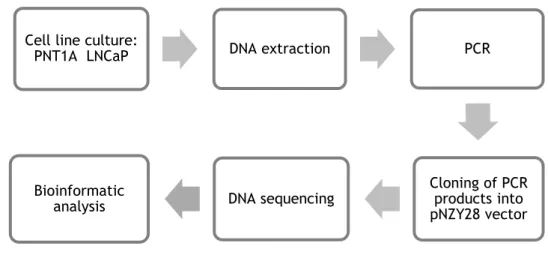
Figure 6: Diagram with the procedures used in the sequence analysis of the promoter region of the STEAP1 gene. ... 25
Figure 7: Arrangement of the primers used to amplify the promoter region and the first exon of the STEAP1 gene ... 26
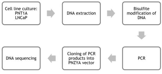
Figure 8: Diagram with the procedures used to determine the methylation pattern of the STEAP1 gene. ... 29
Figure 9: Bisulfite conversion of DNA. ... 30
Figure 10: Arrangement of the primers used to amplify the bisulfite treated DNA. ... 30
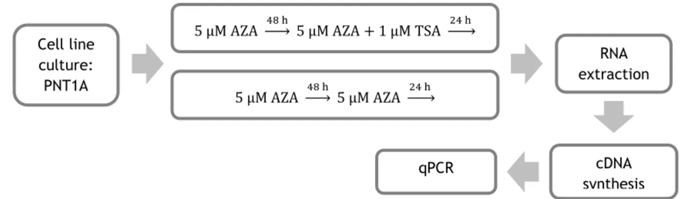
Figure 11: Diagram with the procedures used to evaluate the effect of treatment with DNMT and HDAC inhibitors on STEAP1 mRNA expression in PNT1A cells. ... 32
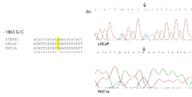
Figure 12: (a) Multiple sequences alignment of the STEAP1 gene sequence with the sequences obtained from PNT1A and LNCaP cells sequencing. (b) Partial nucleotide sequence showing the variant -1863 G/C in LNCaP cells. ... 36
Figure 13: (a) Multiple sequences alignment of the STEAP1 gene sequence with the sequences obtained from PNT1A and LNCaP cells sequencing. (b) Partial nucleotide sequence showing the variant -1195 A/C in LNCaP cells. ... 36
xvii Figure 14: (a) Multiple sequences alignment of the STEAP1 gene sequence with the sequences obtained from PNT1A and LNCaP cells sequencing. (b) Partial nucleotide sequence showing the variant -640 del G in LNCaP cells. ... 37
Figure 15: (a) Multiple sequences alignment of the STEAP1 gene sequence with the sequences obtained from PNT1A and LNCaP cells sequencing. (b) Partial nucleotide sequence showing the variant -563 A/G in LNCaP cells. ... 37
Figure 16: Region of the STEAP1 gene with a relevant CpG island. ... 41
Figure 17: Bisulfite methylation analysis of STEAP1 in PNT1A and LNCaP cells. ... 42
Figure 18: qPCR analysis of the effect of treatment with AZA and TSA (DNMT and HDAC inhibitors respectively) on STEAP1 mRNA expression in PNT1A cells. (**p<0.01; ***p<0.001 relative to control). ... 43
xix
List of tables
Table 1: Genetic alterations found in human PCa. ... 14
Table 2: Epigenetic alterations found in human PCa. ... 17
Table 3: Expression of STEAP1 protein in normal and cancer tissues ... 20
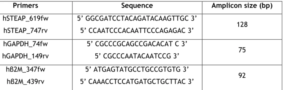
Table 4: Primers sequences and respective amplicon sizes used for amplification of the promoter region of STEAP1 gene. ... 26
Table 5: Reagents and volumes used in each DNA sequencing reaction. ... 28
Table 6: Primer sequences and respective amplicon size used for amplification of the bisulfite treated DNA. ... 31
Table 7: Primers sequences and respective amplicon size used in qPCR analysis... 33
Table 8: Summary of putative TF binding to each allelic variant identified using the TF binding site prediction program Alggen Promo software 3.0. On the nucleotide sequence of potential binding site is highlighted the nucleotide that is altered in each variant. ... 40
xxi
List of abbreviations
aa Amino acids
Akt Protein kinase B
AR Androgen receptor
AZA 5-Aza-2’-deoxycytidine
BPH Benign prostate hyperplasia
BSP Bisulfite sequencing PCR
C/EBPβ CCAAT/enhancer-binding protein β CDKN1B Cyclin dependent kinase inhibitor 1B gene
DHT Dihydrotestosterone
DNMT DNA methyltransferases
DRE Digital rectal exam
E. coli Escherichia coli
GSTP1 Glutathione s-transferase π
H&E Hematoxylin and eosin
HAT Histone acetyl transferases
HDAC Histone deacetylases
LEF-1 Lymphoid enhancing factor-1
LH Luteinizing hormone
NKX3.1 NK3 homeobox 1 gene
PAP Prostatic acid phosphatase
PCa Prostate cancer
PCR Polymerase chain reaction
pDNA Plasmid DNA
xxii
PIA Proliferative inflammatory atrophy
PIN Prostatic intraepithelial neoplasia
PIP3 Phosphatidylinositol 3, 4, 5-triphosphate
PSA Prostate specific antigen
PTEN Phosphatase and tensin homolog gene
qPCR Real-time quantitative PCR
ROS Reactive oxygen species
SNP Single-nucleotide polymorphism
STEAP1 Six transmembrane epithelial antigen of the prostate 1
TF Transcription factor
TRUS Transrectal ultrasound
1
1. Introduction
Partially submitted:
Barroca-Ferreira J, Pais JP, Santos MM, Gonçalves AM, Gomes IM, Sousa I, Rocha SM, Passarinha LA, Maia CJ. Targeting STEAP1 protein in human cancer: current trends and future challenges. Partial submitted to Current Cancer Drug Target
2
1.1. Anatomy and physiology of the prostate
The prostate is an accessory gland of the male reproductive system and is located in the pelvic region just below the bladder (1). This gland has the shape and size of a walnut and its main function is the production of an alkaline fluid containing acid phosphatase, proteases, sucrose and citric acid that allows sperm motility and protection (2, 3). The prostate gland is surrounded by a capsule of collagen and smooth muscle that gives rise to septa which extend to its interior dividing the gland in lobes (4). The prostate grows during puberty to full size due to increasing levels of androgens. After the age of 55 years, the growth is reinitiated due to the growth of nonmalignant cells in the periurethral zone (1).
In according to McNeal’s description, the prostate can be divided into five different zones (Figure 1): the central zone, the peripheral zone, the preprostatic zone, the transition zone and the fibromuscular zone (5). The peripheral zone is the largest region, which comprises nearly 75% of the glandular tissue and appears to be more susceptible to develop cancer (1, 5, 6). The central zone comprises approximately 25% of the glandular tissue. The preprostatic zone or periurethral region is composed of glandular and non-glandular tissue that surrounds the urethra (5, 6). The transition zone represents less than 5% of the gland mass, but is the origin local of the benign prostatic hyperplasia (BPH), which is considered the most common disorder of the prostate (5). The anterior fibromuscular zone is a non-glandular structure that constitutes the anterior surface of the prostate (5, 6).
Figure 1: Human prostate anatomy according to McNeal’s description. a. Central zone; b. Fibromuscular zone; c. Transitional zone; d. Peripheral zone; e. Periurethral gland zone (Adapted from (7)).
3 The prostate is dependent on androgens for the development and maintenance of its structural and functional integrity. Testosterone is the most abundant androgen in circulation and it is produced by Leydig cells in the testes. Others androgens, such as dehydroepiandrosterone (DHEA) and androstenedione (4-DIONE), are produced in the adrenal cortex and converted into testosterone in peripheral tissues. In target tissues like prostate, the testosterone is converted to dihydrotestosterone (DHT) due to high activity of 5-alpha-reductase (8, 9). Thus, DHT is considered the main androgen required for complete prostate morphogenesis. The main mechanism of action of DHT is mediated by its binding to the androgen receptor (AR), which in turn, bind to DNA to activate the transcription of genes involved in cell proliferation, survival, lipid metabolism and differentiation (10, 11).
The prostatic tissue is composed of stromal and epithelial cells (Figure 2). Within the epithelial cells, two different types can be distinguished morphologically: columnar luminal cells and basal cells (12). The columnar luminal cells express the AR and are dependent on androgens to survive. These cells constitute the exocrine compartment of the prostate epithelium, secreting prostate specific antigen (PSA) and prostatic acid phosphatase (PAP) (12, 13). The basal cells do not have secretory activity and express very low levels of AR. Although these cells are androgen independent they are androgen responsive; they do not depend on androgens to survive but their growth and differentiation are stimulated by androgens. This basal layer lies beneath the columnar luminal cells layer and it is believed to have stem cells with the capacity to give rise to all types of prostatic epithelial cells (12–14). There is a third type of epithelial cells dispersed within the luminal and basal cells, the neuroendocrine cells. Although the function of this type of cells is still unknown, it is believed that they may be involved in the proliferation of the adjacent cells by paracrine secretion of neuropeptides. The neuroendocrine cells do not depend on androgens to survive and may play a role in prostate carcinogenesis (12, 13). The stromal cells contain fibroblasts and smooth muscle that provide structural and biochemical support to the prostate epithelium (3, 13, 15). These two types of cells produce the extracellular matrix that helps to generate a microenvironment that controls the growth of the adjacent epithelial cells (3, 15). Some studies have shown that AR is expressed in smooth muscle cells, but not in fibroblasts. Thus, it is believed that androgens act through paracrine signaling pathways on smooth muscle to maintain the fully differentiated growth-quiescent epithelium (3, 15). Ablation of androgens results in prostate involution and loss of epithelial cells by apoptosis. The re-administration of androgens reverse this process inducing the prostate return to normal size and function through rapid proliferation and differentiation of stem cells. The homeostasis between the epithelial and stromal compartments is regulated by a complex signaling pathway that involves the AR and other paracrine factors capable of maintaining the balance between proliferation and apoptosis (8, 16).
4
Figure 2: Histologic arrangement of the normal prostate (H&E stain) (Retrieved from http://www.proteinatlas.org/learn/dictionary/normal/prostate/detail+1/magnification+1
5
1.2. Prostate cancer
1.2.1. Epidemiology and Risk factors
Cancer is one of the leading causes of death in both more and less developed countries. The increasing incidence due to the growth and aging of the population associated with the increasing adoption of risk factors, such as smoking, overweight, physical inactivity and poor diet, made it a burden to the world society (17).
Prostate cancer (PCa) is the second type of cancer most frequently diagnosed in men, and it is the fifth leading cause of cancer death in men worldwide (18). In Portugal, PCa is the most frequently type of cancer diagnosed, representing more than 20% of the diagnosed cancers and is the fourth leading cause of cancer death in men (19, 20). The decrease trend in death rates should be mainly due to early detection and improvement of the available treatments (17).
Risk factors can be divided into two types: the non-modifiable factors and the external factors (modifiable factors). Non-modifiable factors are genetic susceptibility, age, ethnicity or family history, whereas external factors include lifestyle factors such as diet and physical activity (10, 21). Age is one of the most important risk factors for PCa. In men younger (until 50 years old), the incidence of PCa is very low (<0.1%). On the other hand, in men with more than 65 years old the incidence is much higher, representing approximately 85% of the cases (10, 21). Another important risk factor is ethnicity since the incidence of PCa among African-American men is approximately 60% higher than in white men. The lowest rates of PCa are found in Asian, but when these migrate to the US their risk of developing PCa increases, which indicates that external factors are also involved in the development of PCa (10, 22). Besides age and race, family history of the disease is another major risk factor for PCa. The relative risk of developing it increases with the number of family members affected and with the degree of relatedness, and is inversely related to the age at which family members were affected. For example, in men who have a first-degree relative (father or brother) with PCa, the relative risk of developing the disease increase two to threefold depending on the age at which the disease appeared (10, 21, 23). Several PCa susceptibility genes have been identified, such as the RNAse L gene in locus HPC1, the ELAC2 gene in locus HPC2, the MSR1 gene on chromosome 8 and the BRCA genes. One of the most important is the RNAse L gene, which encodes an endoribonuclease involved in the induction of apoptosis and regulation of cell cycle and cell differentiation among others. This gene has typically autosomal dominant hereditary with high penetration (22, 24).
Although several epidemiological studies of the possible association between external factors and PCa risk have been conducted, the results have been inconclusive. One of the external factors is the western lifestyle, which is characterized by diets rich in red meat, dairy products and high intake of fat that may lead to increased risk of developing PCa (10, 22, 25). Preparation of meat at high temperatures leads to the production of heterocyclic
6 amines and other carcinogens. This, together with the production of hydrogen peroxide from the fatty acids oxidation process, could induce damage in the prostate genome (10, 22, 26). Another factor that seems to be related to PCa risk is obesity. The increase of body mass index is associated with decreased risk of developing localized PCa and increased risk of developing aggressive PCa (21, 27, 28). Other external factors that may be associated with the increased PCa risk are infections, smoking and radiation exposure, but these associations remain unclear (10, 21). On the other hand, there are several protective factors such as vegetables and physical activity (22, 26). Vegetables, especially tomatoes have been associated with a lower risk of PCa due to the high content of antioxidants, such as carotenoids that seem to be associated with decreased oxidative DNA damage and reduction of serum PSA levels (26, 29, 30). Cruciferous vegetables are rich in phytochemicals like sulforaphane, and these phytochemical induces the expression of carcinogen detoxification enzymes that prevent DNA and cell damage from carcinogens (26, 31). Regarding physical activity, it is well known that it has many benefits including reduction of PCa risk, although the association between these remains unclear (21, 32).
1.2.2. Diagnosis and treatment
Screening of PCa is based on serum PSA (kallikrein-related peptidase 3; KLK3) levels, digital rectal examination (DRE) and the patient’s symptoms. PSA, a kallikrein-related serine protease, which is responsible for the liquefaction of the seminal coagulum, is produced by both nonmalignant and malignant cells. Despite being prostate specific, the increase of PSA levels in serum are not cancer specific, and may result due to BPH or prostatitis. Therefore, a considerable number of false positives may occur, which decrease the specificity of PSA as a biomarker for PCa (1, 33). However, the predictive value of PSA screening can be improved by complementary tests, such as PSA velocity, PSA density, and free-PSA (34). PSA velocity is based on the rate of change in PSA levels over time, which increases the sensitivity and specificity of the PSA test. Even so, its predictive value is limited by intrasubject variability in PSA measurements (22, 34). PSA density correlates serum PSA levels with prostate volume. PCa cells release more PSA per volume unit than BPH tissue. The division of PSA levels by the volume of the prostate improves the specificity of PSA test because it allows the distinction between BPH and PCa (34, 35). PSA circulates in the blood in an inactive form, mainly aggregated with a protease inhibitor, while free PSA is quickly eliminated from the organism by glomerular filtration. A lower percentage of free-PSA is more associated with PCa than BPH, allowing an improvement of PSA test specificity (34–36).
Regarding DRE, it allows to assess whether there are alterations in prostate size and consistency. In most cases of patients with PCa the size of the prostate increase in the peripheral zone, which is coincident with the area examined in DRE (1).
7 The American Cancer Society recommends that men with 50 years or older should discuss with a health care about the risks and possible benefits of testing for PCa and make an informed decision. In the case of men at high risk, these should talk to a health care 5 to 10 years earlier. The test for PCa is based on serum PSA levels with or without DRE. When there is an increase in PSA levels associated with abnormal DRE, the patient is forwarded to a possible diagnosis of PCa. In order to confirm the diagnosis of PCa, the patients will be subjected to transrectal ultrasound (TRUS)-guided needle biopsy (1, 34). Histopathological diagnosis of PCa in needle biopsy specimens is performed by hematoxylin and eosin (H&E) staining and/or immunohistochemistry against cytokeratin, p63 and racemase (AMACR) proteins (37, 38).
After a diagnosis of PCa, it is necessary to classify the tumor in order to help choose a better treatment. Gleason score system, the most commonly used, is based on the histological pattern of PCa cells in prostatic tissue sections. This system is used to measure the histological aggressiveness, in which the histological pattern is classified according to its glandular pattern and differentiation degree. The dominant and secondary patterns are scored from 1 to 5, in which grade 1 represents a well-differentiated tumor and grade 5 an undifferentiated. The score of the dominant and the secondary patterns are summed to give a total score of 2 to 10. The biologic behavior is generally determined by the area with low differentiation, which is the area with the highest histologic grade (1, 39). The clinical stage of the tumor is one of the most important factors in the choice of treatment and it is generally classified using the tumor-nodes-metastasis (TNM) system. This system divides the tumors in three main stages: Primary tumor (T), Regional lymph nodes (N) and distant metastasis (M). T stage has 4 categories describing how the tumor has been identified, the size of the primary tumor and whether it has invaded nearby structures. N stage describes whether the cancer has spread to nearby lymph nodes while M stage describes whether the cancer has spread to distant parts of the body like bones and lymph nodes (34, 40).
Patients with localized PCa (those who do not appear to have metastasis after staging analyses) have three main treatment options: radical prostatectomy, radiation therapy and active surveillance (1, 34). Radical prostatectomy is usually performed in patients who have tumors confined to the prostate gland (stage T1 and T2) and can undergo surgery. The great advantages of radical prostatectomy is that it provides excellent control of the primary tumors without increasing morbidity (34, 41). For patients with PCa confined to the prostate or surrounding tissues that cannot perform surgery, radiation therapy is an alternative, which can be administered as external beam therapy, brachytherapy or a combination of both. The results of this type of treatment will depend on the stage and dosimetry of radiation. In brachytherapy, the radioactive source is implanted into the prostate or surrounding area. The patients that undergo brachytherapy generally have low Gleason score, low PSA level and tumors on stage T1 or T2 (34, 42). Active surveillance consists in regularly following the patient and initiating therapy if there are signs of tumor progression. In men with indolent or nonprogressive tumors, active surveillance prevents the morbidity of therapy (1, 43).
8 For patients with tumor extension to nearby structures or metastatic disease, other treatment options are hormonal therapy and chemotherapy in addition to the already mentioned above. The growth of PCa is essentially androgen-dependent since these stimulate proliferation and inhibit apoptosis. The main goal of hormonal therapy is to reduce androgen levels by surgery or therapy with anti-androgens or luteinizing hormone (LH) agonists (1, 9, 44). LH agonists lead to inhibition of testosterone secretion in the testis. Anti-androgens act mainly by inhibiting the signaling pathways triggered by AR activation. When PCa evolves to an androgen independent stage, the available treatment is essentially palliative (44).
1.2.3. Molecular pathways of carcinogenesis
PCa arise from precursor preneoplastic lesions that give rise to localized cancer, and then may progress rapidly until development of metastasis (45).
The main preneoplastic lesions are prostatic intraepithelial neoplasia (PIN) and proliferative inflammatory atrophy (PIA), but it is not known if these lesions are part, or not, of the same pathway (Figure 3) (46).
9 PIN is characterized by abnormal proliferation of the epithelium without stromal invasion, and can be classified into low grade or high grade (47, 48). In low-grade PIN, the nuclei of the cells are enlarged and the nucleoli are discreet or small, while in high-grade PIN the nuclei are large, the nucleoli are prominent and there is an increase of chromatin content similar to that found in carcinoma cells (49, 50). The normal orientation of epithelium proliferation is altered in PIN with increased proliferation in luminal surface instead of the basal cell compartment, which is also a characteristic of other preneoplastic lesions (48, 49). There are several evidences that support high-grade PIN as a precursor of PCa, such as epidemiological studies, pathology and molecular alterations (50). The incidence of PIN, like PCa, also increases with age. In younger men, low-grade PIN is most frequently found, while high-grade PIN is more likely with advanced age. Also in concordance with PCa, PIN spreads through the prostate in multiple different patterns and its most common location is in the peripheral zone. In addition, PCa and PIN also have similar proliferative and apoptotic indices, are both multicentric and high-grade PIN is often present in areas that are in continuity with PCa (48–50). Some of the most frequent genetic alterations present in PCa are also present in high-grade PIN, particularly the loss of chromosome 8p and gain of chromosomes 8q, 7, 10 and Xq (48, 50, 51). Another characteristic of PIN is the higher microvessel density than in normal prostatic tissue, but smaller than in PCa, thus representing an intermediate state (48, 52). The progressive phenotypic and genetic abnormalities present in PIN represent an intermediate stage between normal prostatic epithelium and PCa. These progressive alterations are marked by the loss of secretory differentiation, including PSA, PAP and secretory proteins (48).
The detection of PIN can only be conclusive through biopsy. Serum PSA concentration, PSA density and the ratio of free to total PSA have a poor correlation with PIN, and due to its microscopic size cannot be detected by TRUS (48).
It has been proposed that PIA as a precursor of PIN and PCa since the cells present a similar phenotype (53). Prostate lesions with inflammation are often associated with atrophy of the epithelium and this type of lesion occurs preferentially in the peripheral zone (7). In a similar way as PIN and PCa, PIA is characterized by an increase of proliferation of epithelial cells in the luminal compartment instead of the basal cells (54). Some of the molecular pathways altered in PCa are also altered in PIA lesions, including downregulation of the tumor-suppressor genes such as NK3 homeobox 1 (NKX3.1), cyclin dependent kinase inhibitor 1B (CDKN1B), and phosphatase and tensin homolog (PTEN) (7, 26, 53).
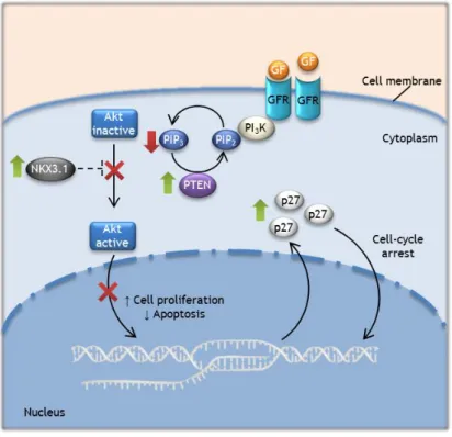
PCa is a heterogeneous disease characterized by several alterations in key regulatory pathways involved in cell cycle regulation, DNA replication and DNA repair (55). Some of the most frequent altered molecular pathways involve the already mentioned tumor suppressor genes NKX3.1, CDKN1B, and PTEN, which are responsible for the regulation of prostate cells growth. However, they are downregulated not only in PCa but also in PIA and PIN lesions (26, 51). NKX3.1 gene encodes a prostate restricted homeobox protein whose function seems to be essential for normal prostate development and can suppress the growth of prostate epithelial
10 cells. This gene appears to be a prostate specific tumor suppressor gene. Decreased NKX3.1 expression seems to be more important for the initiation of the PCa than for the progression to an invasive status (7, 26, 51). PTEN gene encodes a phosphatase that has activity on lipids and proteins. This enzyme is responsible for the dephosphorylation and inactivation of phosphatidylinositol 3,4,5-trisphosphate (PIP3), a second messenger produced by active
phosphatidylinositide 3-kinase (PI3K) in response to activation of several receptors by growth
factors. PIP3 will recruit several proteins to the plasma membrane, such as protein kinase B
(Akt). After Akt activation, several important signaling pathways will be activated, which are involved in inhibition of apoptosis and activation of cell proliferation (Figure 4) (51, 56). CDKN1B encodes the cyclin-dependent kinase inhibitor p27, which plays an important role in inhibition of cell cycle. The loss of PTEN expression leads to increased levels of PIP3, and
consequently, activation of the PI3K-Akt signaling pathway. The continuous activation of this
pathway inhibits the expression of CDKN1B gene, and consequently, the expression of p27 (26, 45, 51). On the other hand, Inhibition of PI3K-Akt signaling pathway increases NKX3.1
expression, which in turn promotes p53 activation and inhibits AR promoter activity. Thus, the loss of PTEN expression leads to NKX3.1 downregulation which allows AR overexpression and activation of its target genes that may be involved in onset and progression of PCa (51, 57).
Figure 4: Molecular pathways in normal prostate cells. PTEN, NKX3.1, and p27 proteins regulate the proliferation and apoptosis of prostate epithelial cells. PTEN and possibly NKX3.1 inhibits the PI3K-Akt signaling pathway, which leads to an increase in p27 levels and apoptosis and decreased proliferation.
11 Considering that PCa cells retain the AR signaling pathway, androgen ablation results in tumor regression. However, with the tumor progression and accumulation of molecular alterations, there is a gain of function in the AR signaling pathway. At this stage of PCa, which is androgen independent, the tumor cells are resistant to androgen ablation due to their acquired ability to activate the AR signaling pathways involved in cell proliferation and survival without requiring physiological levels of androgens (8). In androgen independent PCa, the AR signaling pathway remains essential for the growth and survival of the tumor. In fact, most of the tumors at this stage have high levels of AR expression and continue to express AR target genes (8). One of the alterations found in PCa is the AR gene amplification, which leads to increased sensitivity of PCa cells to low levels of androgens (58). Other mechanisms involved in androgen-independent phenotype include mutations that change the ligand specificity of the receptor, allowing the activation of the AR by other non-androgen molecules, such as steroid hormones, antiandrogens or even growth factors like insulin-like growth-factor-1 (IGF-1), keratinocyte growth factor (KGF) and epidermal growth factor (EGF) (9, 59–63). Several studies have found that these alterations in the AR gene occur in androgen-independent PCa metastasis, but not in primary PCa. However, in primary PCa, the AR signaling pathway already shows alterations, including mutations in the genes that encode the nuclear receptor coactivators and corepressors NCOA2 and NCOR2, respectively (64, 65).
12
1.3. Genetic and epigenetic mechanisms involved in
carcinogenesis of prostate cancer
The transformation of a normal cell into a neoplastic is a multistep process in which the cells gradually acquire alterations in proto-oncogenes, tumor suppressor genes and other genes involved in cellular functions. These alterations lead eventually to the disruption of the network that tightly regulates the homeostatic balance between cell death and proliferation. The molecular mechanisms involved in prostate carcinogenesis remain poorly understood, but it is clear that genetic and epigenetic alterations contribute to this process. Genetic alterations can lead to the expression of abnormal proteins, and consequently the disruption of the signaling pathways that may promote cancer onset and/or progression. Alterations in the cell epigenome may lead to changes in the transcriptional control that will deregulate the cellular mechanisms through inappropriate silencing or activation of cancer-related genes. Genetic, as well as epigenetic changes, are inheritable at the cellular level contributing to the growth of cancer cells. Alterations such as mutations, rearrangements, amplifications or hypomethylation often lead to overexpression or expression of constitutively activated proteins that will induce cellular transformation. On the other hand, alterations like mutations, deletions, allelic loss or hypermethylation are associated with silencing and/or loss of function of proteins. Transformation of normal prostate cells into PCa cells is characterized by a decreased expression or function of genes involved in cell-cycle control, cell adhesion, DNA damage repair and apoptosis, and by an increased expression or gain of function of genes related to cell proliferation, invasion, metastasis, and angiogenesis (reviewed by (51, 55, 66)).
1.3.1. Genetic mechanisms
Cancer development is characterized by the accumulation of genetic alterations that lead to aberrant gene expression. Genetic changes play an important role in tumourigenesis and also in intra- and inter-tumor heterogeneity. In accordance with other types of cancer, PCa is also characterized by the inactivation of tumor suppressor genes and activation of oncogenes, inhibiting apoptosis and allowing cell proliferation. Several genomic alterations have been identified in PCa, from single nucleotide mutations to chromosomal rearrangements, which lead to disruption of signaling pathways that control cellular functions. The most frequent genomic alterations present in PCa are point mutations, gene deletions, gene amplifications and chromosomal rearrangements (26, 55, 67).
One of the most common genetic alterations found in PCa cells is the fusion genes between the androgen-regulated gene TMPRSS2 and the genes of the ETS transcription factors family ERG or ETV1 (8, 68). TMPRSS2 is a type II transmembrane serine protease prostate specific, which is regulated by androgens. The ETS transcription factors family are involved in
13 cell proliferation and invasiveness, being then considered oncogenes (7, 68, 69). The TMPRSS2:ERG fusion appears to be an early event in prostate carcinogenesis that is sometimes already present in high-grade PIN, but is absent in PIA and BPH (69). AR activation induces chromosomal proximity between TMPRSS2 and ERG loci allowing the fusion between the 5’ end of the TMPRSS2 gene and the 3’ end of the ERG or ETV genes. The region between these two genes is often deleted as a consequence. The resulting overexpression of the TMPRS2 and ERG genes are thought to be important for the progression and invasiveness of PCa (8, 69).
Other common chromosomal abnormalities are the gains at 7p, 7q, 8q and Xq and the losses at 6q, 8p, 10q, 13q and 16q (46, 55, 70). One of the genes affected by these losses is the NKX3.1 homeobox gene, which is located in chromosome 8p21 and has one of the two alleles frequently deleted in PCa (26). The loss of heterozygosity at chromosome 10q is associated with the loss of PTEN gene expression. The haploinsufficiency of NKX3.1 and PTEN genes can be associated with abnormal proliferation of prostate cells (26, 45). The gains at chromosome Xq lead to aberrant AR activity due to AR gene amplification, increasing the sensitivity of PCa cells to very low levels of androgens. This alteration is more frequently found in androgen-independent tumors after hormonal therapy than in primary PCa. Although the cells with AR amplification have increased sensitivity, they still require androgens for proliferation (9, 26).
Another frequent genetic alteration found in the AR gene is somatic mutations in the ligand–binding domain. These mutations in the AR gene decrease the specificity of the AR to testosterone and DHT, allowing inappropriate activation by other steroid hormones, or even androgen antagonists. Therefore, the malignant cells can continue to activate the AR signaling pathway and promote proliferation when the levels of androgens are low by using other circulating steroid hormones. A common example is the missense mutation at position 877, which results in the exchange of an alanine for a threonine (T877A). This alteration in the ligand-binding domain allows the AR activation by antagonists. These mutations have a higher incidence in androgen-independent PCa previously treated with hormone therapy (9, 26, 55).
14
Table 1: Genetic alterations found in human PCa.
Gene Location Alterations Notes References
Tumor suppressor genes
NKX3.1 8p21.2 Allelic losses (↓ expression) Encodes a prostate restricted homeobox protein (7, 26, 71) CDKN1B 12p13.1-p12 Allelic losses (↓ expression) Encodes cyclin-dependent kinase inhibitor p27 (7, 26, 71) PTEN 10q23.3
Allelic losses and mutations (↓ expression and/or
function)
Encodes a phosphatase with activity on lipids and
proteins
(7, 26, 71)
TP53 17p13.1 Mutations
Has many tumor suppressor functions like
cell cycle arrest
(7, 26, 71) Oncogenes AR Xq12 Mutations and amplification (↑ expression or altered function)
Encodes the Androgen
Receptor (7, 26, 71)
MYC 8q24.21 Amplification
Transcription factor that regulates genes involved
in cell proliferation, senescence, apoptosis,
and cell metabolism
(7, 71)
ERG 21q22.3 Chromosomal
rearrangement
ETS transcription factors
family (7, 71) ETV1-4 7p21.3, 19q13.12, 1q21-q23, 17q21.31 Chromosomal rearrangement Encodes ETS-like transcription factors 1–4 (7, 71)
15
1.3.2. Epigenetic mechanisms
In addition to the genetic alterations, the disruption of epigenetic mechanisms may conduct to deregulation of gene expression and is also part of the oncogenic process. Epigenetic mechanisms can be defined as heritable modifications that do not affect the DNA sequence (72). The main epigenetic mechanisms are DNA methylation and histone modifications, which have as main function to ensure proper regulation of gene expression by changing the chromatin structure (51, 73).
Histones are essential in the regulation of chromatin packaging by post-translational modifications that include acetylation and methylation among others. These alterations that occur in the N-terminal histone tails promote alterations in chromatin condensation and DNA accessibility. Histone acetylation is associated with a more relaxed chromatin state, which allows transcriptional activity by permitting the access of transcription factors. Histone acetylation is regulated by two enzymes: histone acetyltransferase (HAT) and histone deacetylases (HDACs) (55, 74, 75). Histone hyperacetylation is associated with transcriptionally active chromatin, while histone hypoacetylation is associated with transcriptionally inactive chromatin (76).
In PCa, increased expression of HDACs is frequent resulting in histone hypoacetylation (51, 55). A high expression of the histone deacetylase 1 (HDAC1) gene is associated with lower expression of its target genes (77). Some of the target genes of HDAC1 include Bax, p21, p27, maspin and p53 (51). Aberrant recruitment of HDACs to the promoter region of tumor suppressor genes could contribute to tumor development and progression. An example of epigenetic inactivation by hypoacetylation of the promoter is the cyclin-dependent kinase inhibitor p21, whose function is to inhibit cell-cycle progression. HDACs are also involved in deacetylation of non-histone proteins. Under stress conditions, p53 is phosphorylated and acetylated to promote protein stability and activation. HDAC1 is able to deacetylate p53, blocking its tumor suppressor activity and allowing tumor progression (75).
DNA methylation consists of the addition of a methyl group by covalent bonding at the 5’ position of the cytosine that precedes a guanine. Usually, these CG dinucleotides are concentrated in large clusters, called CpG islands, which are mainly located in the promoter region and/or in the first exon (73). The methylation of these regions leads to gene silencing while unmethylation promotes active gene transcription. The methylation pattern is maintained by DNA methyltransferases (DNMTs), which catalyze the transfer of a methyl group from S-adenosyl-methionine to cytosine (70, 73). Any abnormalities in DNA methylation, which is essential for normal cell function, may lead to the development of several cancers. (73)
Tumor cells are characterized by a methylation pattern that differs from normal cells. In tumor cells, hypermethylation is observed in promoters of specific genes, particularly tumor suppressor genes, and a global hypomethylation contributes to genomic instability and activation of oncogenes (73). In normal cells, DNA hypermethylation is generally observed in
16 satellite sequences and repetitive genomic sequences, being these regions silenced to ensure genomic stability and integrity. The disruption of this mechanism may lead to tumor development and progression (73). One of the genes found unmethylated in PCa was the retrotransposable element 1 (LINE-1). These repetitive sequences constitute approximately 5-10% of the human genome, which are hypermethylated in normal tissues. LINE-1 hypomethylation was found in more than 50% of PCa cases and in more than 60% of PCa with lymph node metastases (70, 78). The most common example of hypermethylation in PCa is the Glutathione S-transferase π (GSTP1) gene a caretaker gene. This gene encodes a phase II detoxification enzyme that is hypermethylated in 90% of PCa, but also in PIA (5-10%) and PIN (70%) lesions. In normal prostate epithelium, the expression of this enzyme allows the detoxification of electrophilic compounds, including carcinogens. Therefore, it is believed that GSTP1 silencing is involved as an earliest event on PCa development that will turn prostate cells more susceptible to mutations (9, 51, 70). DNA methylation alterations in PCa seems to have two phases, the first that will promote cell transformation, and a second that will promote malignant cancer progression. This is supported by the fact that hypermethylation of GSTP1, APC, RASSF1, COX2, and MDR1 can be detected in localized and metastatic PCa while hypermethylation of estrogen receptor (ER), MLH1 and p14/INK4 only can be detected in a later phase of PCa progression (78, 79). It has been suggested that epigenetic changes in PCa are more common and arise earlier than genetic alterations and therefore may be future biomarkers for PCa (70, 80). An example of one of the most common alterations for which methods of detection are already being developed, in various types of samples, is the hypermethylation of the promoter region of the GSTP1 gene (81–83)
DNA hypomethylation of specific genes in tumor cells is less common, and the majority of the hypomethylated promoters belong to tissue-specific genes (73). For example, the Urokinase plasminogen activator (PLAU) gene encodes a multifunctional protein that can promote tumor invasion and metastasis. In normal prostate cells, as well as in hormone responsive PCa cells, PLAU gene is weakly expressed while in hormone-independent and highly invasive PCa cells, it is highly expressed due to hypomethylation of its promoter region (66, 84).
Some of the most frequently epigenetic alterations present in PCa are described in table 2.
17
Table 2: Epigenetic alterations found in human PCa.
Gene Location Alterations Notes References
GSTP1 11q13 Hypermethylation
(↓ expression)
Encodes an enzyme that catalyzes the conjugation of reduced glutathione to electrophilic
substrates
(70, 74)
NKX3.1 8p21.2 Hypermethylation
(↓ expression)
Encodes a prostate restricted homeobox protein
(55, 85)
APC 5q21-q22 Hypermethylation (↓ expression)
Adenomatous polyposis coli (APC) gene encodes a tumor suppressor
protein
(70, 74)
RASSF1A 3p21.3 Hypermethylation
(↓ expression)
Ras association domain family member 1 (RASSF1A) gene encodes a protein similar to the
RAS effector proteins
(70, 74) PTGS2 1q25.2-q25.3 Hypermethylation (↓ expression) prostaglandin-endoperoxide synthase 2 (PTGS2), also known as
cyclooxygenase, is a key enzyme in prostaglandin biosynthesis
(74)
LINE-1
22q11.1-q11.2 Hypomethylation Retrotransposon element 1
(70)
PLAU 10q22.2 Hypomethylation (↑
expression)
Urokinase plasminogen activator gene encodes a secreted serine
protease
18
1.4. Six transmembrane epithelial antigen of the prostate 1
1.4.1. General characteristics
The Six transmembrane epithelial antigen of the prostate 1 (STEAP1) gene was first identified by Hubert and colleagues as a gene overexpressed in PCa using a subtractive hybridization between benign prostatic tissue and PCa xenografts model (86). The STEAP1 gene is one of the four members of the STEAP family, which includes the genes encoding the STEAP1-4 proteins. Also, a very similar gene to STEAP1 is encoded by the human genome, called STEAP1B.
STEAP1 is located at the long arm of chromosome 7q21 and has 10.4 kb, comprising 5 exons and 4 introns (Figure 5). This gene encodes two different mRNA transcripts of 4.0 kb and 1.4kb, but only the last is translated into a protein with 339 amino acids (aa) with approximately 40 kDa (86). STEAP proteins have in common a six-transmembrane domain, an intramembrane heme binding site and intracellular N- and C-termini. The role of the STEAP1 protein remains unclear due to the lack of FNO-like domain and Rossman fold, which are involved in oxidoreductase activity of iron and copper by STEAP2, STEAP3, and STEAP4 (87, 88). Although STEAP1 does not promote iron or copper reduction or uptake, its co-localization in endosomes with transferrin and transferrin receptor 1, specialized proteins in iron uptake, suggests that it may still play a role in metal homeostasis (87). STEAP1 protein is mainly localized on the plasma membrane of epithelial cells, particularly at cell-cell junctions, and its predicted structure supports the idea that this protein may act as an ion channel or transporter protein (86). In fact, it was reported that STEAP1 may allow the transport of small molecules between adjacent cells in culture, indicating that STEAP1 may be involved in intercellular communication (89, 90).
Regarding the STEAP1B gene, it is located on the same chromosome as STEAP1, but on the short arm (chr:7p15). STEAP1B gene shares high homology with STEAP1 gene possibly due to gene duplication during genome evolution. This gene encodes two different mRNA transcripts, STEAP1B1, and STEAP1B2. STEAP1B1 encodes a protein with 332 aa, while STEAP1B2 encodes a protein with 245 aa. However, it remains to be demonstrated the expression of these proteins. Nevertheless, in silico analysis showed that both proteins have some similarity with STEAP1, but with fewer transmembrane domains. In concordance with STEAP1, STEAP1B also lacks the FNO-like domain and Rossman fold. (91, 92).
19
Figure 5: Location of STEAP1 gene at the long arm of chromosome 7, STEAP1 gene organization and schematic of STEAP1 protein structure (Adapted from (93)).
1.4.2. Expression in human tissues
STEAP1 is overexpressed in all stages of PCa, including metastasis, but its expression is most pronounced in androgen sensitive stages than in androgen-independent stages (86). STEAP1 is also overexpressed in other types of tumors, such as breast, lung, bladder, colon, pancreas, ovary and Ewing sarcoma, where the role of STEAP1 has also been studied (86, 89, 94). STEAP1 expression in normal tissues is almost restricted to prostate, but is also expressed at lower levels in other tissues such as bladder, fetal and adult liver, kidney, pancreas and skeletal muscle, as summarized in Table 3 (86, 87). Regarding PIN lesions, STEAP1 expression also shows high levels, suggesting that STEAP1 deregulation is an earlier event in PCa development (95). The STEAP1 levels found in BPH are very low and similar to those that are found in non-neoplastic adjacent tissue of PCa (95). The expression levels of STEAP1 protein in the different types of normal and cancer tissues are described in table 3.
Concerning STEAP1B gene, both mRNAs transcripts are expressed in prostate cell lines PNT1A, PNT2, LNCaP, and PC3. It was shown that STEAP1B2 mRNA is overexpressed in neoplastic cells when compared to non-neoplastic cells, indicating that this gene may also be deregulated in cancer. On the other hand, STEAP1B1 mRNA does not seem to be differentially expressed between neoplastic and non-neoplasic cells (91).
chr7
7q21
E1 E2 E3 E4 E5
20
Table 3: Expression of STEAP1 protein in normal and cancer tissues.
Protein
Tissue Normal Cancer Reference
Bladder Low/Not detectable Moderate/High (89, 96)
Bone marrow Not detectable - (86, 87)
Breast Low Moderate/High (94)
Colon Low Low (86, 87)
Heart Not detectable - (86, 87)
Liver Not detectable - (86, 87)
Lung Not detectable Moderate (86, 87, 89)
Kidney Low/Not detectable Moderate/High (86, 87, 96)
Pancreas Low - (86, 87)
Placenta Not detectable - (86, 87)
Prostate Moderate High (86, 87)
Skeletal
muscle Not detectable - (86, 87)
Stomach Low - (86, 87)
Thymus Not detectable - (86, 87)
1.4.3. Biological functions and its regulation in normal and cancer cells
STEAP1 protein localization at cell junctions and its predicted secondary structure as a channel protein suggest that it may play a role in intercellular communication through the diffusion of ions and small molecules between cells (89, 90, 93). In fact, it has been demonstrated that ion channels contribute to the regulation of several biological processes, such as proliferation, differentiation, and apoptosis. In addition, the malignancy and invasiveness of PCa androgen-independent cells are associated with an altered expression of several ion channels in the plasma membrane, enhancing the apoptotic resistance (97).
In Ewing tumors, STEAP1 protein seems to promote cell growth by increasing intracellular reactive oxygen species (ROS) levels. In fact, STEAP1 overexpression is associated with increased proliferation and invasiveness through the increased cellular ROS levels. The oxidative stress, which may result from STEAP1 overexpression, may enhance tumor aggressiveness through the activation of genes involved in cell proliferation and invasiveness, such as MMP-1, ADIPOR1, and DTX3L. Also, high levels of ROS are associated with the activation of metastatic signaling pathways, which is associated with cancer aggressiveness (98).
The hypothesis that STEAP1 is involved in cancer cell proliferation is supported by the fact that the treatment of PCa cells with STEAP1 monoclonal antibodies is able to inhibit the
21 cell growth (89). Moreover, it was also verified that the knockdown of STEAP1 expression on tumor cells is associated with an antitumor effect due to the disruption of intercellular communication between tumor cells and adjacent tumor-associated stromal cells (90).
Regarding the regulation, it was already shown that STEAP1 expression is regulated by androgens, estrogens, and zoledronic acid. In respect to androgens and estrogens, it was demonstrated that STEAP1 gene is down-regulated in rat prostate and mammary gland, as well as in LNCaP cells and MCF-7 breast cancer cells (94, 99). STEAP1 down-regulation by androgens is mediated by the AR signaling pathway and seems to be dependent on de novo protein synthesis (99). On the other hand, the STEAP1 down-regulation by estrogens does not seem to be mediated by the estrogen receptor signaling pathway (94, 99). Concerning the effect of zoledronic acid on STEAP1 expression in PCa cells, STEAP1 expression decrease in a dose-dependent manner (100). Zoledronic acid is one of the most commonly used bisphosphonates for the prevention and treatment of skeletal complications in PCa patients with bone metastasis (101).
The mechanisms that lead to overexpression of STEAP1 in human tumors remain poorly understood. The regulation of STEAP1 expression by transcriptional and post-translational mechanisms was already assessed through STEAP1 mRNA and protein stability. In LNCaP cells, STEAP1 mRNA and protein stability are higher when compared with PNT1A cells, suggesting that post-translational mechanisms may contribute for STEAP1 overexpression. In silico analysis of post-translational modifications in STEAP1 protein reveal some potential sites for N-glycosylation, glycation, phosphorylation and O-β-GlcNAcylation (91). These modifications play a role in common mechanisms of protein function regulation and may confer higher stability to proteins. Alterations in these mechanisms may lead to the development several diseases, including cancer (91, 102). However, these alterations still do not justify the overexpression of STEAP1 in tumor cells, suggesting that other mechanisms may be involved.
22
23 STEAP1 is overexpressed in several human tumors, particularly in PCa, and several investigators have pointed it out as a potential biomarker or therapeutic target. Regarding the regulation of STEAP1 gene, some factors have been identified as involved in regulation of its expression, but the mechanisms that lead to STEAP1 overexpression in PCa remain unknown. Therefore, the aim of this project was to identify molecular mechanisms involved in overexpression of the STEAP1 gene in LNCaP PCa cells. To achieve this goal, it was hypothesized that genetic and/or epigenetic changes may be involved in STEAP1 regulation in PCa. To test this hypothesis, several experimental approaches were delineated in order to:
Identify mutations in the promoter region of the STEAP1 gene in LNCaP cells.
Compare the methylation pattern of the STEAP1 gene between PNT1A (non-neoplastic cells) and LNCaP cells.
Evaluate the effect of treatment with 5-Aza-2’-deoxycytidine (AZA) and Trichostatin A (TSA), DNMT and HDAC inhibitors respectively, on STEAP1 mRNA expression in PNT1A cells.
24