UNIVERSIDADE DE LISBOA
FACULDADE DE CIÊNCIAS
DEPARTAMENTO DE BIOLOGIA ANIMAL
Contribution for the knowledge on curable sexually transmitted
infections with special emphasis on Chlamydia trachomatis
Zohra Gulzar Lodhia
Mestrado em Biologia Humana e Ambiente
Dissertação orientada por:
Doutora Maria José Borrego
Professora Doutora Deodália Dias
Dedication
This dissertation is dedicated to the love of my life, the joy of my soul and the warmth of my heart, my
baby niece Mariam Shanawaz Lodhia. The one that reassures my beliefs and hopes with just a
glance from those amazing almond-shaped eyes and whose toothless smile makes my day, every day, every time.
Little hooman, my goduchas,
Jola Fui
loves you so much and will always be there for you, my beautifulprincess. ©
“You are my one and only, you can wrap your fingers round my thumb and hold
me tight… And you’ll be alright”
©
Acknowledgements
First of all, I would like to thank Allah, to Whom I pray every single day and night, Who has been granting me so many blessings over the years, helping me throughout my struggles and giving me patience & strength.
A special thank you to my advisor, Dr. Maria José Borrego, responsible for the National Reference Laboratory for Sexually Transmitted Infections of INSA, for accepting me in her laboratory, for being such an amazing mentor who taught me so much, for her sympathy, thoughtfulness, patience, advices and for always being there when I needed.
To my internal advisor and masters’ coordinator, Professor Dr. Deodália Dias, for always being available, for her kindness and for always figuring out a solution to every problem.
To Dr. Vítor Borges, researcher at the Bioinformatics Unit of the Infectious Diseases Department at INSA, for his contribution on my dissertation, for his teachings, gentleness and friendship.
To Miguel Pinto, my fellow lab colleague, who has the best playlists of the laboratory and with whom I learned so much on so many subjects (non-scientific included). Thank you for making me
‘meow’ every single song in my head, for the productive conversations and discussions, for helping me
in some aspects of my dissertation, for the friendship, kindness and all the laughter.
To Dr. João Paulo Gomes, responsible for Research Unit of the Infectious Diseases Department at INSA, for his sympathy, professionalism and sense of humor.
To Professor Dr. Marília Antunes, for finding some time in her busy agenda to help me with the statistical analysis of this dissertation.
To my amazing parents, for giving me the possibility to pursue another step in my academic formation, for always being comprehensive, for insisting so much with me when I did not feel like doing a break from my work to go to eat because I was so focused, for supporting, caring and loving so much their baby girl.
To my brother and sister-in-law who are both always so caring and crazy, but mostly supportive, for always wishing me the best, spoiling me with so much love and ‘bussa bussi’ and for putting up with my snapchats when I was in my many ‘fritanços’.
To Dani (Daniela Ferreira), my best friend, the one that is always there when I need, for always helping me to see the sunny side of everything, for cheering me up, for caring and worrying, for reminding me what I am capable of and for always having the best future plans ever.
To my Madrinha Sissi (Sílvia Firmino) for being the best godmother I could have asked for, for always treating me as her ‘piqui’, for always sending me messages asking how things were going and for always reassuring I could count on her, no matter what.
To Abdullah & Munisa, thank you for insisting, calling me on my phone a hundred times and basically forcing me to go out to unwind when most of the times I did not even feel like it, but surely needed even when it was just for a ‘quick coffee break’.
To Fátima Karolia, ‘mommy dearest’, the sister I got to “choose”, for all the support, all the crazy talks and laughs and for “motivational baby Adam” photos, videos and video-calls.
To Saf (Safwaan Zamakda), my cousin, who was always so nice and thoughtful and, although he is literally on the other side of the world, he was always so supportive, always cheering me up and rooting for me.
To Neves (André Lopes), one of the most amazing friends I have, for our lunch breaks, for our conversations and hilarious stories and for holding and helping me when my world was turned upside down and I felt like I could not reach the light and you led me right to it.
To all my friends: a HUGE thank you for all the support, for believing in me, cheering me up, giving me strength, caring, worrying, loving, making me laugh, for the many dinners out and for all the “you are going to make it, do not worry!”.
It was a really exhaustive and long time, fulfilled with so much hard work, so much knowledge and challenges and I could not be any more grateful than I am for all those who were part of this journey and for those who somehow helped me get through it.
Resumo
Introdução e objetivos: As infeções sexualmente transmissíveis (ISTs) abrangem um amplo
conjunto de síndromes com quadros clínicos variados que vão desde a ausência de sintomas até grave morbilidade, incluindo a própria morte. Existem mais de 30 agentes patogénicos de origem bacteriana, viral ou parasitária, que podem ser transmitidos de uma pessoa para outra durante o ato sexual. As ISTs estão entre as condições agudas mais comuns no mundo, sendo Chlamydia trachomatis, Neisseria
gonorrhoeae, Trichomonas vaginalis e Mycoplasma genitalium as quatro principais causas de ISTs
curáveis, sendo responsáveis por uretrite, cervicite, vaginite e proctite. Em Portugal, não há dados sobre a prevalência destas ISTs, desconhecendo-se o seu respetivo impacto para saúde pública. Deste modo, a presente dissertação teve por objetivo: a) o estudo da prevalência dos quatro microrganismos acima referidos no contexto de uma consulta de ISTs (Unidade de consulta de DST/CAD da Lapa); b) a análise da variabilidade genética do gene usado para genotipagem de C. trachomatis, ompA, o qual codifica a principal proteína da membrana externa de C. trachomatis, com vista a caracterizar as diferenças genéticas entre estirpes, as quais podem estar subjacentes a um processo contínuo de evolução e adaptação desta bactéria. Foram usadas as estirpes reunidas pelo laboratório de acolhimento ao longo de um período de 27 anos; c) o estudo da variabilidade intra-estirpes de C. trachomatis, de alvos
potencialmente associados à variação de fase e à adaptação bacteriana no contexto da infeção in vivo.A
variação de fase, que pode ser causada por mutações reversíveis que influenciam a expressão génica e a sua função, é conhecida por ser essencial para a adaptação e virulência de algumas bactérias, mas pouco se sabe sobre o seu eventual papel na biologia e na patogenicidade de C. trachomatis.
Materiais e Métodos: a) Foram avaliadas 1034 amostras biológicas colhidas na consulta DST/CAD
Lapa, durante o período de tempo compreendido entre Setembro de 2016 e Setembro de 2017, relativamente à presença de C. trachomatis, N. gonorrhoeae, T. vaginalis e M. genitalium, utilizando dois sistemas de PCR em tempo real, Cobas® 4800 CT/NG (Roche Sistemas de Diagnóstico) e
S-DiaMGTVTM (Diagenode); b) Foi efetuada genotipagem-ompA de 370 amostras utilizando uma técnica
de nested-PCR, seguida de sequenciação pelo método de Sanger e a análise de similaridade das sequências genéticas obtidas relativamente à sequência ompA de estirpes protótipo de C. trachomatis. Posteriormente, essas amostras foram incluídas na base de dados do laboratório de acolhimento, num total de 2579 estirpes de C. trachomatis reunidas entre 1990 e 2017, tendo sido analisada toda a coleção relativamente ao genótipo ompA, ao género, à localização anatómica da infeção e à distribuição das variantes genotípicas; c) Foram analisadas 167 amostras (96 amostras de DNA selecionadas de entre as constantes na base de dados de C. trachomatis do laboratório, juntamente com um conjunto de 71 amostras para as quais os dados de sequenciação total do genoma tinham sido recentemente disponibilizados) relativamente a 12 tratos homopoliméricos potencialmente variáveis no genoma desta bactéria, através de uma técnica com base na sequenciação de nova geração (NGS), de produtos de amplificação (amplicões).
Resultados: a) Um quinto da população estudada estava infetada por pelo menos uma IST, tendo
sido C. trachomatis a mais frequentemente detetada, seguida por N. gonorrhoeae, M. genitalium e T.
vaginalis. Os indivíduos do sexo masculino revelaram-se mais frequentemente infetados que as
mulheres, com especial ênfase para os indivíduos com idades compreendidas entre os 25 e os 34 anos, e para os indivíduos com mais de um parceiro sexual. No nosso estudo, N. gonorrhoeae foi a segunda IST mais frequente, sendo que os homens contribuíram com mais de dois terços dos casos e que cerca de metade deles eram homens que faziam sexo com homens (HSH); b) A genotipagem-ompA de estirpes clínicas de C. trachomatis evidenciou 12 genótipos-ompA, de entre os quais os genótipos E e F foram os mais frequentes, enquanto os genótipos B e o C foram os mais raros. Os genótipos G e L2 foram
muito mais comuns nos homens do que nas mulheres, tendo sido o endocervix/uretra o local anatómico onde mais frequentemente se efetuou a pesquisa e, consequentemente, se detetou a infeção. Aliás, o endocervix/uretra foi o único local anatómico onde os 12 genótipos foram detetados; c) Os resultados deste estudo revelaram diferentes perfis de variação intra- e inter-paciente para os poli (Ns) estudados, os quais inequivocamente sublinham o papel de um homopolímero em desencadear mecanismos reversíveis de "ON/OFF" da citotoxina de C. trachomatis (CT166) in vivo. Foram ainda identificados outros novos potenciais mediadores de mecanismos de variação de fase em C. trachomatis.
Discussão: a) De acordo com os dados de prevalência de ISTs nos EUA e na Europa, nos quais C.
trachomatis é descrita como a IST bacteriana mais frequente, e com o aumento do número de casos que
têm sido descritos nos últimos anos, era esperado que C. trachomatis fosse a IST mais frequentemente detetada na população estudada. No entanto, as taxas de prevalência podem subestimar a verdadeira dimensão desta infeção dado o seu caráter assintomático. Por outro lado, a sua maior frequência pode também ser apenas um reflexo da melhoria dos sistemas de vigilância das ISTs, em muitos países, muitas vezes suportados por estudos de rastreio em populações assintomáticas; tais estudos são facilitados pela disponibilidade de melhores ferramentas de diagnóstico laboratorial (sensibilidade e especificidade próximas dos 100%). O número de casos positivos de ISTs na região anorretal e na orofaringe, testadas apenas em HSH, justificam a necessidade de alargar o diagnóstico a esses locais anatómicos em homens heterossexuais e em mulheres. Deste modo, e considerando que este estudo envolveu pacientes de apenas uma consulta IST, é de salientar a necessidade de uma avaliação a nível nacional, uma vez que até à data a prevalência destas ISTs na população portuguesa é desconhecida; b) A elevada frequência dos genótipos-ompA E e F, e a menor ocorrência de estirpes variantes nas estirpes destes genótipos, sugere uma melhor adaptação desses genótipos; tal situação favorecerá que não sejam reconhecidos pelos mecanismos de defesa do hospedeiro e que não sejam eliminados pelos sistemas de defesa do hospedeiro, facilitando a sua disseminação e justificando o seu predomínio em termos epidemiológicos, em detrimento doutros genótipos-ompA. No entanto, são necessários estudos envolvendo o genoma total de estirpes clínicas de C. trachomatis, que deverão ser acompanhados pela análise dos respetivos dados clínicos, para uma mais completa compreensão das características genéticas de C. trachomatis e a sua relação com o desenvolvimento de patologia; c) Este estudo enriquece o conhecimento sobre a variabilidade intra-paciente de tratos homopoliméricos potencialmente mediadores de mecanismos de variação de fase, consolidando a hipótese de que a funcionalidade da conhecida citotoxina de C.
trachomatis (CT166), durante o processo infecioso, será regulada por mecanismos de variação de fase,
o que ficou evidenciado pela elevada variabilidade inter e intra-paciente. Por outro lado, foi também possível demonstrar, pela primeira vez, que existem outros tratos homopoliméricos com variabilidade intra-estirpe para além do referido acima. É de destacar a necessidade de se alargar esta abordagem,
utilizando-se um conjunto mais homogéneo e amplo de amostras de C. trachomatis, considerando
também os respetivos dados clínicos e os genes relacionados putativamente com mecanismos de variação de fase, de modo a consolidar algumas das hipóteses suscitadas no decurso da presente dissertação de mestrado.
Palavras-chave:
Infeções sexualmente transmissíveis curáveis; C. trachomatis; Genótipos-ompA; Variação de fase; Homopolímeros.Abstract
Introduction and objectives: Sexually transmitted infections (STIs) refer to a wide variety of
clinical syndromes and infections, which are among the most common acute conditions in the world, causing serious morbidity, and even death. Chlamydia trachomatis, Neisseria gonorrhoeae,
Trichomonas vaginalis and Mycoplasma genitalium are the four main causes of curable STIs, being
responsible for urethritis, cervicitis, vaginoses and proctitis. In Portugal, there is no data on their prevalence, being unknown the impact of these STIs. This dissertation aimed at a) studying the prevalence of these four microorganisms in the context of a sexually transmitted diseases (STDs) consultation; b) analyzing the variability of C. trachomatis genotyping gene, ompA – which codes to the major outer membrane protein –, throughout a timeline of 27 years. The genetic differences detected in this gene may provide important clues about the continuous process of evolution and adaptation of this human pathogen; c) studying C. trachomatis intra-strain variability in vivo, as a means to identify potentially phase-variable targets associated with bacterial adaptation in the context of infection.
Materials and Methods: a) We evaluated 1034 samples collected from patients of the major
Portuguese STD clinic (Unidade de consulta de DST/CAD da Lapa)for the microorganism’s presence
using two real-time PCR systems, Cobas® 4800 CT/NG (Roche Sistemas de Diagnóstico) and
S-DiaMGTVTMkit (Diagenode); b) ompA-genotyping of 370 samples was performed using a nested-PCR
technique, Sanger sequencing and ompA-sequence similarity analysis regarding C. trachomatis prototype strains. Then, we added these samples to the Instituto Nacional de Saúde Doutor Ricardo Jorge database collection, composed by 2579 C. trachomatis specimens, gathered from 1990 to 2017, and analyzed the whole database regarding ompA-genotype, gender, anatomical site of infection and genotypic variants distribution; c) We analyzed 167 samples (96 selected C. trachomatis-positive DNA samples from the laboratory database, along with a set of 71 samples for which whole-genome sequencing data had been recently released) with an amplicon-based next generation sequencing (NGS) technique for 12 selected potentially variable poly (Ns).
Results: a) We found that almost a fifth of our study population was infected by at least one STI,
with C. trachomatis being the most commonly detected, followed by N. gonorrhoeae, M. genitalium and, finally, T. vaginalis. Men were, in general, more infected than women, with especial evidence for individuals aged 25 to 34 years, and for people who had more than one sexual partner; b) ompA-genotyping of C. trachomatis clinical strains evidenced 12 ompA-genotypes, among which E and F were the most represented and B and C were the least frequent. Genotypes G and L2 were much more common among men than among women and the endocervix/urethra was the anatomical site of infection more represented, and the only site where all genotypes could be detected; c) The scrutiny of the selected potentially variable poly (Ns) revealed distinct trends of inter- and intra-patient variation that, not only unequivocally reinforce the role of a reversible poly(N) in the ON/OFF switching of C. trachomatis cytotoxin (CT166) in vivo, but also launch other poly(N)s as potential mediators of phase variation mechanisms in C. trachomatis.
Discussion: a) According to its prevalence in the USA and Europe, and because the number of
detected cases have been rising the past few years, C. trachomatis was expected to be the most frequently detected STI. However, reported prevalence rates underestimate the true burden of this STI due to its asymptomatic character. On the other hand, its frequency may also reflect the expansion of screening, namely among asymptomatic because of the development of better and more sensitive diagnostic tools, and also due to the improvement of reporting systems. The rectum and the oropharynx were only tested in MSM, and the number of positive STIs cases in these anatomical locations highlight the need for their
investigation among heterosexual men and women; b) The high frequency of E and F together with the low occurrence of variant strains suggest a better fitness of these ompA-genotypes that would favor them to go undetected by the host defense mechanisms, facilitating their dissemination, and consequently leading to their predominant epidemiological rates. However, genomic studies involving the whole genome sequence together with clinical data are required for a more complete understanding of the genetic features of C. trachomatis; c) This study constitutes an unequivocal turning point on our knowledge of the intra- and inter-patient heterogeneity affecting C. trachomatis poly (Ns), by identifying novel potential targets of phase variation in the context of C. trachomatis infection. In particular, it consolidates the hypothesis that the functionality of the well-known C. trachomatis cytotoxin (CT166) is regulated by an ON/OFF mechanism of phase variation during infection. A future scale-up of the strategy applied in the present study approach, using an homogeneous and wider range of C. trachomatis-positive DNA samples together with their clinical background, along with other putative phase variation related genes, should be performed in order to consolidate some of the hypothesis raised through the present master’s thesis.
Key-words:
Curable sexually transmitted infections; Chlamydia trachomatis; ompA-genotypes; Phase variation; Homopolymers.Table of Contents
Dedication ... ii
Acknowledgements ... iii
Resumo ... v
Abstract ... vii
Figure Index ... xii
Table Index ... xiv
List of abbreviations, acronyms and symbols ... xv
Introduction ... 1
1. Sexually Transmitted Infections ... 1
2. Chlamydia trachomatis ... 2
2.1. The life cycle ... 2
2.2. Biological and genomic features ... 3
2.3 Epidemiology and pathology ... 5
2.4 Diagnosis ... 8
3. Neisseria gonorrhoeae ... 9
3.1. Epidemiology and human diseases ... 9
4. Mycoplasma genitalium ... 12
4.1. Biological and genomic features ... 12
4.2. Epidemiology and pathology ... 12
5. Trichomonas vaginalis ... 14
5.1. Biological and genomic features ... 14
Objectives ... 17
Materials and Methods ... 18
1. Determining the prevalence of C. trachomatis, N. gonorrhoeae, M. genitalium and T. vaginalis in patients of the major Portuguese STD clinic ... 18
1.1. Biological samples and patient population ... 18
1.2. Real-time PCR ... 19
1.3. Statistical analysis ... 20
2. Evaluation of the distribution of ompA-genotypes among the collection of the Portuguese National Institute of Health (Instituto Nacional de Saúde Doutor Ricardo Jorge, INSA, I.P.) 21 2.1. Biological samples and patient population ... 21
2.2. DNA extraction and ompA-genotyping ... 21
2.3. Statistical analysis ... 23
3. Evaluation of intra-patient C. trachomatis genetic heterogeneity affecting homopolymeric tracts potentially driving phase variation, among C. trachomatis-positive DNA samples selected from the collection of the Portuguese National Institute of Health (Instituto Nacional de Saúde Doutor Ricardo Jorge, INSA, I.P.) ... 24
3.1. Biological samples and patient population ... 24
3.2. Homopolymeric tracts evaluation ... 24
3.3. Amplicon-based NGS and bioinformatics analysis ... 25
Results ... 27
1. Determination of the prevalence of C. trachomatis, N. gonorrhoeae, M. genitalium and T. vaginalis in patients of the major Portuguese STD clinic (Unidade de consulta de DST/CAD da Lapa) ... 27
2. Evaluation of the distribution of C. trachomatis ompA-genotypes among the collection of the Portuguese national institute of health (Instituto Nacional de Saúde Doutor Ricardo Jorge, INSA, IP) ... 34
2.1. ompA-genotype distribution at INSA’s collection ... 34
2.2. Analysis of ompA-genotype variants ... 39
3. Evaluation of intra-patient C. trachomatis genetic heterogeneity affecting homopolymeric tracts potentially driving phase variation, among C. trachomatis-positive DNA samples selected from the collection of the Portuguese National Institute of Health (Instituto Nacional de Saúde Doutor Ricardo Jorge, INSA, I.P.) ... 44
Discussion ... 56
1. Determination of the prevalence of C. trachomatis, N. gonorrhoeae, M. genitalium and T. vaginalis in patients of the major Portuguese STD clinic (Unidade de consulta de DST/CAD da Lapa) ... 56
2. Evaluation of the distribution of C. trachomatis ompA-genotypes among the collection of the Portuguese national institute of health (Instituto Nacional de Saúde Doutor Ricardo Jorge, INSA, IP) ... 61
2.1. Characterization of C. trachomatis clinical strains ... 61
2.2. Genotypes analysis ... 61
2.3. Sub-types and variants analysis ... 62
3. Evaluation of intra-patient C. trachomatis genetic heterogeneity affecting homopolymeric tracts potentially driving phase variation, among C. trachomatis-positive DNA samples selected from the collection of the Portuguese national institute of health (Instituto Nacional de Saúde Doutor Ricardo Jorge, INSA, IP) ... 64
Final Remarks and Future Perspectives ... 66
References ... 67
Figure Index
Figure 1.1 – Schematic representation of the development cycle of C. trachomatis. Figure 2.1 – Schematic representation of homopolymeric tracts counts.
Figure 3.1 – Distribution (in %) of the diagnosed STIs (CT – C. trachomatis; NG – N. gonorrhoeae;
MG – M. genitalium; TV – T. vaginalis).
Figure 3.2 – Prevalence of C. trachomatis (CT) in the studied population and distribution of CT+ by
gender.
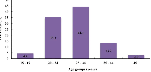
Figure 3.3 – Frequency of C. trachomatis infection according to age groups.
Figure 3.4. – Distribution (in %) of C. trachomatis infections according to patient’s number of sexual
partners.
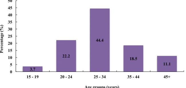
Figure 3.5 –Distribution (in %) of N. gonorrhoeae infected patients according to age groups.
Figure 3.6 – Distribution (in %) of N. gonorrhoeae infections according to patient’s number of sexual
partners.
Figure 3.7 – Distribution (in %) of M. genitalium infected patients according to age groups.
Figure 3.8 – Distribution (in %) of M. genitalium infections according to patient’s number of sexual
partners.
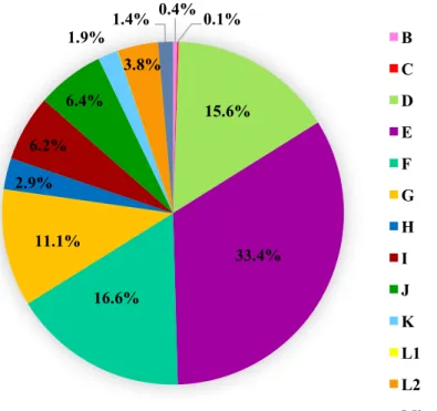
Figure 3.9 – Distribution (in %) of ompA-genotypes in INSA’s C. trachomatis positive sample
collection.
Figure 3.10 – Distribution (in %) of ompA-genotypes per anatomical site of infection. Figure 3.11 – Distribution (in %) of ompA-genotypes per gender.
Figure 3.12 – Distribution (in %) of ompA-genotypes per age. Figure 3.13 – Distribution (in %) of ompA-genotypes per year.
Figure 3.14 – Proportion of ompA prototype and variant strains among INSA’s C. trachomatis
collection.
Figure 3.15 – Frequency (in %) of variable sites per CD and VD of the ompA gene. Figure 3.16 – Distribution (in %) of C. trachomatis ompA-genotypes per anatomical site.
Figure 3.17 – Genetic heterogeneity in the homopolymeric tracts probably driving phase variation of
the virulence factor CT166 (C. trachomatis cytotoxin). Each bar displays the relative percentage of sequence reads with a particular base count for 87 C. trachomatis in vivo populations. Profiles in red color range represent poly (G) count potentially yielding protein truncation (OFF), while green color range represent a functional protein (ON). The color blue represents a profile typically observed in
Only base counts relying on a count coverage >10 were considered. Profiles were marked as ‘variable’
(V) in a given sample if the dominant ‘count’ represented less than 90% of all respective reads (Forward
+ Reverse) counted in that region.
Figure 3.18 – Intra-patient variability and profile of the homopolymeric tracts affecting CT166 in C.
trachomatis strains, according to clade (for more detailed information, see Annex 13 a)).
Figure 3.19 – Intra-patient variability and profile of the homopolymeric tracts affecting CT042 in C.
trachomatis strains, according to clade (for more detailed information, see Annex 13 b)).
Figure 3.20 – Intra-patient variability and profile of the homopolymeric tracts affecting CT694 in C.
trachomatis strains, according to clade (for more detailed information, see Annex 13 c)).
Figure 3.21 – Intra-patient variability and profile of the homopolymeric tracts affecting CT561 in C.
trachomatis strains, according to clade (for more detailed information, see Annex 13 d)).
Figure 3.22 – Intra-patient variability and profile of the homopolymeric tracts affecting CT605 in C.
trachomatis strains, according to clade (for more detailed information, see Annex 13 e)).
Figure 3.23 – Intra-patient variability and profile of the homopolymeric tracts affecting CT541 in C.
trachomatis strains, according to clade (for more detailed information, see Annex 13 f)).
Figure 3.24 – Intra-patient variability and profile of the homopolymeric tracts affecting CT172 in C.
trachomatis strains, according to clade (for more detailed information, see Annex 13 g)).
Figure 3.25 – Profile of homopolymeric tracts affecting CT172 in C. trachomatis strains clade T1,
according to genotype.
Figure 3.26 – Intra-patient variability and profile of the homopolymeric tracts affecting CT445 in C.
trachomatis strains, according to clade (for more detailed information, see Annex 13 h)).
Figure 3.27 – Intra-patient variability and profile of the homopolymeric tracts affecting CT871 in C.
trachomatis strains, according to clade (for more detailed information, see Annex 13 i)).
Figure 3.28 – Intra-patient variability and profile of the homopolymeric tracts affecting CT326 in C.
trachomatis strains, according to clade (for more detailed information, see Annex 13 j)).
Figure 3.29 – Intra-patient variability and profile of the homopolymeric tracts affecting CT823 in C.
trachomatis strains, according to clade (for more detailed information, see Annex 13 k)).
Figure 3.30 – Intra-patient variability and profile of the homopolymeric tracts affecting CT259 in C.
Table Index
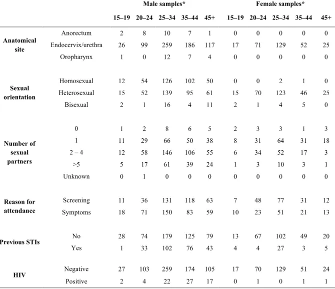
Table 2.1 – Distribution of biological samples per anatomical site, age, sexual orientation, number of
sexual partners, reason for attendance, previous STIs and HIV.
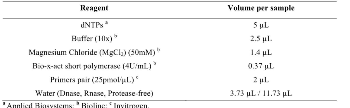
Table 2.2 – PCR mix for S-Dia MGTVTMkit (final volume 25µL).
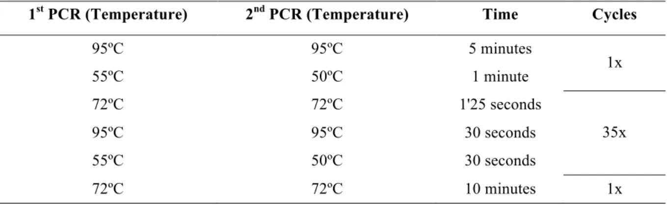
Table 2.3 – kPCR amplification profile for S-DiaMGTVTMkit.
Table 2.4 – Run validity testing.
Table 2.5 – ompA nested-PCR reaction mixture for a final volume of 25µL.
Table 2.6 – C. trachomatis ompA nested-PCR amplification profile. Table 2.7 – Sequencing mix.
Table 2.8 – Sequencing profile.
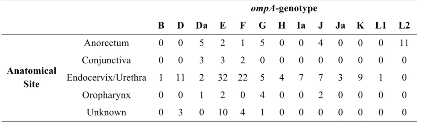
Table 2.9 – Distribution of biological samples per anatomical site and ompA-genotype. Table 2.10 – PCR mix for homopolymeric targets.
Table 2.11 – Amplification profile for homopolymeric regions.
Table 3.1 –Nucleotide sequence variation within ompA for each genotype in relation with the respective
prototype strain.
Table 3.2 – Genetic markers clade-specific for T1 and LGV strains.
List of abbreviations, acronyms and symbols
µL – Microliter; µm – Micrometer; AB – Abnormal Body;
AMR – Anti-microbial resistance; BV – Bacterial Vaginosis;
CAD – Centro de Aconselhamento e Deteção (portuguese); CD – Conserved Domain;
CDC – Centers for Disease Control and Prevention; COMC – Chlamydia Outer Membrane Complex; CT – Chlamydia trachomatis;
DFA – Direct immunofluorescence assay; DNA – Deoxyribonucleic acid;
DST – Doença sexualmente transmissível (portuguese); EB – Elementary Body;
ECDC – European Centre for Disease Prevention and Control; ELISA – Enzyme-linked immunosorbent assay;
EUA – Estados Unidos da América (portuguese);
EU/EEA – European Union and the European Economic Area;
FCUL –Faculty of Sciences, University of Lisbon (from the Portuguese Faculdade de Ciências da
Universidade de Lisboa);
HIV – Human Immunodeficiency Virus;
HSH – Homens que fazem sexo com homens (portuguese); HSV – Herpes Simplex Virus;
IFN-g - Interferon-g;
INSA – National Institute of Health (from the Portuguese Instituto Nacional de Saúde); IST – Infeção Sexualmente Transmissível (portuguese);
LGV – Lymphogranuloma venereum; LPS – Lipopolysaccharide;
MACPF – Membrane Attack Complex/Perforin; MOMP – Major Outer Membrane Protein; MG – Mycoplasma genitalium;
MSM – Men who have Sex with Men; NAAT – Nucleic acid amplification test;
NAH – Non-amplified nucleic acid hybridization assays; NG – Neisseria gonorrhoeae;
NGS – Next-Generation Sequencing; NGU – Non-Gonococcal Urethritis; ORF – Open Reading Frame; PBS - Phosphate-buffered saline; PCR – Polymerase Chain Reaction; PID – Pelvic inflammatory disease; PLD – Phospholipase D;
PMP – Polymorphic Membranes Proteins; POC – Point-of-care;
PROM – Premature Rupture of Membranes; PZ – Plasticity Zone;
RB – Reticulate Body;
RFLP – Restriction Fragment Length Polymorphism; RNA – Ribonucleic acid;
SARA – Sexually acquired reactive arthritis; STD – Sexually Transmitted Disease; STI – Sexually Transmitted Infection;
T3SS – Type III Secretion System;
TARP – Translocated Actin Recruiting Phosphoprotein; TFI – Tubal Factor Infertility;
TOC – Test-of-cure;
TV – Trichomonas vaginalis; UDF – User-defined workflow; UK – United Kingdom;
USA – United States of America;
USPSTF – United States Preventive Services Task-Force; VD – Variable Domain;
WHO – World Health Organization; XDR – Extensive drug resistance.
Introduction
1. Sexually Transmitted Infections
The term “Sexually Transmitted Infections” (STIs) refers to a wide variety of clinical syndromes
and infections, which are among the most common acute conditions in the world [1]. There are more than
30 bacterial, viral, and parasitic pathogens [2, 3] that can be transmitted from one person to another person
during sexual intercourse [2, 4].
The World Health Organization (WHO) estimates that more than 1 million STIs are acquired everyday worldwide, and that 357 million new episodes of four STIs, Chlamydia trachomatis (131 million), Neisseria gonorrhoeae (78 million), Treponema pallidum (6 million) and Trichomonas
vaginalis (142 million) occur every year [5].
About 20 million STIs cases are annually reported to the Centers for Disease Control and Prevention (CDC) of the United States, which are considered an epidemic of tremendous magnitude and a public
health concern [6].
Adolescents and young adults aged between 15 to 24 years old account for the majority of STIs cases because of a combination of biological, behavioral and cultural reasons; moreover, the earlier
people start their sexual life, the higher are the chances of acquiring a STI [6, 7]. Some populations are at
higher risk of getting a STI, i.e. men who have sex with men (MSM), intravenous drug users, people
who have multiple partners, and non-users of condoms [6, 8, 9, 10].
The most predominant STIs symptoms are the urogenital infections, which are often associated with
urethritis and epididymitis in men and cervicitis and pelvic inflammatory disease [11]. However, because
most of these infections are frequently asymptomatic, people do not realize they are infected [12, 13] and
accordingly, fail to seek treatment, which may lead to severe complications, long term sequelae, and a variety of diseases across multiple organ systems. If not treated, some STIs may cause ectopic pregnancy or tubal infertility. Some STIs can also be vertically transmitted, from an infected mother to her infant, inducing serious consequences for the offspring, such as neonatal death, premature delivery, blindness
or severe disability in infants [1, 3, 11], justifying the need for screening every pregnant woman. However,
STIs are frequently associated with social stigma, social stereotyping and vulnerability, and shame, which justify the trying of hiding a (potential) condition and the avoidance of screening (and treating) these infections.
Low incidence rates often reflect differences in healthcare systems, lack of accurate diagnostic tools, or insufficient diagnostic capacity, rather than a genuinely low or declining frequency of these
infections. These reasons also lead to the underestimation of the true prevalence of STIs [9]. Therefore,
increasing the scope and accessibility for screening STIs would provide an opportunity to identify and treat these infections, a public health priority. Laboratory and point-of-care (POC) tests are potentially powerful contributors to the management and control of STIs, facilitating prevention by precluding transmission. Although there are a wide variety of tests that can be applied to diagnose STIs, the CDC
recommends the use of nucleic acid amplifications tests, also known as NAATs [2, 14].
The present Master's thesis focuses on curable STIs main agents, C. trachomatis, N. gonorrhoeae,
T. vaginalis and Mycoplasma genitalium, which are responsible for urethritis, cervicitis, vaginitis and
2. Chlamydia trachomatis
Chlamydia trachomatis is an obligate intracellular gram-negative bacterium and it belongs to the
genus Chlamydia (phylum Chlamydiae, order Chlamydiales, family Chlamydiaceae) [15].
2.1. The life cycle
Throughout an evolutionary process undertaking several millions of years, Chlamydiae evolved developing mechanisms for interacting and colonizing eukaryotic host cells (usually epithelial) in which they go through a biphasic developmental cycle, unique in nature. During this life cycle with duration of 48 – 72h, the bacterium alternates between two highly specialized morphological forms: the
elementary bodies (EBs) and the reticulate bodies (RBs) [16].
The metabolically inert infectious EBs are small (~ 0,3µm), have a coccoid form and the external membrane contains extensive disulfide cross-links, within and between outer membrane proteins, providing it with a “sporelike” structure, i.e. a rigid cell wall that keeps the microorganism stable outside the cell, allowing its survival in the extracellular environment for a short period. On the other hand, RBs are larger (~ 1 µm), noninfectious, metabolically active, structurally flexible and osmotically fragile,
richer in RNA and containing diffuse and fibrillar DNA [14, 17].
The chlamydial infectious process may be divided into five major events (Figure 1.1). It begins with the attachment and entry of the EB into the host epithelial cell, triggering host actin reorganization,
membrane deformation, and internalization as endocytic vacuoles [17, 18, 19]. Subsequently, the EB
differentiates into RB inside the inclusion, and gene transcription starts [20]. The inclusion suffers some
modifications, and the RB replication occurs by polarized cell division processes [21]. Next steps are
inclusion expansion and RB transition into EB. Finally, EBs are released by host cell lysis or extrusion, a phenomenon that occurs 30 – 72h post-infection (depending on species and strain) and that allows
them to attach to neighboring epithelial cells and to initiate a new cycle [16, 20, 22, 23, 24].
However, the developmental cycle might be interrupted under stressful conditions caused by host immunological response, nutrient starvation or antibiotic treatment and, until these stress factors are
Figure 1.1 – Schematic representation of the development cycle of C. trachomatis [20].
2.2. Biological and genomic features
As any gram-negative bacteria, C. trachomatis is provided with a double membrane. The outer membrane is constituted by the chlamydia outer membrane complex (COMC) which includes the major outer membrane protein (MOMP), which accounts for 60% of the membrane dry weight, OmcA, OmcB, PorB, OprB, and OmcB, the type III secretion system (T3SS), the polymorphic membranes proteins
(Pmps), porins, lipids and polysaccharides (LPS) [26, 27, 28, 29].
COMC proteins are the first to interact with the host during infection and they maintain the integrity
of the chlamydial infectious particle [29]. In fact, MOMP is the major chlamydial antigen and a serologic
type classification could be established based on it; during chlamydial replication, MOMP may act as a
residues 64 to 83; VDII: residues 139 to 160; VDIII: residues 224 to 237; and VDIV: residues 288 to
317, flanked and interspaced by five conserved domains (CDs) [30, 31]. The antigenic determinants of
MOMP allowed to elicit 15 serotypes/serovars in C. trachomatis, A to L3, and serotype-specific
monoclonal antibodies were developed to recognize them [11, 32]. The ocular biovars (serovars A, B, Ba,
and C) origin trachoma, a conjunctival infection that can lead to blindness and constitutes the most
common cause of preventable blindness in the world, being endemic in many developing countries [2,
24]. The urogenital biovars (serovars D-K) are sexually transmitted, where E is the most common
followed D and F; serovars D, G, and J tend to predominate among MSM [10, 32, 33, 34, 35, 36, 37]. Serovars D
to K can cause ocular infections in neonates and adults, by secondarily inoculation of the eye with
infected genital secretions [32]. The LGV biovar (L1, L2, and L3) is also sexually transmitted, but the
infection often spreads to the regional draining lymph nodes and to the rectum causing lymphadenitis and proctitis [2, 17, 38].
With the advent of molecular biology techniques, the ompA gene that codes for MOMP evidenced
dissimilarities that allowed to differentiate 15 genotypes [39]. Genotyping became the methodology of
choice to distinguish the particular C. trachomatis types and variants involved in each infection, which is an important tool to understand C. trachomatis epidemiology and pathogenesis; in fact, correlating
ompA genotypes and clinical manifestations, could help to develop strategies for disease control, such
as vaccines. Genotyping would also be useful to differentiate between persistent and new infections, to identify outbreaks and transmission patterns among sexual networks, and would allow the surveillance
of specific types of interest [37, 40, 41]. ompA-genotyping subdivides C. trachomatis strains into three
distinct phylogenetic clades, the B-complex (B/Ba, D, E, L1, and L2), the C-complex (A, C, H, I, J, K,
and L3), and the intermediate complex (F and G) [11]. However, there is no correlation between these
phylogenetic clades and disease or tissue tropism[42].
The genome of obligate intracellular parasites is known to be relatively small when compared to free-living bacteria. While becoming metabolic parasites, they went through a lot of biological processes, in particular genome reduction, as they will benefit from living in the host cytoplasm, where
they can obtain high-energy phosphate compounds [17, 43].
C. trachomatis is also thought to have undergone this genome reduction pathway, likely from
nonpathogenic ancestors that became specialized to infect humans, while maintaining the ability to
infect different tissue types, and cause human diseases of major public health significance [44, 45, 46]. It
comprises a single circular chromosome with approximately 1 Mb and ∼7 kb plasmid, with a highly conserved genome, with the exception of the plasticity zone (PZ), near the replication terminus, that is
dissimilar among C. trachomatis strains [14, 47, 48, 49]. Genes in the PZ include the tryptophan synthase
(trp), the cytotoxin, the membrane attack complex/perforin (MACPF), and the phospholipase D (PLD),
and they enclose strain- and species-variable alleles [50].
The trp operon is a virulence factor, which in the presence of the host interferon IFN-g is able to
synthesize tryptophan [39]. However, only the genital genotypes exhibit an intact operon encoding a
functional tryptophan synthase, whereas ocular strains display mutations in the trpA or trpB genes that result in a nonfunctional synthase, providing a metabolic distinction between strains that is nearly
axiomatic with tissue-specific appetence [51, 52].
The chlamydial cytotoxin is a highly polymorphic gene that is thought to act on the rapid disassembly of the cytoskeleton actin filaments during the bacterial internalization process, but only the genital genotypes encode both the functional glycosiltransferase and the UDP-glucose binding domains
of the cytotoxin open reading frame (ORF), CT166, while ocular genotypes encode only the
UDP-glucose binding domain, and LGV genotypes lack the cytotoxin ORF [14, 53].
On the other hand, genes like MACPF and PLD are present in all genotypes, taking part in the processes of acquisition of metabolites from the host, which are essential for the survival and success of
the developmental cycle [50].
There are genes outside the PZ that are related to virulence and pathogenicity, such as the Pmp
genes, TARP (translocated actin recruiting phosphoprotein) genes, Inc genes and ompA [40]. The T3SS
effector TARP is spatially and temporally associated with the rapid polymerization of actin filaments
required at the site of EB invasion, with a high degree of variability [54]. C. trachomatis uses it to
translocate virulence effector proteins directly into the host cells cytoplasm, where they subvert cellular processes to promote cell invasion, inhibition of phagocytosis, establishment of the inclusion, acquisition of nutrients, modulation of intracellular trafficking, early inhibition, late induction of
apoptosis, and avoidance of innate immune responses, all contributing for chlamydial pathogenesis.[55,
56, 57].Inc genes code for proteins (Inc’s) that are inserted in the inclusion membrane, exposed to the host
cell cytosol, and are also effectors of the T3SS family.
Pmps are coded by a highly heterogenic gene family with a different number of pmp genes intra and
interspecies, which have been characterized as adhesins and autotransporters [40, 45, 58].
LPS are the main lipid present in the outer membrane, functioning as a permeability barrier that avoids bacterial cell-damaging agents such as detergents, proteases, bile salts, and hydrophobic
antimicrobials, being essential to Chlamydia sp. [59].
Due to the genus, species and type epitopes present in MOMP, inducing both humoral and cellular immune response in the host, its encoding gene, ompA, is provided with variability and antigenic
dissimilarities capable of constituting ompA as a good candidate for the development of a vaccine [41].
Phase variation mechanisms are known to play an important role in bacterial adaptation and pathogenesis, relying on the ability of bacteria to rapidly adapt in response to stimuli, usually associated with a reversible switching between ON and OFF state of specific proteins, such as proteins involved in
biosynthesis/expression of the bacterial capsule and LPS [60, 61]. These mechanisms normally occur due
to expansion/contraction of genomic homopolymeric tracts, described as repetitive regions of DNA
single base, leading to different phenotypes [62, 63, 64]. Although it is still not well-established if C.
trachomatis employs these mechanisms to promote adaptation and virulence, this subject warrants
further investigation as recent findings have revealed intra-strain heterogeneity targeting
homopolymeric tracts with potential to shape C. trachomatis virulence [60].
2.3 Epidemiology and pathology
C. trachomatis is the most common bacterial STI, infecting about 131 million people, worldwide,
each year[1]. The number of cases has grown during the last decade; in fact, the CDC reports an increase
of 2.8% in 2014 (1 441 789 cases reported in the United States), in relation to 2013 and the ECDC
reported an increase of 67% from 2004 to 2013 among consistently reporting countries [7, 65, 66]. However,
this rise may be a consequence of the widespread testing of asymptomatic individuals, and of the
C. trachomatis prevalence is usually higher in people younger than 24 years [4, 17, 69]. In Europe, in 2013, two thirds of the C. trachomatis cases were reported in young adults aged between 15 to 24 years, affecting both men and women, although more frequently reported in women, and were mostly (88%)
heterosexually transmitted [7, 43, 66, 67]. Multiple/high number of sexual partners, having a sexual partner
with a STI, inconsistent condom use, and/or have another STI, are some of the risk factors for getting a
C. trachomatis infection [4, 12].
Despite the estimations described above, C. trachomatis true incidence and prevalence are likely to be significantly higher; in fact, the asymptomatic nature – which allows the infection to go unnoticed until more severe symptoms develop – and the differences in testing methods, testing coverage, screening programs, and surveillance systems, many cases might be neither diagnosed nor reported. In fact, 83% of the data available for Europe are based in four countries – Denmark, Norway, Sweden and
the United Kingdom – the countries where C. trachomatis reporting seem most effective [66]. Thus, a
large burden of disease is expected and the health associated risks justify the CDC recommendation for annually screen all sexually active women under 25 years, as well as older women at high risk of
infection, to prevent sequelae, and to test and treat their sexual partners [4, 7].
According to the USPSTF (United States Preventive Services Task-Force) [70], there is insufficient
evidence to recommend routine C. trachomatis screening in sexually active young men, because of several factors (e.g., feasibility, efficacy, and cost-effectiveness); however, the screening of sexually active young men should be considered in clinical settings where a high prevalence of C. trachomatis is to be expected (e.g., adolescent clinics, correctional facilities, STD clinics, and MSM).
Although 70 – 95% of the C. trachomatis infected women are asymptomatic, symptoms might occur which include urethritis, dysuria, vaginal discharge, postcoital bleeding, cervicitis, mucopurulent cervical discharge, cervical friability, cervical edema, endocervical ulcers, mid-cycle spotting, poorly
differentiated abdominal pain or lower abdominal pain and proctitis [71]. If left untreated, C. trachomatis
infection can progress and damage the upper reproductive tract, leading to pelvic inflammatory disease (PID), tubal factor infertility (TFI), chronic pelvic pain, ectopic pregnancy, endometritis, salpingitis or
sexually acquired reactive arthritis (SARA) [4, 7, 10, 17, 43, 67, 71, 72, 73]. Repeated C. trachomatis infections
might increase the risk of PID and tubal damage [12]; however, there has been a decline in the
hospitalization rates for PID and ectopic pregnancy, since the introduction of C. trachomatis control
programs, in women [67, 74, 75].
In men, more than 50% of the C. trachomatis infections are asymptomatic; when present, symptoms might appear as penile tip irritation, watery and viscous excretions, urethral discharge, proctitis and epididymitis. C. trachomatis seems to neither exerts deleterious effects on spermatozoa nor impairs male
fertility, unlike to the described for women [76].
It is assumed that 20 – 54% of the uncomplicated urogenital infections will clear spontaneously in
1 year, in average [24, 77].
For both genders, the clinical signs and symptoms are not exclusive of the C. trachomatis infection as they can be caused by other STIs like T. vaginalis, N. gonorrhoeae or M. genitalium (common STIs
coinfections) and candidiasis and a laboratory diagnosis is required for confirming the case [2, 7, 35, 38, 67,
71, 78]. Moreover, everyone who receives a diagnosis of C. trachomatis should be tested for HIV and
Since 2004, in particular among MSM, outbreaks of lymphogranuloma venereum (LGV), an invasive infection, have been reported. LGV is caused by C. trachomatis L1, L2 and L3 strains, among
which the L2b variant was referred as the most frequent [79, 80, 81, 82]. The main site of infection is the
rectum and, although it might be asymptomatic, major symptoms include tenesmus, constipation, anorectal pain, mucopurulent discharge, bleeding per rectum, diarrhea, abdominal pain and proctocolitis
(often confused with inflammatory bowel disease) [68, 71, 83, 84]. If not treated early, it can progress to
chronic colorectal fistulas and strictures, lymphatic obstruction and, uncommonly, meningoencephalitis,
hepatitis and death [85, 86].
Rectal chlamydial infections in women might become more frequent, as data from the UK shows
that 15% - 17% of heterosexual couples reported anal sex practices [68]; however, no LGV outbreaks
have been reported. Moreover, no association could be established between patients reporting anal intercourse and rectal chlamydia infection, suggesting that the infection could be caused by dispersion
from the genital tract to the rectal site [24].
Pharyngeal chlamydial infections can also occur and its detection rates in MSM range from 0.5% to
2.3% [87, 88]. They are usually asymptomatic, but symptoms like mild sore throat can occur [89].
In women, untreated maternal chlamydial infections have been associated with premature rupture of membranes (PROM), preterm delivery, perinatal mortality and postpartum endometritis in addition
to neonatal morbidity [13]. Neonatal infections are acquired during birth, when the baby passes through
a C. trachomatis infected birth canal (risk range from 50% to 75%) [71, 90]. Infants may develop
conjunctivitis – occurring in the first 3 weeks of life, where C. trachomatis replicates extensively in epithelial conjunctival cells, causing considerable cell damage – and/or pneumonia, being the nasopharynx the most frequent site of perinatally acquired C. trachomatis infection, with approximately 70% of infected infants having positive cultures at that site, occurring within the first 3 months of life
[17, 38, 67, 71, 91]. Neonatal C. trachomatis infections may remain asymptomatic and persistence for as many
as 3 years has been demonstrated; this hypothesis may cause confusion with C. trachomatis acquisition
because of child sexual abuse [17]. An effective measure to prevent the transmission of mothers’ C.
trachomatis infection to the newborn would be screening and treating before delivery, which would
highly reduce the risk of future complications [12, 17, 91].
For urogenital C. trachomatis infections (even for pregnant), azithromycin, 1g single dose, is considered the first-choice treatment; single-dose therapies tend to maximize adherence to treatment. Alternative treatment is doxycycline, 100mg two times daily for seven days. For LGV, because of its
invasiveness, the recommended treatment is 100mg of doxycycline, orally, twice a day for 21 days [4, 67,
71]. To minimize the risk of transmission to sex partners, sexual intercourse abstention for 7 days after
single-dose therapy or until completion of the regimen and resolution of symptoms, is required [4, 85, 92].
Test of cure (3 – 4 weeks after completion of therapy to avoid the presence of nonviable organisms that can lead to false-positive results) are not recommended, except for pregnant, as antibiotic resistance is considered rare; the fact that C. trachomatis is an intracellular parasite living inside a cell vacuole largely
precludes exchanges of antimicrobial resistance genetic material [13]. Thus, most post-treatment
infections do not result from treatment failure, but rather from reinfection with an untreated sex partner
2.4 Diagnosis
C. trachomatis urogenital infection can be diagnosed in women by testing first-catch urine and
endocervix (or vagina) swab specimens. In men, C. trachomatis urethral infection can be diagnosed by
testing urethral swabs or, first option, first-catch urines [4, 38, 93]. However, anatomic sampling sites vary,
according to sexual practices (anorectal and oropharynx samples might be required) and clinical presentation (conjunctivitis, arthritis, etc).
Bacterial isolation, in tissue culture, standardized in the 1970s, is no longer used for C. trachomatis diagnosis because it depended on maintaining organisms viable. This process required strict transportation (4ºC, 24h maximum) and laboratory storage (-70ºC or less) conditions which allied to the time and associated cost of cell culture justified the abandon of this method for diagnosis routine purposes [2, 32].
In the early 1980s, new techniques were developed, such as antigen detection assays, direct immunofluorescence assays (DFAs), and solid phase enzyme-linked immunosorbent assays (ELISAs) for a faster and easier detection of chlamydial particles in urogenital exudates. However, these methods lacked sensitivity, even when compared to culture and, most of all, suboptimal specificity, which
justified not recommending them for C. trachomatis diagnosis [2].
The major advancement for C. trachomatis diagnosis was the employment of nucleic acid amplification methods. Initially there way non-amplified nucleic acid hybridization assay tests with a sensitivity similar to culture, but they were replaced by nucleic acids amplification tests (NAATs) that revolutionized C. trachomatis screening. NAATS employ enzymatic methods to exponentially amplify DNA (or RNA) targets into billions of copies and use sequence-specific probes with binding dyes for the detection of amplified DNA products, and become the methodology of choice for C. trachomatis diagnosis nowadays, combining specificity and sensitivity near to 100%. NAATS made possible to study a broader range of biological samples (including extragenital), less invasive and/or self-collected
(i.e. urine, vulvar exudates) [2, 4, 38].
When using NAATs C. trachomatis viability is no longer a requirement, transport conditions are less strict, accommodating some delay between collection and laboratory processing without significant
loss of sensitivity (nor specificity) [38]. Sample pooling is a possibility in resource-limited settings, and
most of the commercially available tests were designed for detecting both C. trachomatis and N.
gonorrhoeae simultaneously [2]. Additionally, NAATs are well suited to automation, which results in increased standardization and quality assurance of nucleic acid extraction, amplification and detection, as well as significantly increased throughput. Therefore, these tests became considered to have superior performance characteristics compared to any other test for the detection of C. trachomatis infections and, as such, they are the assay type recommended by the WHO and the CDC for both diagnosis and screening [2, 4].
3. Neisseria gonorrhoeae
The genus Neisseria contains two species primarily pathogenic to humans, N. gonorrhoeae and N.
meningitidis, and approximately 30 usually nonpathogenic species such as N. lactamica, N. sicca, N. cinerea, N. flavescens, N. subflava, and N. mucosa. These organisms mainly inhabit the upper
respiratory tract as commensals, but they can be found, infrequently, in the lower urogenital tract [2].
3.1. Epidemiology and human diseases
Gonorrhea is one of the earliest known human diseases with biblical references dating back to the
Old Testament [94]. The causative agent of this infection, N. gonorrhoeae, is a gram-negative
diplococcus, aerobic, non-flagellated, non-sporulating and it is a fastidious organism that requires
complex nutritionally enriched culture medium for in vitro growth [2, 94, 95]. It has a marked tropism for
human mucosal surfaces, affecting only humans, being transmitted almost exclusively through sexual contact [2].
N. gonorrhoeae is the second most common bacterial STI, after chlamydial infection [4, 7, 13, 96]. The ECDC refers an increase of 31% of reported cases between the years of 2008 and 2011, among the 28 EU/EEA Member States; in the year 2013, 52 995 cases were reported, with an overall rate of 17 cases
per 100 000 population [66]. Between 2013 and 2014, the CDC describes an increase of 5.1%, with 350
062 cases of gonorrhea reported in the US only, and a rate of 110.7 cases per 100 000 population [7]. In
most EU/EEA Member States, in 2013, about half of all gonorrhea cases (43%) were reported in MSM; in fact, since 2008, the overall rate has raised by 79%, mostly because of the increasing number of cases in men, especially MSM, a population recommended for screening by the ECDC at all anatomical exposure sites [66, 97].
In Portugal, the incidence of N. gonorrhoeae infection was estimated from 0.27 to 0.7 cases per 100 000 population but, because it is estimated that many cases remain undiagnosed or are not reported, the
true burden of disease is likely higher [95].
N. gonorrhoeae prevalence is higher among adolescents and young adults, with ages from 15 to 24
years, accounting for 39% of all gonorrhea cases, in particular among those with a new sex partner, multiple sex partners, inconsistent condom use, having a sex partner with concurrent partners, or having
a sex partner with a STI [4, 7, 66]. In contrast to chlamydial infections, gonorrhea is reported three times
more often in men than in women, with 28.9 notifications per 100 000 in men in contrast to 9.7 per 100
000 in women; this might be explained by the increasing number of cases among MSM [7, 66].
After exposure, typical incubation period for men varies between two to five days [95, 98]. Gonococcal
infections tend to cause a stronger inflammatory response than C. trachomatis but may remain
asymptomatic until the development of complications [13]. The most common clinical presentations
include acute urethritis, urethral discharge and dysuria; more rare complications may include penile edema, penile lymphangitis, periurethral abscess, acute and chronic prostatitis, seminal vesiculitis,
urethral strictures and fistulae. Up to 60 – 80% of male patients have minimal or no symptoms at all [99,
100, 101].
For women, typical incubation period is a bit longer, varying from five to ten days following exposure, and most of them (>85%) are asymptomatic. When symptoms occur, the most common
manifestations are cervicitis (with vaginal discharge), cervical bleeding, pruritis, and dysuria [98]. If left
peritoneal and pelvic adhesions and, rarely, abdominal peritonitis or perihepatitis; infertility develops in
approximately 15% of women with gonorrhea [94, 99, 100, 101, 102]. During pregnancy, gonorrhea may cause
complications, such as chorioamnionitis, premature rupture of membranes, preterm birth and
spontaneous abortions [103, 104].
Infections of the rectum and the pharynx are predominantly found in MSM, but, according to the
sexual behaviors adopted, it can be found in both genders [2, 94]. Rectal infections are largely
asymptomatic but, occasionally, patients may complain of rectal and anal pain or discharge. Pharyngeal
infections are mainly asymptomatic too, but mild sore throat and pharyngitis may occur [2]. Ocular
infections caused by N. gonorrhoeae are most commonly detected in neonates (ophthalmia neonatorum) and are acquired upon the passage through the birth canal of infected mothers. In adults, conjunctivitis by N. gonorrhoeae is due to auto-inoculation, and if it is left untreated, can lead to scarring and blindness
[94, 98].
Repeated gonococcal infections increase the duration of erosion, the presence of local immune target cells, and infectivity; by altering host immune defenses, there is an increased risk of both acquisition
and transmission of HIV [105, 106]. In fact, the risk of acquiring an HIV infection increases 8-fold for MSM
who had two prior rectal C. trachomatis or N. gonorrhoeae infections [105].
When not treated, gonorrhea may evolve to complications such as meningitis and endocarditis. As
described for C. trachomatis, every N. gonorrhoeae patient should also be tested for other STIs [4].
N. gonorrhoeae diagnosis is established by direct detection (microscopy of stained smears), culture
or molecular biology techniques. In women, primary collection site is the endocervical canal or the vagina; in heterosexual men, specimens should be collected from the urethra. In the case of oral and/or
anal sex, the rectum and the oropharynx should be sampled for testing [107].
Microscope observation of smears allow to identify gonococci as extracellular and, very often, intracellular diplococci in polymorphonuclear leukocytes. This is a cheap method, providing rapid results in symptomatic men with urethral discharge, with high sensitivity (95%) and specificity (97%)
[2, 96]. In women, the same methodology can only detect 40 – 60% of the cases, which may reflect a lower
number of gonococci in women cervical infections. Beyond the required experience of the laboratory technician, this methodology has a low sensitivity and cannot be used in the case of asymptomatic men, nor for pharyngeal or rectal gonorrhea, where commensal Neisseria species exist and could provide false positive results [2, 94].
Culture, still considered the “gold-standard”, offers high sensitivity, up to 100% specificity and enables antimicrobial resistance (AMR) testing. Nevertheless, this method is relatively slow (relies on the growth kinetics of the organism), and requires strict conditions of specimen’s collection,
transportation and storage [2, 94].
The first molecular tests that were developed for N. gonorrhoeae were non-amplified nucleic acid hybridization (NAH) assays that relied simply on the binding of specific complementary nucleic acid probes and subsequent signal amplification to detect binding. Thus, throughout the last two decades, the clinical laboratory industry developed NAATs to detect N. gonorrhoeae with higher sensitivity than any prior diagnostic method, for which endocervical and vaginal swabs for women, urethral swabs for men,
and urine specimens for both genders can be used, as well as pharyngeal and rectal specimens [2, 96].
NAATs are less demanding regarding specimen collection, transport and storage, detecting also nonviable gonococci, where noninvasive, self-collected samples can be effectively used. NAATs are