Sexually Transmitted Bacteria in Semen of Male Partners
of Infertile Couples in Tunisia: The Effect on Semen
Parameters and Spermatozoa Apoptosis Markers
Hanen Sellami1, Abir Znazen1*, Afifa Sellami2, Hela Mnif3, Nour Louati3, Soumaya Ben Zarrouk2, Leila Keskes2, Tarek Rebai2, Radhouane Gdoura4, Adnene Hammami1
1Department of Microbiology and research laboratory ‘‘Microorganismes et Pathologies Humaines’’, Habib Bourguiba University Hospital of Sfax, Sfax, Tunisia,
2Histology Embryology Research Unit, Faculty of Medicine of Sfax, University of Sfax, Sfax, Tunisia,3Sfax Regional Center of Blood Transfusion, Sfax, Tunisia,4Unit Research of Toxicology-Microbiology Environmental and Health, Sciences Faculty of Sfax, University of Sfax, Sfax, Tunisia
Abstract
This study was undertaken to determine the prevalence ofChlamydia trachomatis,Mycoplasmas, andUreaplasmasin semen samples of the male partners of infertile couples and to investigate whetherChlamydia trachomatiscould initiate apoptosis in human spermatozoa. A total of 85 males partners of infertile couples undergoing routine semen analysis according to World Health Organization guidelines were included. Specimens were examined for the presence ofChlamydia trachomatis,
Neisseria gonorrhoeae,Mycoplasma hominis,Mycoplasma genitalium,Ureaplasma urealyticum andUreaplasma parvumby Real time PCR (qPCR). Semen specimens were analysed for the appearance of apoptotic markers (sperm DNA fragmentation, activated caspase 3 levels, mitochondrial membrane potential (DYm)) using flow cytometry.C. trachomatis,N. gonorrhoeae,
U. urealyticum,M genitaliumwere detected in semen samples of 13 (15.2%), 5 (5.8%), 5 (5.8%) and 3 (3.5%) male partners of infertile couples, respectively.M. hominisandU. parvumwere detected in semen sample of only one patient (1.1%). The semen of infertile men positive forC. trachomatisshowed lower mean of semen count and lower rapid progressive motility (category [a]) of spermatozoa compared to uninfected men with statistically significances (p= 0.02 and p= 0.04, respectively). Flow cytometry analyses demonstrated a significant increase of the mean rate of semen with lowDYm and caspase 3 activation of infertile men positive forC. trachomatis compared to uninfected men (p= 0.006 andp= 0.001, respectively). DNA fragmentation was also increased in sperm of infertile men positive forC. trachomatis compared to uninfected men but without statistical significances (p= 0.62). Chlamydialinfection was associated to loss ofDYm and caspase 3activation. Thus,C.trachomatis infection could be incriminated in apoptosis induction of spermatozoa. These effects may explain the negative direct impact ofC. trachomatisinfection on sperm fertilizing ability.
Citation:Sellami H, Znazen A, Sellami A, Mnif H, Louati N, et al. (2014) Molecular Detection ofChlamydia trachomatisand Other Sexually Transmitted Bacteria in Semen of Male Partners of Infertile Couples in Tunisia: The Effect on Semen Parameters and Spermatozoa Apoptosis Markers. PLoS ONE 9(7): e98903. doi:10.1371/ journal.pone.0098903
Editor:Anil Kumar, University of Missouri-Kansas City, United States of America
ReceivedMarch 7, 2014;AcceptedMay 8, 2014;PublishedJuly 14, 2014
Copyright:ß2014 Sellami et al. This is an open-access article distributed under the terms of the Creative Commons Attribution License, which permits unrestricted use, distribution, and reproduction in any medium, provided the original author and source are credited.
Funding:The work was financed by the research laboratory ‘‘MPH’’ Habib Bourguiba University Hospital of Sfax Tunisia. The funders had no role in study design, data collection and analysis, decision to publish, or preparation of the manuscript.
Competing Interests:No competing interests to declare.
* Email: [email protected]
Introduction
Sexually transmitted infections are of major concern to researchers and clinicians in the field of reproductive medicine. It is estimated that 15% of male infertility is related to genital tract infection [1]. Men can harbor subclinical infections in the genital tract over extended periods of time and several sexually transmitted infection pathogens, such asC. trachomatishave been detected in semen from asymptomatic men [2]. According to a World Health Organization (WHO) [3] report, C. trachomatis is responsible for the most common sexually transmitted bacterial infection worldwide, affecting more than 90 million people and has been known for some time to have a significant effect on human reproduction [4]. The role of C. trachomatis infections in male infertility is controversial [5–6]. A number of studies have specifically looked at the relationship betweenChlamydialinfection
and semen quality. While some authors have shown that C. trachomatis infection is associated with poor semen quality [7–8], others have claimed that it does not [9–10]. Some reports indicated thatC. trachomatisinfection is associated with a decrease in sperm concentration and motility and also with altered semen pH and reduced volume of the ejaculate [11,12,13–14]. Conversely, other studies have revealed no association between
C. trachomatisinfection of the male genital tract and altered sperm quality [9,15,16,17,18,19,20–21]. In summary, the available evidence is conflicting and still makes it impossible to establish a clear relationship between C. trachomatis infection and semen quality.
the cell populations in tissues upon physiological and pathological conditions [22]. Apoptosis markers characterized in somatic cells were noted in human spermatozoa in several studies. These include, principally, plasma membrane externalization of phos-phatidylserine (PS) and DNA fragmentation. Such markers are observed with higher frequency in ejaculates of infertile men compared with fertile controls [23–24]. In addition, key compo-nents of the somatic cell apoptosis pathways, such as presence and activation of caspases, have been described in purified populations of ejaculated sperm from the high and low-motility fractions [24– 25]. Moreover, mitochondria play a major role in the control of apoptosis [26]. Marchettiet al(2002) demonstrated that analysis of DYm is a sensitive test to determine sperm quality when compared with the analysis of the basic sperm parameters, generation of reactive oxygen species, and presence of DNA fragmentation [27]. Severalin vitroandin vivostudies tried to establish a relationship between apoptosis markers in spermatozoa and Chlamydial
infection. In vitro, some authors have demonstrated that C. trachomatis is able to interact with sperm cells, affecting their function and inducing apoptosis [28,29–30]. Apoptosis of human sperm can be induced byin vitroincubation of human sperm cells with Chlamydial LPS, which has a 550 fold greater spermicidal activity thanEscherichia coliLPS [31–32]. In addition,C. trachomatis
serovar E can attach to human spermatozoa and influence its function leading to premature capacitation [33]. It has been shown thatChlamydialLPS interact with CD14 on the sperm surface, thus leading to increased production of reactive oxygen species and resulting in caspase-mediated apoptosis [29]. Despite all thisin vitro
studies, a clear association between C. trachomatis and sperm damage has not yet been corroborated byin vivostudies. Gallegoset al(2008) reported that patients withC. trachomatisandMycoplasmas
genitourinary infections have increased sperm DNA fragmentation in comparison with fertile controls [34]. Lastly, we showed that inoculation of fertile male Swiss mice in the meatus urethra withC. trachomatiscould lead to alteration of semen parameters, induction of apoptosis in spermatozoa, and decrease of the reproductive performance of male mice [35]. Taken together, these data support a role of C. trachomatis in sperm apoptosis induction. However, most studies indicate that apoptosis-inducing mecha-nism is unknown.
In the present Study, we aimed to determine the prevalence ofC. trachomatis, Mycoplasmas, and Ureaplasmas in semen samples of the male partners of infertile couples and mainly to investigate whether
C. trachomatiscould initiate apoptosis in human spermatozoa.
Materials and Methods
Subjects
A total of 85 infertile men attending obstetrics and gynecology clinics in Sfax (South of Tunisia) for diagnostic semen analysis were selected to the study. All men were undergoing semen analysis as part of a work-up for infertility investigations after failing to conceive with their partner after one year of unprotected intercourse. The mean duration of infertility was 4 years (range 1– 15). The mean age of patients was 36.7 years (range 23–57). This study was approved by our institutional review board ‘‘Habib Bourguiba University hospital ethics committee’’ with the given number 8–12. All subjects signed a written informed consent. Consent form was also approved by our ethic committee
Sperm seminological variables
Prior to semen analysis, the men were asked to abstain from sexual intercourse or masturbation for 3–5 days before attending the clinic. All samples for analysis were produced on site and
collected into standard containers that had previously been shown not to have any cytotoxic effects on human spermatozoa according to the methods outlined by WHO. Immediately after semen production, samples were placed in an incubator and liquefied at 37uC for up to 30 minutes before analysis. Semen analysis was performed according to the WHO criteria [36] to determine the following variables: sperm concentration, vitality, total progressive motility (category [a+b]), rapid progressive motility (category [a]) Peroxidase staining, a practical and reliable method recommended by WHO [36] for determining leukocytes in the semen, was employed to count and differentiate leukocytes (white blood cells) from immature germ cells. Leukocytospermia was indicated by a concentration of leukocytes$106/ml.
Spermiocultures analysis
Samples were seeded quantitatively using a calibrated loop on agar plates: blood agar, chocolate agar with isovitalex (1%) incubated in 5% CO2 at 37uC for 48 hours. Microorganisms were identified by Gram staining and Bio-Me´rieux Api systems (Bio-Me´rieux, Marcy l’Etoile, France).
Spermiocultures were considered positive when the number of colonies was$104CFU ml21in case of Gram positive cocci and
$105CFU ml21in case of Gram negative rods.
Bacterial quantification in semen specimens by qPCR
For each male patient, 200ml of semen specimens were used for bacterial quantification by Real time PCR.
Extraction of DNA by Cetyltrimethylammonium bromide (CTAB)-phenol-chloroform/isoamyl alcohol method. The precipitates from each 200ml of semen specimens were harvested by centrifugation at 14,000 g for 20 minutes. The precipitates were treated with 5ml of proteinase K (20 mg/ml) at 55uC for 2 h in 600ml of digestion buffer (30ml of 10% sodium dodecyl sulphate and 570ml of TE buffer [10 mM Tris-HC1 (pH: 8), 1 mM EDTA]).
After homogenisation, the samples were incubated in a solution of CTAB-NaCl (100ml of 5 M NaCl and 80ml of 10% CTAB) for 10 minutes at 65uC, and then mixed with 750ml of chloroform-isoamyl alcohol (24:1 [vol/vol]) and centrifuged for 15 minutes at 14,000 g in an Eppendorf centrifuge. The aqueous phase was separated, mixed with 750ml of phenol chloroform/isoamyl alcohol (25:24:1 [vol/vol/vol]) and centrifuged for 15 minutes at 14,000 g in an Eppendorf centrifuge. The obtained aqueous phase was mixed with an equal volume of isopropanol.
The samples were left at280uC for 1 h and then centrifuged for 15 minutes at 14,000 g. The DNA pellet was washed up once with 70% ethanol, air dried, and dissolved in a final volume of 100ml of TE buffer.
Primers and probes for Qpcr. Initially, the extracted DNA was tested for humanb-globin gene to check that there were no PCR inhibitors in the samples. Primers b-GPCO (59 -ACA-CAACTGTGTTCACTAGC- 39) andb-GPCPO (59 -GAAACC-CAAGAGTCTTCTCT- 39) were used to amplify a 209-bp fragment of the humanb-globin gene [37]. Samples found to be negative by PCR forb-globin were retested after dilution 10-fold in distilled water. Samples shown to beb-globin positive were then examined for bacterial quantification by Real time PCR.
Por A pseudogene for N. gonorrhoeae and 146 bp of the Urease gene of U. parvum and U. urealyticum.
Real-time PCR included initial denaturation at 95uC for 2 min, followed by 40 cycles of 95uC for 30 s and annealing temperature according to microorganisms for 30 s (C. trachomatis 60uC, M. genitalium, M. hominis and N. gonorrhoeae 55uC, U. parvum and U. urealyticum50uC).
In all experiments, each PCR run included a negative extraction control (sterile water) and a negative PCR control, containing 5ml Diethylpyrocarbonate (DEPC) treated H2O instead of DNA extract, to detect any possible contaminating DNA. Samples and controls were run in duplicate.
Positive recombinant plasmid control. To facilitate bac-terial quantification, a plasmid containing the target gene for all bacteria was constructed.
DNA was extracted from C. trachomatis, N. gonorrhoeae, M. genitalium,M. hominis,U. parvumandU. urealyticumreferences strains and the target sequence for all genes selected for Real Time PCR were amplified with the same primers in (Table 1).
The final 25ml reaction mixture contained 1X PCR buffer (Promega, Lyon, France), 0.2 mM each primer, 0.2 mM each
dNTP, 2.5 mM MgCl2, 1.25 U Go Taq DNA polymerase
(Promega), and 5mL of DNA extract. PCR was performed in Gene-Amp PCR System 9700 (Applied Biosystems, Foster City, California) according to the following procedure: 4 min at 95uC, 35 cycles at 95uC for 30 s, 55uC for 1 min, 72uC for 20 min. PCR products were then purified with QIAquick Gel Extraction Kit (Qiagen) and cloned into a vector using a cloning kit (pGEM-T vector; Promega, Madison, WI, USA), in accordance with the manufacturer’s instructions. Isolation of recombinant plasmid DNA was performed using the QIAprepSpin Miniprep kit (Qiagen), and the presence of the correct insert was confirmed by sequencing using the commercial BigDye Terminator v3.1 kit
(Applied Biosystems) on a 3730XL sequencer (Applied Biosys-tems). The obtained sequences were processed by the ABI 3100 Genetic Analyzer and were compared with the sequences available in GenBank by using the BLAST server from the NCBI website (http://www.ncbi.nlm.nih.gov/BLAST). Plasmids were then line-arized and quantified with a NanoDrop ND-1000 Spectropho-tometer. Copy numbers of the cloned gene was calculated using the following equation reported by [38] to generate standards ranging from 1 to 106molecules and stored at220uC.
Evaluation of Viability of sperm using 7-amino-actinomycin-D Dye
The percentage of dead sperms cells (cells with 7-AAD positive) and viable sperm cells (cells with 7-AAD negative) were assessed using 7-AAD Dye. 7-AAD penetrates only dead cells. From each sperm sample, 1 ml of a sperm solution in PBS containing 26106cells/ml was stained with 10ml of 7-amino-actinomycin-D (7-AAD) (Immunotech, a Beckman Coulter Company, Marseille– France). The samples were incubated in the dark at room temperature for 20 minutes before flow cytometric analysis. After the incubation period, 1 ml PBS was added and the sample was analyzed by flow cytometry.
Evaluation of Mitochondrial Membrane Potential (DYm)
JC-1 possesses the unique ability to differentially label mitochondria with low and high DYm. In mitochondria with highDYm, JC-1 forms multimeric aggregates that emit in the high orange wavelength of 590 nm when excited at 488 nm. In mitochondria with low DYm, JC-1 forms monomers; these monomers emit in the green wavelength (525–530 nm) when excited at 488 nm.
The DYm was analyzed using MitoProbe JC-1 Assay kit (Molecular Probes, Eugene, OR). For staining, 2mM stock
Table 1.Primers and probes used for detection and quantification of C. trachnomatis, N. gonorrhoeae. U. urealyticum, M. genitalium, U. parvum and M. hominis by qPCR.
Bacteria Primers and probes Oligonucleotide sequence (59R39) Target gene Product size (bp) Ref
C. trachnomatis Forward AACCAAGGTCGATGTGATAG Cryptic plasmid 149 [73]
Reverse TCAGATAATTGGCGATTCTT
Probe ROX-CGAACTCATCGGCGATAAGG-BHQ2
N. gonorrhoeae Forward CCGGAACTGGTTTCATCTGATT PorA 101 [74]
Reverse GTTTCAGCGGCAGCATTCA
Probe FAM-CGTGAAAGTAGCAGGCGTATAGGCGGACTT-BHQ1
M. genitalium Forward GAGAAATACCTTGATGGTCAGCAA MgPa 80 [75]
Reverse GTTAATATCATATAAAGCTCTACCGTTGTTATC
Probe HEX-ACTTTGCAATCAGAAGGT-BHQ1
M. hominis Forward TTTGGTCAAGTCCTGCAACGA 16S
rRNA-encodinggene
101 [76]
Reverse CCCCACCTTCCTCCCAGTTA
Probe ROX-TACTAACATTAAGTTGAGGACTCTA-BHQ1
U. urealyticum Forward CATTGATGTTGCACAAGGAG Urease (UreD Subunit) 146 [77]
Reverse CGTGATTTTAATGTATCGGCTTTC
Probe FAMTTGACCACCCTTACGAGBHQ1
U. parvum Forward CATTGATGTTGCACAAGGAG Urease (UreD Subunit) 147 [77]
Reverse CGTGATTTTAATGTATCGGCTTTC
Probe HexTTGTCCGCCTTTACGAGBHQ1
solution of JC-1 in dimethylsulfoxide (DMSO) was prepared. From each sperm sample, 1 ml of a sperm solution in PBS containing 26106cells/ml was stained with 10ml of JC-1 stock solution. The samples were incubated at 37uC in the dark for 20 minutes before flow cytometric analysis. In this way, 2 sperm subpopulations were identified:
1) Represented spermatozoa with high DYm (orange fluores-cence).
2) Represented spermatozoa with low DYm (green fluores-cence).
As suggested by the protocol, in order to confirm the JC-1 sensitivity to changes in membrane potential, carbonylcyanide 3-chlorophenylhydrazone (CCCP = 50mM final concentration) was used as membrane potential disruptor (negative control).
Flow cytometric detection of activated caspase 3
Activated Caspase 3 levels were detected in spermatozoa using fluorescein- labeled inhibitor of caspases (FLICA), which is cell permeable, non cytotoxic, and binds covalently to active Caspase 3. The inhibitor was used with the appropriate controls according to the kit instructions provided by the manufacturer (Carboxy-fluorescein FLICA Apoptosis Detection Kit, AbCys, France). Briefly, 3.106sperm were resuspended in 300ml PBS. A 150-fold stock solution of the inhibitor was prepared by dissolving the lyophilized caspase-inhibitor in 50ml dimethyl sulfoxide (DMSO) and was further diluted 1:5 in PBS to yield a 30-fold working solution (per aliquot: 2ml of the stock solution plus 8ml PBS). All test aliquots and controls (with 300ml PBS) were incubated at 37uC in the dark for 1 h with 10ml of the working solution. Sperm samples were then washed resuspended in 400ml of Wash Buffer and kept in ice until flow cytometry analysis.
A negative control (sample with 300ml PBS) and a positive control (sample treated with 10mM H2O2 for 1 hour at 37uC)
were used in all experiments.
TUNEL assay
For the evaluation of DNA fragmentation, a commercial kit (In situ Cell Death Detection Kit, Fluorescein, Takara, Japon) based on an enzymatic reaction of labelling free 39-OH termini was used. In brief, 3.106cells were washed with phosphate- buffered saline (1xPBS, pH 7.4) then fixed with 200ml of 4% paraformaldehyde for 1 h at room temperature in the dark. After wards, sperm cells were washed with 1xPBS and permeabilised using 0.1% Triton X-100 in 0.1% sodium citrate for 15 min on ice. After washing with PBS, sperm DNA was labelled by incubating spermatozoa with 50ml of the TUNEL reaction mixture (Tdt enzyme and FITC-labelled nucleotides) in a humidified atmosphere for 60 min at 37uC in the dark, with mixing each 15 min. Washed and labelled sperm cells were then resuspended in 1xPBS for flow cytometry analysis. A negative control (sample without the addition of Tdt enzyme) and a positive control (sample treated with DNase I (3 U/ ml, Invitrogen) for 10 min at room temperature to generate DNA strand breaks) were also assessed by TUNEL assay.
Flow Cytometry and data analyses
Flow cytometric analysis was carried out using an EPICS XL flow cytometer (Beckman Coulter) equipped with a 15mW argon-ion laser for excitatargon-ion at 488 nm. At least 10,000 events per sample were analysed. Light-scattering and fluorescence data were obtained at a flexed gain setting in logarithmic mode. Debris was excluded by establishing a region around the population of interest on the basis of light scatter characteristics (forward-angle light
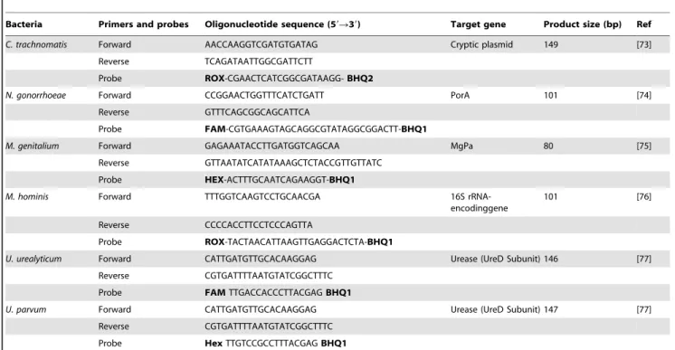
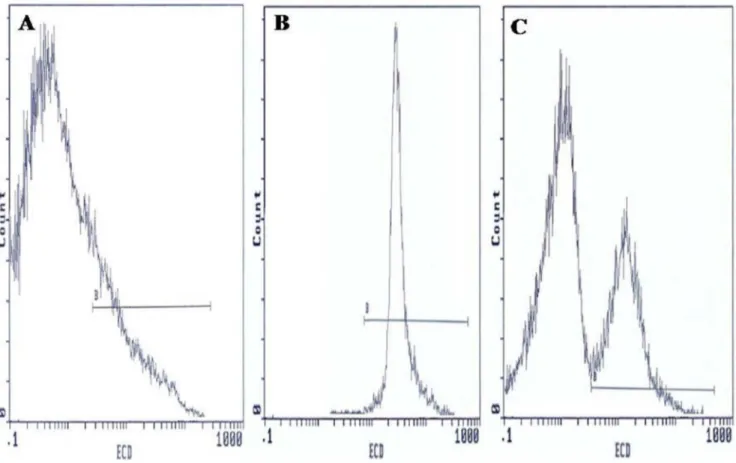
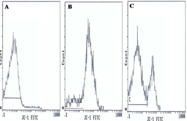
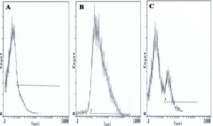
scatter (FSC) vs. side-angle light scatter (SSC). The percentage of labelled sperm was characterized by identifying a region that included .90% of events in the frequency histogram of the positive controls both in the assessments of Viability, DYm, Caspase 3 activation and DNA fragmentation. Data were expressed as percentage of stained cells from histograms using System II software. Typical examples of histograms obtained by flow cytometry for the detection of fluorescence are shown in Figure 1 (sperm viability), Figure 2 (DYm), Figure 3 (Caspase 3 activation) and Figure 4 (TUNEL assay).
Statistical analysis
The SPSS 18.0 software (SPSS Inc, Chicago, Ill) was used for statistical analysis. Testx2was used to compare frequencies. Non-parametric test (Mann-Whitney) from SPSS software was used to compare distribution sperm parameters and flow cytometry data of infected and uninfected men. Correlation between semen parameters means, DYm, DNA fragmentation and caspase 3 activation andC. trachomatisinfection was assessed using T-test. All tests were considered statistically significant when p,0.05.
Results
Spermiocultures analysis
Spermioculture analysis was positive in 6 cases (7%). Group B
Streptococcus(GBS) was found in 3 samples (3.5%),Enterococcus sppin 1 sample (1.1%), Staphylococcus aureus in 1 sample (1.1%) and
Corynebacterium sppin 1 sample (1.1%).
Frequency of urogenital bacteria in semen samples using qPCR
Among 85 semen samples, 13 (15.2%) were positive for C. trachomatis and 5 (5.8%) for N. gonorrhoeae. U. urealyticum, M. genitalium, U. parvum and M. hominis were detected in 5 patients (5.8%), 3 patients (3.5%), 1 patient (1.1%) and 1 patient (1.1%) respectively. The distribution of detected species in patients is shown in table 2.
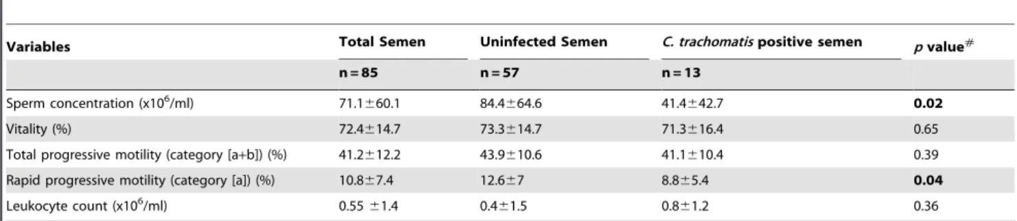
C. trachomatisinfection and semen quality
The mean values (6SD) for semen parameters of the 85 included patients are shown in Table 3. The sperm vitality and total motility of spermatozoa in the male partners of infertile couples withC. trachomatisDNA in semen specimens were lower but not significantly that those of uninfected male partners (71.3% vs 73.3%,p= 0.65 and 41.1% vs 43.9 %,p= 0.39, respectively) (Table 3). The sperm concentration and rapid progressive motility (category a) of spermatozoa inC. trachomatisDNA positive semen were significantly lower than those of uninfected semen (41.46106/ml vs 84.46106/ml, p= 0.02 and 8.8% vs 12.6%,
p= 0.04, respectively) (Table 3). The leukocyte count in the male partners of infertile couples with C. trachomatis DNA in semen specimens was higher but not significantly than those uninfected semen (0.86106/ml vs 0.46106/ml,p= 0.36) (Table 3).
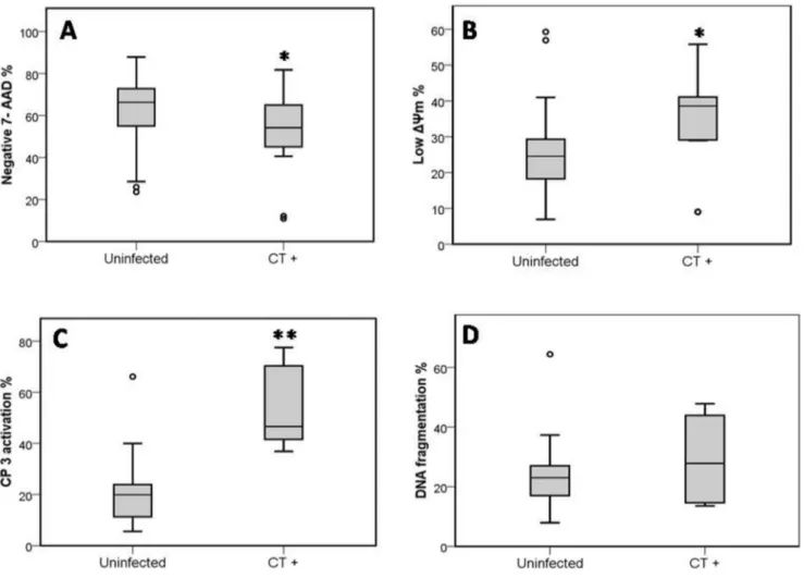
Sperm viability using 7-amino-actinomycin-D Dye
51.3621.13% in patients positive forC.trachomatis qPCR with a statistically significant difference (p= 0.014) (Table 4).
Mitochondrial Membrane Potential (DYm)
Analysis of the state of mitochondrial respiration in human spermatozoa was assessed using JC-1 to determine the DYm as shown in Figure 2. Flow cytometry results are expressed as percentage of sperm cells with low DYm (green fluorescence). Figure 2 presents frequency distribution histograms of negative control (Fig. 2A), positive control (Fig. 2B), and one semen of male partners of infertile couples positive for C. trachomatis qPCR (Fig. 2C). The mean percentage of spermatozoa with lowDYm (6SD) was higher in male partners of infertile couples positive for
C. trachomatisqPCR than those of uninfected patients (33.7613.3% vs. 24.569.7%) and the difference was statistically significant (p= 0.006) (Table 4).
Caspase 3 activation
The results of flow cytometry are expressed as percentage of activated caspase 3 sperm cells. Figure 3 presents frequency distribution histograms of negative control (Fig. 3A), positive control (Fig. 3B), and one semen of male partners of infertile couples positive for C. trachomatis qPCR (Fig. 3C). Mean percentage of spermatozoa with activated caspase 3 (6SD) was higher in male partners of infertile couples positive for C. trachomatisqPCR than those of uninfected patients (54.5618.1% vs. 20.8614%) and the difference was statistically significant (p=,0.001) (Table 4).
DNA fragmentation
TUNEL coupled flow cytometry results are expressed as percentage of DNA fragmented sperm cells. Figure 4 presents frequency distribution histograms of negative control (Fig. 4A), positive control (Fig. 4B), and one semen of male partners of infertile couples positive forC. trachomatisqPCR (Fig. 4C). Mean percentage of spermatozoa with DNA fragmentation (6SD) was higher in male partners of infertile couples positive for C. trachomatisqPCR than those of uninfected patients (29.2617.2% vs. 25.1614.3%). But the increase in sperm DNA fragmentation remains statistically not significant (p= 0.62) (Table 4).
Distributions of percentages of different apoptotic markers among patients positive for C. trachomatis qPCR compared to uninfected patients are shown in figure 5.
Discussion
The importance of genital tract microorganisms as an etiologic factor in male infertility is still a controversial topic [39]. The purpose of this study was to determine the prevalence of several common sexually transmitted pathogens among male members of infertile couples. Asymptomatically infected individuals may carry lower amount of organisms [40]. Besides, real time PCR is easier and has higher sensitivity and specificity. Thus, real time PCR may be the technique of choice for bacterial detection and quantification in semen specimens of asymptomatic male partners. Our study demonstrated thatC. trachomatisseems to be the most widespread sexually transmitted pathogen among male partners of Figure 1. Flow cytometric of sperm viability using 7-amino-actinomycin-D Dye.Histograms show: (A) Negative control with 10% 7-AAD positive cells. (B) Positive control with 98.5% 7-AAD positive cells. (C) Semen sample of one male partner of infertile couples positive forC. trachomatisqPCR with 56.5% 7-AAD negative cells and 43.5% 7-AAD positive cells. B: window adjusted to detect the percentage of cells with 7-AAD positive.
infertile couples in Sfax (South of Tunisia), as shown by its high prevalence. Our findings confirm previous reports among male partners of infertile couples in Tunisia [41], with lower frequency (15.2% vs 43.3%). This difference might be explained by the use of different methods for the detection of this bacterium. We have used a quantitative real time PCR, which is more specific than in-house PCR-microtiter plate hybridization method. The prevalence ofN. gonorrhoeaein our study was (5.8%) among male partners of infertile couples. This prevalence was higher than that previously reported in recent studies conducted in other country such as in Canada [42] and in our country [41]. This prevalence of N. gonorrhoeae (5.8%) was nearly similar to that reported in other recent studies [43] in Jordan (6.5%). In addition, the qPCR used in our study was reported to be highly sensitive and specific by two authors [44–45]. The results of this study, also revealed that the prevalence ofM. genitalium(3.5%) in infertile men is nearly similar with that reported by Gdoura et al(2008) (4.8%) in our country and Al-Sweihet al(2012) in Jordan (3.2%) [41–46]. Surprisingly, the prevalence of U. urealyticum (5.8%) found in our study was considerably lower than previously reported in our country by Gdoura et al(2008) [41]. In the literature, the prevalence of U. urealyticum in the semen samples of male infertile patients varies from 5% to 42% [47,48–49]. This wide range might be explained by the diversity of detection methods used for characterizing the studied populations. Most of the previous reported studies have discussed the role of Ureaplasma in male infertility without discriminating between U. urealyticumand U. parvum [47–50]. In
our study, we used a quantitative real time PCR for facilitating the detection and quantification ofU. urealyticum,U. parvum,M. hominis, andM. genitaliumin semen specimens. By this method,U. parvum
was detected in only one patient (1.1%). The prevalence of this species in our study was lower than that reported by Knoxet al
(2003) (19.2%) and was nearly similar to that reported by Gdoura
et al(2008) in our country (2.9%) [41–48]. In the literature,M. hominis has been associated with bacterial vaginosis, pelvic inflammatory disease in women [51]. However, its role in nongonoccocal urethritis and in infertility was rarely investigated [52]. The prevalence of M. hominis in our study was (1.1%) comparable to that reported by Rosemondet al(2006) (0%) but less than that found by Gdouraet al(2008) (9.6%) [41–53]. The role of
A final conclusion from all studies is difficult to establish due to the diversity of population on one hand and variability in sensitivity and specificity of used techniques on the other hand. Moreover, during infertility assessment, infertile couples are not systematically screened for this infection, hence clinically silent C. trachomatis
infection may be revealed by complications. In fact, the mean duration of infertility in our study was 4 years and patients consulted at different stages of the infection. Lastly, we showed that inoculation of fertile male Swiss mice in the meatus urethra withC. trachomatiscould lead to alteration of semen parameters (the sperm motility, viability, morphology and sperm concentration) [35]. Our study are concordant with our latter experimental study, the sperm concentration and rapid progressive motility (category a) of spermatozoa in the male partners of infertile couples withC. trachomatisDNA in semen specimens showed a significant decrease in comparison with those without infection. Moreover, the sperm vitality and total motility of spermatozoa in the male partners of infertile couples withC. trachomatisDNA in semen specimens was lower but without significances compared to patients without infection. The leukocytes count in the male partners of infertile couples withC. trachomatis DNA in semen specimens was higher but without significances compared with those without infection. Thus, C. trachomatis infection could lead to a decrease in sperm quality.
Apoptosis is a mode of programmed cellular death based on a genetic mechanism that induces a series of cellular, morphological and biochemical alterations, leading the cell to suicide without eliciting an inflammatory response. Mature sperm cells have been reported to express distinct markers of apoptosis-related cell damage [60–61]. Externalization of PS to the sperm outer
membrane brochure is considered to mark terminal apoptosis. Activated caspase-3, loss of the integrity of theDYm and DNA fragmentation are other markers of terminal apoptosis expressed by a varying proportion of ejaculated sperm [25–62]. It has been hypothesized that sperm cell death is associated with male infertility [63–64]; however, the exact mechanisms of its involve-ment remain to be elucidated [65]. Sperm apoptosis and dysfunction have also been reported after sperm exposure toC. trachomatisbothin vivoandin vitro.In vitrostudies have shown that the coincubation of human sperm with C. trachomatis serovar E causes a significant decline in the percentage of motile sperm and results in premature sperm death [33]. This sperm death has been demonstrated to be primarily caused by LPS [32]. Moreover, it has been shown thatChlamydialLPS interact with CD14 on the sperm surface, leading to increased production of reactive oxygen species and resulting in caspase-mediated apoptosis by using a fluorogenic substrate [29]. Lastly, Sattaet al(2006) observed that the experimental C. trachomatis infection causes sperm PS externalization and DNA fragmentation [30].In vivostudies have reported a higher frequency of sperm cells with fragmented DNA in infertile subjects withC. trachomatisgenitourinary infection than in control fertile subjects, using the sperm chromatin dispersion test [34]. Moreover, our experimental mouse model has also showed a significant increase of apoptotic and necrotic sperma-tozoa percentages in infected mice when compared with the control group [35]. In line with these findings, our data demonstrated a direct role ofC. trachomatisin apoptosis. In order to elucidate the implication of apoptosis in infected semen withC. trachomatis DNA, we studied in the first part of our study the viability of spermatozoa using 7-AAD vital stain dye. We found a Figure 3. Flow cytometric caspase 3 detection histograms.(A) Negative control with 0.85% FITC labelled cells. (B) Positive control with 95.8% FITC labelled cells. (C) Semen sample of one male partner of infertile couples positive forC. trachomatisqPCR with 32.5% FITC labelled cells. D: window adjusted to detect the percentage of cells exhibiting caspase 3 activation.
significant decrease of the mean percentage of viable spermatozoa (7-AAD negative) in male partners of infertile couples with C. trachomatis DNA in comparison with uninfected male partners of infertile couples. C. trachomatis infection was more correlated negatively with the viability measured using 7-AAD dye than with the viability measured using eosin staining. 7-AAD Dye is more
objective than eosin staining. In the second part of our study we studied the state of mitochondrial membrane potential in semen using the lipophilic fluorescent probe JC-1. JC-1 probe has been validated in the assessment of stallion and bull spermatozoa using Flow Cytometry [66–67] and provides a more rigorous estimate of metabolic function than Mito Tracker or Rhodamine 123 [67]. In Figure 4. TdT (terminal deoxynucleotidyl transferase)-mediated dUTP nick-end labeling (TUNEL) assay of spermatozoa.Histograms show: (A) negative control with 2.35% TUNEL positive cells. (B) Positive control (spermatozoa treated with DNaseI) with 90.5% TUNEL positive cells. (C) Semen sample of one male partner of infertile couples positive forC. trachomatisqPCR with 20.5% TUNEL positive cells. B: window adjusted to detect the percentage of TUNEL positive cells.
doi:10.1371/journal.pone.0098903.g004
Table 2.Frequency of urogenital bacteria detected by qPCR and spermiocultures analysis in semen samples of 85 infertile male patients.
Species Patients N = 85 Frequency (%)
qPCR
C. trachomatis 13 15.2
N. gonorrhoeae 5 5.8
M. genitalium 3 3.5
M. hominis 1 1.1
U. urealyticum 5 5.8
U. parvum 1 1.1
Spermiocultures
Group BStreptococcus 3 3.5
Staphylococcus aureus 1 1.1
Enterococcus spp 1 1.1
Corynebacterium spp 1 1.1
our study, we found a significant increase of the mean percentage of spermatozoa with lowDYm in male partners of infertile couples withC. trachomatisDNA in semen specimens in comparison with male partners of infertile couples without C. trachomatis DNA in semen specimens. At our knowledge, our study represents the first study to characterize the state ofDYm in spermatozoa of infertile couples withC. trachomatisDNA. In line with our findings, Mabelet al(2010) have reported a significant reduction in the percentage of sperm with intactDYm byin vitroincubation of human sperm cells with E. coli bacteria and the supernatant obtained from these bacteria [68]. In addition, this study demonstrates that contact withE. colibacteria affects sperm mitochondrial function and also confirm the first in vitro study reported by Villegas et al (2005), demonstrating that soluble factors released byE. colicontribute to increase in apoptotic markers in human spermatozoa [69]. Ourin vivostudy confirms thesein vitrofindings and leads to suggest thatC. trachomatis infection could affect sperm mitochondrial function. Caspase activity has been shown to be present in human sperm [25–70]. Furthermore, in infertile men a higher percentage of sperm with activated caspases was found, confirming the existence of a caspase-dependent apoptotic pathway in ejaculated human sperm [71]. In the third part of our study, we studied the activation of caspase 3 in spermatozoa of infertile men. We noticed also a significant increase of caspase 3 activation in male partners of infertile couples with C. trachomatis DNA in semen specimens in comparison to male partners of infertile couples without C. trachomatis DNA in semen specimens. Ourin vivoresult corrobo-rated with that of Eleyet al(2005), who demonstrated that thein vitro co-incubation of sperm with C. trachomatis LPS results in
cellular death which is in part due to apoptosis and is caspase 3 mediated [29]. In the last part of our study we studied the sperm DNA fragmentation using (TUNEL) assay. Induction of DNA fragmentation of sperm’s nuclei has been widely suggested by several authors because their possible impact on fertility goes beyond fertilization and pregnancy outcome [34–72]. In fact, Gallegos et al (2008) assessed sperm DNA integrity with sperm dispersion test have found that men with C. trachomatis and
Mycoplasma infections had significantly greater sperm DNA fragmentation than fertile control subjects [34]. These results suggest thatC. trachomatisandMycoplasmamay affect sperm DNA. In line with this study, we noticed a slight increase in sperm DNA damage in male partners of infertile couples withC. trachomatis
DNA in semen specimens in comparison with male partners of infertile couples without C. trachomatis DNA in semen speci-mens.The limitations of our study were firstly the low number of our population (only 85 infertile men) and secondly the absence of a control groups composed of fertile men. Thus, we have limited our comparison between semen from infected and uninfected infertile men withC. trachomatis.
In conclusion, using a quantitative Real time PCR our study indicated that this PCR provides a sensitive measure to detect humanC. trachomatis, genitalMycoplasmas, and genitalUreaplasmas
DNA, which is useful for epidemiologic studies of these pathogens. Our results also demonstrated that C. trachomatis seems to be widespread among male partners of infertile couples in Sfax (South of Tunisia). This study supports thatC. trachomatisinfection could lead to a decrease in sperm quality and apoptosis induction. In fact, C. trachomatis infection was found to increase the DYm Table 3.Seminological variables of semen ofC. trachomatispositive patients compared to uninfected patients.
Variables Total Semen Uninfected Semen C. trachomatispositive semen pvalue#
n = 85 n = 57 n = 13
Sperm concentration (x106/ml) 71.1
660.1 84.4664.6 41.4642.7 0.02
Vitality (%) 72.4614.7 73.3614.7 71.3616.4 0.65
Total progressive motility (category [a+b]) (%) 41.2612.2 43.9610.6 41.1610.4 0.39
Rapid progressive motility (category [a]) (%) 10.867.4 12.667 8.865.4 0.04
Leukocyte count (x106/ml) 0.55
61.4 0.461.5 0.861.2 0.36
Note: Values are means (6Standard Error). #
Unless indicated, variables were tested T-Test. doi:10.1371/journal.pone.0098903.t003
Table 4.7-AAD,DYm, caspase 3 activation and sperm DNA fragmentation of semen ofC. trachomatispositive patients compared to uninfected men.
Parameters Uninfected Semen C. trachomatispositive semen pvalue#
n = 57 n = 13
Negative 7-AAD (%) 63.2613.9 51.3621.1 0.014
LowDYm (%) 24.569.7 33.7613.3 0.006
CP 3 activation (%) 20.8614 54.5618.1 ,0.001
DNA fragmentation (%) 25.1614.3 29.2617.2 0.62
Values are means (6Standard Error).
#Unless indicated, variables were tested by T-Test.
7-AAD: 7-amino-actinomycin-D. DYm: Mitochondrial membrane potential. CP3: Caspase3.
dysfunction in spermatozoa and caspase 3 activation. However, sperm DNA damage was not significantly associated to C. trachomatisinfection. This leads us to suggest that caspase 3 could be implicated during C. trachomatis infection but does not cause directly DNA damage.
Author Contributions
Conceived and designed the experiments: HS AZ RG AS. Performed the experiments: HS SB AZ HM NL. Analyzed the data: HS AZ RG AS HM. Contributed reagents/materials/analysis tools: HM TR AH LK. Wrote the paper: HS AZ RG AS. Contributed to specimens collection: LK.
References
1. Keck C, Gerber-Scha¨fer C, Clad A, Wilhelm C, Breckwoldt M (1998) Seminal tract infections: impact on male fertility and treatment options. Hum Reprod Update 4: 891–903.
2. Hamdad-Daoudi F, Petit J, Eb F (2004) Assessment ofChlamydia trachomatis infection in asymptomatic male partners of infertile couples. J Med Microbiol 53: 985–990.
3. Rowe PJ, Comhaire FH, Hargreave TB, Mahmoud AMA (2000) WHO manual for the standardized investigation, diagnosis and management of the infertile male.Cambridge University Press, Cambridge.
4. Paavonen J, Eggert-Kruse W (1999)Chlamydia trachomatis: impact on human reproduction. Hum Reprod Update 5: 433–447.
5. Gdoura R, Keskes-Ammar L, Bouzid F, Eb F, Hammami A, et al. (2001) Chlamydia trachomatisand male infertility in Tunisia. Eur J Contracept Reprod Health Care 6: 102–107.
6. Ochsendorf FR (2008) Sexually transmitted infections: impact on male fertility. Andrologia 40: 72–75.
7. Cengiz T, Aydog˘anli L, Baykam M, Mungan NA, Tunc¸bilek E, et al. (1997) Chlamydialinfections and male infertility. Int Urol Nephrol 29: 687–693.
8. Al-Mously N, Cross NA, Eley A, Pacey AA (2009) Real-time polymerase chain reaction shows that density centrifugation does not always removeChlamydia trachomatisfrom human semen. Fertil Steril 92: 1606–1615.
9. Habermann B, Krause W (1999) Altered sperm function or sperm antibodies are not associated withChlamydialantibodies in infertile men with leucocytospermia. J Eur Acad Dermatol Venereol 12: 25–9.
10. Hosseinzadeh S, Eley A, Pacey AA (2004) Semen quality of men with asymptomaticChlamydialinfection. J Androl 25: 104–109.
11. Idahl A, Boman J, Kumlin U, Olofsson JI (2004) Demonstration ofChlamydia trachomatisIgG antibodies in the male partner of the infertile couple is correlated with a reduced likelihood of achieving pregnancy. Hum Reprod 19: 1121–1126. 12. Mazzoli S, Cai T, Addonisio P, Bechi A, Mondaini N, et al. (2010)Chlamydia trachomatisinfection is related to poor semen quality in young prostatitis patients. Eur Urol 57: 708–714.
13. La Vignera S, Vicari E, Condorelli RA, D’Agata R, Calogero AE (2011) Male accessory gland infection and sperm parameters (review). Int J Androl 34:e330– e347.
Figure 5. Distributions of percentages of different apoptotic markers among patients positive forC. trachomatisqPCR compared to uninfected patients.(A) Mean percentage of Sperm Vitality, evaluated with 7-amino-actinomycin-D Dye (7-AAD). (B) Mean percentage of Sperm mitochondrial membrane potential (DYm), evaluated with JC-1. (C) Mean percentage of Caspase 3 activation, evaluated with fluorescein-labeled inhibitor of caspases (FLICA). (D) Mean percentage of Sperm DNA fragmentation, evaluated with (TUNEL).Uninfected: Sperm of uninfected patients (negative for all PCRs performed and for spermioculture analysis).CT+: sperm of patients positive forC. trachomatisqPCR. * Indicates significant
14. Pajovic B, Radojevic N, Vukovic M, Stjepcevic A (2013) Semen analysis before and after antibiotic treatment of asymptomaticChlamydiaandUreaplasma-related pyospermia. Andrologia 45: 266–271.
15. Weidner W, Floren E, Zimmermann O, Thiele D, Ludwig M (1996)Chlamydial antibodies in semen: search for ‘‘silent’’Chlamydialinfections in asymptomatic andrological patients. Infection 24: 309–313.
16. Ochsendorf FR, Ozdemir K, Rabenau H, Fenner T, Oremek R, et al. (1999) Chlamydia trachomatisand male infertility:Chlamydia-IgA antibodies in seminal plasma areC. trachomatisspecific and associated with an inflammatory response. J Eur Acad Dermatol Venereol 12: 143–152.
17. Vigil P, Morales P, Tapia A, Riquelme R, Salgado AM (2002)Chlamydia trachomatisinfection in male partners of infertile couples: incidence and sperm function. Andrologia 34: 155–161.
18. Eggert-Kruse W, Rohr G, Kunt B, Meyer A, Wondra J, et al. (2003) Prevalence ofChlamydia trachomatisin subfertile couples. Fertil Steril 80: 660–663. 19. Motrich RD, Cuffini C, Oberti JP, Maccioni M, Rivero VE (2006)Chlamydia
trachomatisoccurrence and its impact on sperm quality in chronic prostatitis patients. J Infect 53: 175–183.
20. De Barbeyrac B, Papaxanthos-Roche A, Mathieu C, Germain C, Brun JL, et al. (2006)Chlamydia trachomatisin subfertile couples undergoing anin vitrofertilization program: a prospective study. Eur J Obstet Gynecol Reprod Biol 129: 46–53. 21. Gdoura R, Kchaou W, Chaari C, Znazen A, Keskes L, et al. (2007)Ureaplasma
urealyticum, Ureaplasma parvum, Mycoplasma hominis and Mycoplasma genitalium infections and semen quality of infertile men. BMC Infect Dis 7: 129. 22. Gewies A, Grimm S (2003) Cathepsin-B and cathepsin-L expression levels do
not correlate with sensitivity of tumour cells to TNF-alpha-mediated apoptosis. Br J Cancer 89: 1574–1580.
23. Gorczyca W, Traganos F, Jesionowska H, Darzynkiewicz Z (1993) Presence of DNA strand breaks and increased sensitivity of DNA in situ to denaturation in abnormal human sperm cells: analogy to apoptosis of somatic cells. Exp Cell Res 207: 202–205.
24. Schuffner A, Morshedi M, Vaamonde D, Duran EH, Oehninger S (2002) Effect of different incubation conditions on phosphatidylserine externalization and motion parameters of purified fractions of highly motile human spermatozoa. J Androl 23:194–201.
25. Paasch U, Grunewald S, Agarwal A, Glandera HJ (2004) Activation pattern of caspases in human spermatozoa. Fertil Steril 81: 802–809.
26. Rasola A, Bernardi P (2007) The mitochondrial membrane transition pore and its involvement in cell dead and disease pathogenesis. Apoptosis 12: 815–833. 27. Marchetti C, Obert G, Deffosez A, Formstecher P, Marchetti P (2002) Study of
mitochondrial membrane potential, reactive oxygen species, DNA fragmenta-tion and cell viability by flow cytometry in human sperm. Hum Reprod 17: 1257–1265.
28. Hosseinzadeh S, Brewis IA, Eley A, Pacey AA (2001) Co-incubation of human spermatozoa withChlamydia trachomatisserovar E causes premature sperm death. Hum Reprod 16: 293–299.
29. Eley A, Hosseinzadeh S, Hakimi H, Geary I, Pacey AA (2005) Apoptosis of ejaculated human sperm is induced by coincubation withChlamydia trachomatis lipopolysaccharide. Hum Reprod 20: 2601–2607.
30. Satta A, Stivala A, Garozzo A, Morello A, Perdichizzi A, et al. (2006) ExperimentalChlamydia trachomatisinfection causes apoptosis in human sperm. Hum Reprod 21: 134–137.
31. Galdiero F, Sommese L, Gorga F, Galdiero E, Rizzo A, et al. (1994) Toxic effect on human spermatozoa byChlamydia trachomatispurified lipopolysaccharide. FEMS Microbiol Lett 115: 197–200.
32. Hosseinzadeh S, Pacey AA, Eley A (2003)Chlamydia trachomatis-induced death of human spermatozoa is caused primarily by lipopolysaccharide. J Med Microbiol 52: 193–200.
33. Hosseinzadeh S, Brewis IA, Pacey AA, Moore HDM, Eley A (2000) Coincubation of human spermatozoa withChlamydia trachomatis in vitrocauses increased tyrosine phosphorylation of sperm proteins. Infect Immun 68: 4872– 4876.
34. Gallegos G, Ramos B, Santiso R, Goyanes V, Gosalvez J, et al. (2008) Sperm DNA fragmentation in infertile men with genitourinary infection byChlamydia trachomatisand mycoplasma. Fertil Steril 90: 328–334.
35. Sellami H, Gdoura R, Mabrouk I, Frikha-Gargouri O, Keskes L, et al. (2011) A proposed mouse model to study male infertility provoked by genital serovar E, Chlamydia trachomatis. Journal of Andrology 32: 86–94.
36. World Health Organisation (1999) WHO Laboratory Manual for the Examination of Human Semen and Sperm Cervical Mucus Interaction. 4th edition. Cambridge, United Kingdom: University Press.
37. Vogels WHM, Van Voost Vader PC, Schrorder FP (1993)Chlamydia trachomatis infection in a high-risk population: comparison of polymerase chain reaction and cell culture for diagnosis and follow-up. J Clin Microbiol 31: 1103–1107. 38. Whelan JA, Russell NB, Whelan MA (2003) A method for the absolute
quantification of cDNA using real-time PCR. J Immunol Methods 278: 261– 269.
39. Dejucq N, Jegou B (2001) Viruses in the mammalian male genital tract and their effects on the reproductive system. Microbiol Mol Biol Rev 65: 208–231. 40. Witkin SS (2002) Immunological aspects of genitalChlamydiainfections. Best
Pract Res Clin Obstet Gynaecol.16: 865–874.
41. Gdoura R, Kchaou W, Ammar-Keskes L, Chakroun N, Znazen A, et al. (2008) Assessment of Chlamydia trachomatis, Ureaplasma urealyticum,Ureaplasma parvum, Mycoplasma hominis, and Mycoplasma genitalium in semen and first void urine
specimens of asymptomatic male partners of infertile couples. J Androl 29: 198– 206.
42. Domes T, Lo KC, Grober ED, Mullen JB, Mazzulli T, et al. (2011) The utility and cost of Chlamydia trachomatisandNeisseria gonorrhoeaescreening of a male infertility population. Fertil Steril 97: 299–305.
43. Abusarah EA, Awwad ZM, Charvalos E, Shehabi AA (2013) Molecular detection of potential sexually transmitted pathogens in semen and urine specimens of infertile and fertile males. Diagn Microbiol Infect Dis 77: 283–286. 44. Jalal H, Delaney A, Bentley N, Sonnex C, Carne CA (2013) Molecular epidemiology of selected sexually transmitted infections. Int J Mol Epidemiol Genet 4: 167–174.
45. Datcu R, Gesink D, Mulvad G, Montgomery-Andersen R, Rink E, et al. (2013) Vaginal microbiome in women from Greenland assessed by microscopy and quantitative PCR. BMC Infect Dis 13: 480.
46. Al-Sweih NA, Al Fadli AH, Omu AE, Rotimi VO (2012) Prevalence ofChlamydia trachomatis,Mycoplasma hominis,Mycoplasma genitaliumandUreaplasma urealyticum infections and seminal quality in infertile and fertile men in Kuwait. J Androl 33: 1323–1329.
47. De Jong Z, Pontonnier F, Plante P, Perie N, Talazac N, et al. (1990) Comparison of the incidence ofUreaplasma urealyticumin infertile men and in donors of semen. Eur Urol 18: 127–131.
48. Knox CL, Allan JA, Allan JM, Edirisinghe WR, Stenze DL, et al. (2003) Ureaplasma parvumandUreaplasma urealyticumare detected in semen after washing before assisted reproductive technology procedures. Fertil Steril 80: 921–929. 49. Wang Y, Liang CL, Wu JQ, Xu C, Qin SX, et al. (2006)Ureaplasma urealyticum
infections in the genital tract affect semen quality? Asian J Androl 8: 562–568. 50. Bornman MS, Mahomed MF, Boomker D, Schulenburg GW, Reif S, et al. (1990) Microbial flora in semen of infertile African men at Garankuwa hospital. Andrologia 22: 118–121.
51. Yoshida T, Maeda S, Deguchi T, Ishiko H (2002) Phylogeny-based rapid identification of mycoplasmas andureaplasmas from urethritis patients. J Clin Microbiol 40: 105–110.
52. Pannekoek Y, Trum JW, Bleker OP, Veen FVD, Spanjaard L, et al. (2000) Cytokine concentrations in seminal plasma from subfertile men are not indicative of the presence ofUreaplasma urealyticumorMycoplasma hominisin the lower genital tract. J Med Microbiol 49: 697–700.
53. Rosemond A, Lanotte P, Watt S, Sauget AS, Guerif F, et al. (2006) Existe-t-il un be´ne´fice au de´pistage systematique deChlamydia trachomatis,Mycoplasma hominiset Ureaplasma urealyticumdans les pre´le`vements genito-urinaires re´alise´s au cours d’un bilan d’infertilite´? Pathol Biol 54: 125–129.
54. Rybar R, Prinosilova P, Kopecka V, Hlavicova J, Veznik Z, et al. (2012) The effect of bacterial contamination of semen on sperm chromatin integrity and standard semen parameters inmen from infertile couples. Andrologia 44: 410– 418.
55. Liu J, Wang Q, Ji X, Guo S, Dai Y, et al. (2014) Prevalence ofUreaplasma Urealyticum,Mycoplasma Hominis,Chlamydia Trachomatis Infections, and Semen Quality in Infertile and Fertile Men in China. Urology 83:795–799. 56. Jakiel G, Robak-Chołubek D, Wieczorek P, Bokiniec M (2004) Evaluation of
some parameters of human semen with positiveChlamydialreaction. Ann Univ Mariae Curie Sklodowska Med 59: 61–4.
57. Custo GM, Saitto LV, Frongillo RF (1989)Chlamydialinfection and male fertility: an epidemiological study. Arch Androl 23: 243–248.
58. Land JA, Van Bergen JE, Morre´ SA, Postma MJ (2010) Epidemiology of Chlamydia trachomatisinfection in women and the cost- effectiveness of screening. Hum Reprod Update 16: 189–204.
59. Veznik Z, Pospisil L, Svecova D, Zajicova A, Unzeitig V (2004)Chlamydiaein the ejaculate: their influence on the quality and morphology of sperm. Acta Obstet Gynecol Scand 83: 656–660.
60. Sakkas D, Mariethoz E, St John JC (1999) Abnormal sperm parameters in humans are indicative of an abortive apoptotic mechanism linked to the Fas mediated pathway. Experimental Cell Research 251: 350–355.
61. Shen HM, Dai J, Chia SE, Lim A, Ong CN (2002) Detection of apoptotic alterations in sperm in subfertile patients and their correlations with sperm quality. Human Reproduction 17: 1266–1273.
62. Evenson DP, Larson KL, Jost LK (2002) Sperm chromatin structure assay: its clinical use for detecting sperm DNA fragmentation in male infertility and comparisons with other techniques. J Androl 23: 25–43.
63. Sakkas D, Seli E, Bizzaro D, Tarozzi N, Manicardi GC (2003) Abnormal spermatozoa in the ejaculate: abortive apoptosis and faulty nuclear remodelling during spermatogenesis. Reprod Biomed Online 7: 428–432.
64. Taylor SL, Weng SL, Fox P, Duran EH, Morshedi MS, et al. (2004) Somatic cell apoptosis markers and pathways in human ejaculated sperm: potential utility as indicators of sperm quality. Mol Hum Reprod 10: 825–834.
65. Agarwal A, Said TM (2005) Oxidative stress, DNA damage and apoptosis in male infertility: a clinical approach. BJU Int 95: 503–507.
66. Garner DL, Thomas CA (1999) Organelle-specific probe JC-1 identifies membrane potential differences in the mitochondrial function of bovine sperm. Mol Reprod Dev 53: 222–229.
67. Gravance CG, Garner DL, Baumber J, Ball BA (2000) Assessment of equine sperm mitochondrial function using JC-1. Theriogenoyly 53: 1691–1703. 68. Schulz M, Sanchez R, Soto L, Risopatron J, Villegas J (2010) Effect ofEscherichia
69. Villegas J, Schulz M, Soto L, Sanchez R (2005) Bacteria induce expression of apoptosis in human spermatozoa. Apoptosis 10: 105–110.
70. Weng SL, Taylor SL, Morshedi M, Schuffner A, Duran EH, et al. (2002) Caspase activity and apoptotic markers in ejaculated human sperm. Mol Hum Reprod 8: 984–991.
71. Paasch U, Grunewald S, Fitzl G, Glander HJ (2003) Deterioration of plasma membrane is associated with activated caspases in human spermatozoa. J Androl 24: 246–252.
72. Reichart M, Kahane I, Bartoov B (2000)In vivoandin vitroimpairment of human and ram sperm nuclear chromatin integrity by sexually transmittedUreaplasma urealyticuminfection. Biol Reprod 63: 1041–1048.
73. Jalal H, Stephen H, Curran MD, Burton J, Bradley M, et al. (2006) Development and validation of a rotor-gene real-time PCR assay for detection, identification, and quantification ofChlamydia trachomatisin a single reaction. J Clin Microbiol 44: 206–213.
74. Hjelmevoll SO, Olsen ME, Sollid JU, Haaheim H, Unemo M, et al. (2006) A fast real time polymerase chain reaction method for sensitive and specific detection of theNeisseria gonorrhaeaepor A pseudogene. J Mol Diagn 8: 574–581. 75. Jensen JS, Bjo¨rnelius E, Dohn B, Lidbrink P (2004) Use of TaqMan 59nuclease real-time PCR for quantitative detection ofMycoplasma genitaliumDNA in males with and without urethritis who were attendees at a sexually transmitted disease clinic. J Clin Microbiol 42: 683–692.
76. Pascual A, Jaton K, Ninet B, Bille J, Greub G (2010) New Diagnostic Real-Time PCR for Specific Detection ofMycoplasma hominisDNA. Int J Microbiol 2010: 317512.