UNIVERSIDADE FEDERAL DO PARÁ INSTITUTO DE CIÊNCIAS BIOLÓGICAS
PROGRAMA DE PÓS-GRADUAÇÃO EM NEUROCIÊNCIAS E BIOLOGIA CELULAR
FRANCISCO BRUNO TEIXEIRA
CARACTERIZAÇÃO DAS ALTERAÇÕES NO CÓRTEX MOTOR DE RATOS ADULTOS SUBMETIDOS À EXPOSIÇÃO CRÔNICA COM MERCÚRIO
INORGÂNICO.
CARACTERIZAÇÃO DAS ALTERAÇÕES NO CÓRTEX MOTOR DE RATOS ADULTOS SUBMETIDOS À EXPOSIÇÃO CRÔNICA COM MERCÚRIO
INORGÂNICO.
Dissertação de Mestrado apresentada ao Programa de Pós-Graduação em Neurociências e Biologia Celular da Universidade Federal do Pará como requisito parcial para obtenção do grau de Mestre em Neurociências e Biologia Celular.
Área de Concentração: Biologia Celular Orientador: Prof. Dr. Rafael Rodrigues Lima
CARACTERIZAÇÃO DAS ALTERAÇÕES NO CÓRTEX MOTOR DE RATOS ADULTOS SUBMETIDOS À EXPOSIÇÃO CRÔNICA COM MERCÚRIO
INORGÂNICO.
Dissertação de Mestrado apresentada ao Programa de Pós-Graduação em Neurociências e Biologia Celular da Universidade Federal do Pará como requisito parcial para obtenção do título de Mestre em Neurociências e Biologia Celular.
Aprovado em: __/__/____ Banca Examinadora
_____________________________________________ - Orientador Prof. Dr. Rafael Rodrigues Lima
Instituto de Ciências Biológicas – UFPA
_____________________________________________ Prof.ª Dr.ª Maria Elena Crespo López
Instituto de Ciências Biológicas – UFPA
_____________________________________________ Prof. Dr. Walace Gomes Leal
A Deus, por todas as graças que concede em minha vida.
Ao meu papai João de Deus Teixeira (in memoriam), meu exemplo de vida. Obrigado por todo o apoio concedido, pelos ensinamentos, pela dedicação, pela confiança e pelo amor que guardo em meu coração. Todo seu esforço será lembrado em gerações de nossa família. Você é o meu herói!
À minha mamãe Maria Zioneuda, minha guerreira. Obrigado pelo colo, pelo carinho, pelo incentivo e pelo abraço que nunca faltou durante minha vida acadêmica e que recebo sempre que retorno para casa. Você é a minha rainha!
Aos meus irmãos, Junior, Felipe e Nataniele, meus companheiros. Agradeço pela ajuda, pelos conselhos, pelo suporte e pelos momentos de descontração que me ofereceram.
A todos os professores que passaram seus conhecimentos para esta pessoa, que com toda admiração que possui, tentará trilhar seus passos.
Ao prof. Rafael Lima, meu orientador e conselheiro acadêmico. Agradeço pela oportunidade que me concedeu durante o período de graduação, fazendo despertar em mim, o prazer pela pesquisa científica e pela vida acadêmica.
A todos os integrantes do LABEF (Laboratório de Biologia Estrutural e Funcional) que me ajudaram a realizar essa obra.
Ao programa de Pós-graduação em Neurociências e Biologia Celular pela oportunidade de mestrado.
À Universidade Federal do Pará, símbolo que carregarei em meu braço direito eternamente.
Enfim, a todos que de forma direta ou indireta me ajudaram a escalar mais um morro em minha vida profissional, seja em sugestões, críticas, elogios, comentários, abraços, carinhos, julgamentos... Meu muito obrigado!
O mercúrio é um metal tóxico, que pode se apresentar no meio ambiente nas formas elementar, orgânica e inorgânica. O mercúrio inorgânico possui menor lipossolubildade, e logo menor absorção no organismo e passagem na barreira hematoencefálica. Por este motivo, modelos de exposição que utilizam o mercúrio inorgânico em ratos e que busquem avaliar seus efeitos no sistema nervoso central são escassos, principalmente em indivíduos adultos. Diante disso, investigamos se o cloreto de mercúrio (HgCl2), em um modelo de
exposição crônica e em baixas concentrações é capaz de promover alterações motoras associadas a variáveis no balanço oxidativo, citotoxicidade celular e apoptose no córtex motor de ratos adultos. Para esta finalidade, ratos foram expostos por 45 dias em uma dose de 0.375 mg/kg/dia. Após este período, os animais foram submetidos à avaliação motora e em seguida coletado o córtex motor para mensuração de mercúrio depositado no parênquima neural, avaliação e quantificação de citotoxicidade celular e apoptose e avaliação do balanço oxidativo. Além disso, animais foram perfundidos para avaliação da densidade de neurônios maduros e astrócitos do córtex motor. Nossos resultados verificaram que a exposição crônica ao mercúrio inorgânico promoveu diminuição do equilíbrio e da coordenação motora fina. Além disso, verificamos que este modelo de exposição promoveu a morte celular por citotoxicidade e indução de apoptose no córtex motor; diminuição do número de neurônios e de astrócitos, formação de depósitos de mercúrio e estresse oxidativo evidenciado pelo aumento da lipoperoxidação e da concentração de nitritos e diminuição da capacidade antioxidante total. Assim, nossos resultados fornecem evidências que a exposição ao mercúrio inorgânico, mesmo diante de sua baixa capacidade de atravessar barreiras biológicas, ainda assim é capaz de induzir alterações motoras associadas a morte celular por citotoxicidade e apoptose e estresse oxidativo no córtex motor de ratos adultos.
Mercury is a highly toxic heavy metal, which can be found in organic and inorganic elemental forms in the environment. The inorganic mercury has lower liposolubility and consequently, lower absorption in the body, and lower passage through the blood brain barrier. For this reason, exposure models using inorganic mercury in rats to evaluate its effects in the central nervous system are rare, mainly in adults. Therefore, we investigate the potential of low concentration of mercury chloride (HgCl2), in a chronic exposure model to
promote motor changes associated to variables in the oxidative balance, cellular cytotoxicity and apoptosis in the motor cortex of adult rats. For this purpose, rats were exposed for 45 days to a dose of 0.375 mg/kg/day. After this period, the animals were submitted to motor evaluation and then were collected for measurement of total deposited mercury in neural parenquima, assessment and quantification of cellular cytotoxicity and apoptosis and evaluation of the oxidative balance. Furthermore, animals were perfused to evaluate the density of mature neurons and astrocytes of the motor cortex. It was observed that chronic exposure to inorganic mercury decreased balance and fine motor coordination. In addition, we found that this exposition model led to cytotoxicity and cell death by apoptosis, formation of deposits of mercury and oxidative stress evidenced by the increase of lipoperoxidation and the concentration of nitrites and decrease in total antioxidant capacity. Thus, our results provide evidence that a exposition to inorganic mercury, even before his lower capacity to cross the biology barriers, It is still capable to inducing motor changes associated to cell death and apoptosis and oxidative stress in the motor cortex of the adult rats.
LISTA DE ABREVIATURAS E SIGLAS
(CH3)2Hg Dimetilmercúrio
ATSDR Agency for Toxic Substances and Disease Registry EDHF Fator Hiperpolarizante Derivado do Endotélio ERO Espécies Reativas do Oxigênio
Hg Mercúrio
Hg0 Mercúrio Elementar
Hg2+ Íon Mercúrico
Hg22+ Íon Mercuroso
HgCl2 Cloreto de Mercúrio
MeHg Metilmercúrio
SUMÁRIO
1. INTRODUÇÃO
1.1 MERCÚRIO... 10
1.2 ESPÉCIES DE MERCÚRIO... 11
1.3 BIODISPONIBILIDADE DO MERCÚRIO... 13
1.4 O MERCÚRIO NA AMAZÔNIA... 14
1.5 TOXICOLOGIA DO MERCÚRIO INORGÂNICO... 15
1.6 TOXICOCINÉTICA DO CLORETO DE MERCÚRIO... 17
1.7 O MERCÚRIO E O SISTEMA NERVOSO CENTRAL... 18
1.8 HIPÓTESE E DELINEAMENTO DA PERGUNTA EXPERIMENTAL 19 2. OBJETIVOS 2.1 OBJETIVO GERAL... 21
2.2 OBJETIVOS ESPECÍFICOS... 21
3. ARTIGOS... 22
4. APÊNDICE 1... 22
5. APÊNDICE 2... 38
6. CONCLUSÕES... 70
7. REFERÊNCIAS... 71
8. ANEXO 1 – NORMAS DA REVISTA... 81
9. ANEXO 2 – PARECER CEPAE... 94
INTRODUÇÃO
1.1. MERCÚRIO
O mercúrio (Hg) (do latim hydrargirus, prata líquida) é um metal pesado e está ranqueado como o terceiro elemento ou substância mais tóxica do planeta segundo a US Government Agency for Toxic Substances and Disease Registry (RICE et al., 2014). O mercúrio é um elemento que se encontra naturalmente na crosta terrestre. Ao longo do tempo, este elemento tem sido distribuído por todo o meio ambiente através de processos naturais, tais como a atividade vulcânica, incêndios, ressurgência oceânica, processos biológicos e movimento de rios, lagos e córregos (OMS, 2003, BERHOFT, 2012, RICE et al., 2014).
Desde a revolução industrial no final do século XVIII, as ações antrópicas tornaram-se um contributo significativo para a distribuição de mercúrio e seus compostos no meio ambiente (ATSDR, 1999; OMS, 2003). Desde o ano 2700 antes de Cristo, registros históricos demonstram que o elemento químico mercúrio é utilizado pela espécie humana. Neste período já era conhecido sua capacidade de conjugação com o ouro, entretanto, somente após a revolução industrial, o mercúrio passou a ser utilizado como matéria prima para diversos produtos industrializados, como na fabricação de lâmpadas e produtos farmacêuticos, o que levou ao aumento gradativo do despejo deste metal nos diversos ecossistemas. (ATSDR, 1999; CLIFTON, 2007, AL-BATANONY et al, 2013).
O solo e a água contaminados por mercúrio propiciam a entrada deste elemento na cadeia alimentar de animais (GOLDMAN & SHANON, 2001; DARVIDSON et al., 2004). Uma vez na cadeia alimentar, o mercúrio pode se bioacumular e permanecer por vários anos, causando efeitos adversos à saúde humana (HARADA et al., 2001). O mecanismo exato por qual o mercúrio entra na cadeia alimentar provavelmente varia entre os diversos ecossistemas (RICE et al., 2014).
MEIO AMBIENTE DO JAPÃO, 2013)e em outros países, como no Iraque na década de 70 com o óbito de 459 pessoas (SKERFVING & COPPLESTONE, 1971, CLARKSON et al., 1981).
1.2. ESPÉCIES DE MERCÚRIO
O mercúrio pode se apresentar no meio ambiente em três espécies: (i) mercúrio elementar ou mercúrio metálico (por exemplo, vapor de mercúrio - Hg0); (ii) mercúrio orgânico (por exemplo, o metilmercúrio – MeHg), o qual é a
forma mais comum de intoxicação em humanos e (iii) o mercúrio inorgânico (por exemplo, o cloreto de mercúrio - HgCl2). O comportamento biológico, a
farmacocinética e as manifestações clínicas após exposição humana varia entre essas formas de mercúrio e suas estruturas químicas (ATSDR, 1999, OMS, 2003, BERNHOFT, 2012).
O mercúrio em sua forma elementar é a espécie predominantemente liberada para a atmosfera na forma de vapor por processos naturais. Em temperatura ambiente, o mercúrio elementar se encontra em fase líquida e é a forma mais comumente conhecida pela população por estar presente em termômetros, lâmpadas incandescentes, esfigmomanômetros e outros produtos, os quais, durante a sua fabricação expõem os funcionários à inalação de vapores de mercúrio (GUZZI & LAPORTA, 2008, ATSDR, 1999, BERHOFT, 2012, AL-BATANONY et al, 2013; RICE et al., 2014).
O mercúrio orgânico, ou composto organomercurial, são combinações resultantes da ligação entre o mercúrio e o carbono. Há um grande número de compostos orgânicos de mercúrio, no entanto, o organomercurial mais comumente encontrado no ambiente é o metilmercúrio (ATSDR, 1999; OMS, 2003). O metilmercúrio pode ser encontrado em ecossistemas aquáticos, onde é absorvido facilmente por microrganismos, adentrando-se assim na cadeia alimentar de peixes, onde este elemento se bioacumula. Por este fato, os peixes são a fonte primária de exposição de metilmercúrio nos seres humanos (CLARKSSON & MAGOS, 2006, RICE et al., 2014). Devido estas razões, é a espécie de mercúrio mais recorrente em estudos na literatura.
sangue onde será distribuído por todo o corpo. As fontes potenciais de mercúrio orgânico incluem a exposição a emissões de combustível fósseis, a incineração de lixo médico, termômetros, esfigmomanômetros, barômetros, luzes incandescentes, baterias e vários produtos comerciais que incluem produtos dermatológicos, sabões germicidas e vários medicamentos, (GOLDMAN & SHANON, 2001; GUZZI & LA PORTA, 2008).
Os compostos inorgânicos de mercúrio ocorrem quando o mercúrio se combina com elementos, tais como o cloro, o enxofre ou o oxigênio. Estes compostos são também chamados de sais de mercúrio. A maioria dos compostos de mercúrio inorgânico são pós ou cristais brancos, com exceção do sulfeto de mercúrio que se apresenta na cor vermelha (também conhecido como cinábrio) (ATSDR, 1999).
O mercúrio inorgânico se apresenta sob a forma catiônica, podendo se apresentar em sais monovalentes (íon mercuroso - Hg22+) ou bivalentes (íon
mercúrico – Hg2+). Entre os vários compostos, encontram-se o cloreto de
mercúrio, o cloreto mercuroso, e o sulfeto de mercúrio (ATSDR, 1999; OMS, 2003; BERHOFT, 2012).
Alguns compostos de mercúrio inorgânico são usados como antissépticos, fungicidas, e desinfetantes tópicos. O cloreto de mercúrio tem sido usado em cremes de clareamento da pele. Além disso, os compostos inorgânicos de mercúrio são usados em pequenas quantidades, como conservantes em algumas formulações de medicamentos e vacinas. O sulfeto de mercúrio é um dos corantes vermelhos mais utilizados em tintas para tatuagem (ATSDR, 1999; HUANG et al., 2011).
A forma natural de formação de mercúrio inorgânico se dá pela transformação de mercúrio elementar (vapor de mercúrio) em sais de mercúrio. O fenômeno ocorre quando uma pequena fração de mercúrio elementar é retirada da atmosfera durante os eventos de precipitação, podendo ocorrer uma oxidação do Hg0 para Hg2+ por reações oxidantes atmosféricas com
As exposições ocupacionais são as principais causas de intoxicação com mercúrio inorgânico (HARADA et al., 2001; AL-BATANONY et al, 2013). No entanto, o contato com termômetros e afins, em solos contaminados e a alimentação com animais próxima ao topo da cadeia alimentar como peixes advindos de áreas contaminadas também são fontes de exposições com mercúrio inorgânico (OMS, 2003; HUANG et al., 2011, MARTÍN-DOIMEADIOS et al., 2014).
A literatura sobre a intoxicação e a exposição com mercúrio inorgânico em humanos se mostra principalmente em relatos de casos clínicos. Os sintomas mais comumente relatados são náusea, desconforto abdominal, insônia, agressividade, fraqueza muscular e diarreia e como sinais geralmente se apresentam a febre, hipertensão arterial, hipocalemia, distúrbios renais, taquicardia e leucocitose (TRIUNFANTE et al., 2009; BENZ et al., 2011; BEASLEY et al., 2013).
1.3. BIODISPONIBILIDADE DO MERCÚRIO
O mercúrio é transportado no ambiente pelo ar e pela água, além de poder ser transportado por organismos biológicos sendo transmitido ao longo dos níveis tróficos da cadeia alimentar. Como mencionado anteriormente, existe uma grande variedade de formas químicas de mercúrio, isto concede a este elemento a capacidade de se converter dentro destas várias formas dependendo do ambiente em que se encontra e isso proporciona para o mercúrio a sua disseminação e permanência no ambiente (RICE et al., 2014).
O vapor de mercúrio elementar (Hg°) do solo e da água consegue entrar na atmosfera, onde pode ser transportado e redistribuído ao longo da superfície da Terra. A ressurgência oceânica e as erupções vulcânicas ajudam a trazer minerais de camadas profundas da Terra para a superfície. Desta forma, o mercúrio entra no ar na forma de vapor e posteriormente é convertido para forma iônica de Hg2+ (solúvel em água). Essa forma é rapidamente
A ingestão de mercúrio elementar ou inorgânico por microrganismos aquáticos, principalmente bactérias anaeróbias sulforredutoras, resulta em um processo de metilação das formas solúveis de mercúrio em metilmercúrio, um composto bastante estável no ambiente. A partir disso, o mercúrio pode se bioacumular na cadeia alimentar de animais marinhos, perfazendo-se assim um ciclo biogeoquímico alternativo (MALM, 1998; MARTÍN-DOIMEADIOS et al., 2014).
O mercúrio apresenta uma intensa capacidade de acúmulo biológico, assim a introdução deste metal no ambiente marinho promove o seu acúmulo nos diversos níveis tróficos da cadeia alimentar, onde organismos que ocupam níveis mais elevados tendem a apresentar maior concentração de mercúrio em sua constituição (BRUINS et al., 2000, CLARKSON & MAGOS, 2006). Nos ecossistemas marinhos, os peixes podem armazenar grandes quantidades de mercúrio e promover fenômenos de intoxicação nos indivíduos que os consomem (HARADA et al., 2001; MINISTÉRIO DE SAÚDE DO JAPÃO, 2013; RICE et al., 2014).
1.4. O MERCÚRIO NA AMAZÔNIA
A utilização do mercúrio na Amazônia foi introduzida na década de 80, período este que consistiu em processos desordenados de extrativismo mineral, principalmente na região do Baixo Tapajós. O mercúrio era utilizado em processos gavimétricos, que consistia na separação do ouro conjugado ao mercúrio de outros minerais. Este processo de exploração e aproveitamento do ouro ficou conhecido como garimpagem. O excesso de mercúrio utilizado neste processamento é liberado por procedimentos de lavagens ou por evaporação, fazendo que o mercúrio entre em contato direto com o ambiente, favorecendo o fenômeno de biotransformação na água e a sua dispersão na atmosfera (BOISCHIO & CERNICHIARI, 1998, BOISCHIO & HENSHEL, 2000).
população ribeirinha local, visto que a principal fonte nutritiva proteica se dá pelo consumo desses animais neste ecossistema (MALM et al., 1998; HARADA et al., 2001; PINHEIRO et al., 2007, 2008).
Além da atividade garimpeira, fatores como o desmatamento, a erosão do solo e as represas de rios em hidrelétricas também contribuem para a liberação e o acúmulo de mercúrio na região Amazônica (MALM 1998; ROULET et al., 1998; BERZAS NEVADO et al. 2010). Pelas razões relatadas anteriormente, o mercúrio se tornou a fonte principal de poluição ambiental na Amazônia (BERZAS-NEVADO et al., 2010; CRESPO-LÓPEZ et al.,2011).
A região da bacia do rio Tapajós e do rio Madeira foram locais contaminados por mercúrio, sobretudo pela atividade garimpeira. As populações ribeirinhas destas regiões participaram de vários estudos de monitoramento quanto à contaminação de mercúrio em seres humanos. Análises realizadas em amostras de cabelo destas populações têm demonstrado diferenças de concentrações entre os indivíduos analisados, sendo maiores os valores encontrados naqueles que se alimentam mais comumente de peixes carnívoros (BOISCHIO & CERNICHIARI, 1998; MALM, 1998; ROULET et al., 1998; BOISCHIO & HENSHEL, 2000; BARBOSA et al., 2001; HARADA et al., 2001; PINHEIRO et al., 2006; PINHEIRO et al., 2007; SAMPAIO DA SILVA et al., 2009; CRESPO-LÓPEZ et al., 2011; MARTÍN-DOIMEADIOS et al., 2014)
Uma linha de pesquisa que percorreu durante a maioria dos estudos relatados na literatura sobre a relação entre o mercúrio e a Amazônia focou-se principalmente na exposição de peixes e dos seres humanos ao metilmercúrio. No entanto, recentemente foi aberta uma discussão sobre os possíveis riscos do mercúrio inorgânico quando MARTÍN-DOIMEADIOS et al., (2014) relataram a presença desta espécie de mercúrio em peixes comumente encontrados na culinária da Amazônia advindos de regiões contaminadas por mercúrio (Rio Tapajós e hidrelétrica de Tucuruí).
1.5. TOXICOLOGIA DO MERCÚRIO INORGÂNICO
em sua composição o mercúrio inorgânico foi o principal contribuinte para estes números (GUTIERREZ et al., 2006).
O rim e o fígado são os principais órgãos-alvo para acumulação do mercúrio inorgânico. O cloreto de mercúrio induz lesão renal aguda e danos na hemodinâmica glomerular em humanos e em animais (FREITAS et al., 2012; PEIXOTO & PEREIRA, 2007; GADO & ALDAHMASH, 2013; JOSHI et al., 2014). No fígado pode ser observada morte celular acompanhada com esteatose (TREBUCOBICHET al., 2013). Por estas características, o HgCl2 é
comumente utilizado em doses agudas e concentradas como um indutor de nefrotoxicidade e hepatoxicidade em animais de experimentação (ASLANTURK et al., 2014).
No trato gastrointestinal o HgCl2 causa corrosão nas paredes dos
órgãos o que implica no aumento da permeabilidade de absorção deste mesmo elemento no organismo (BERHOFT, 2012).
No sistema cardiovascular, estudos experimentais demonstram que a exposição com cloreto de mercúrio provoca disfunção endotelial e vasoconstrição (DA CUNHA et al., 2000; GOLPON et al., 2003; WIGGERS et al., 2008; OMANWAR et al., 2011). A exposição ao mercúrio provoca estresse oxidativo nos vasos sanguíneos por perda seletiva do óxido nítrico (NO) mediador do relaxamento e pela persistência e/ou regulação positiva do fator hiperpolarizante derivado do endotélio (EDHF) (OMANWAR et al., 2013).
Com relação ao sistema reprodutor, a literatura demonstra que o sistema masculino é mais suscetível em comparação ao feminino. A exposição crônica com cloreto de mercúrio causa toxicidade testicular devido à presença de necrose, desintegração de espermatófitos da membrana basal, edema grave no tecido intersticial dos testículos e diminuição da concentração plasmática de testosterona (EL-DESOKY et al., 2012; KALENDER et al., 2013). A exposição crônica com cloreto de mercúrio em ratas causa poucas modificações na liberação de hormônios no plasma sanguíneo e não afeta a ovulação e a implantação do zigoto na parede uterina, no entanto, este tóxico consegue atravessar a barreira placentária e causar danos ao feto (HEATH et al., 2009, CHEMIHI et al., 2012).
citados anteriormente, provavelmente está associada, pelo menos em parte, aos seus efeitos pró-oxidantes e, consequentemente, as suas capacidades de contribuírem para a geração de espécies reativas de oxigênio (EROs) e a inibição das defesas antioxidantes resultando em estresse oxidativo (ASCHNER & ASCHNER, 1989, CLARKSON & MAGOS, 2006; RICE et al., 2014).
1.6. TOXICOCINÉTICA DO CLORETO DE MERCÚRIO
A via oral é o principal caminho pelo qual o mercúrio inorgânico possui acesso ao organismo. Apenas 2% do contingente ingerido desta espécie de mercúrio são absorvidos inicialmente, embora se acredite que seu efeito corrosivo sobre o trato gastrointestinal possa aumentar a permeabilidade e, consequentemente, aumentar a sua absorção com a exposição prolongada. Devido a este efeito, a absorção deste elemento pode alcançar níveis de até 16% (OMS, 2003; BERHOFT, 2012). No organismo, a meia-vida do cloreto de mercúrio é de aproximadamente 41 dias (CLARKSSON, 1993; SYVERSEN & KAUR, 2012).
Embora se acredite que ocorra penetração do cloreto de mercúrio sobre a pele, os dados disponíveis na literatura sobre esta via de exposição ainda são insuficientes para fazer a comparação quantitativa com a via oral (BERHOFT, 2012), mesmo possuindo relatos de casos clínicos de intoxicação com esta substância pela via tópica (BENZ et al., 2011).
Após ser absorvido, o cloreto de mercúrio distribui-se na corrente sanguínea, concentrando-se principalmente no plasma sanguíneo, diferentemente das outras espécies de mercúrio que se concentram nos eritrócitos. Este fato ocorre devido os sais mercuriais serem hidrossolúveis em contraste com o mercúrio orgânico que é lipossolúvel (CLARKSON & MAGOS, 2006; OMS, 2003).
A quantidade de cloreto de mercúrio que atravessa a barreira hematoencefálica é muito baixa, devido a sua baixa solubilidade em lipídios. Além disso, o HgCl2 não atravessa eficientemente a barreira placentária, no
desenvolvimento serem mais suscetíveis aos efeitos deste tóxico devido à barreira hematoencefálica ainda estar em processo de maturação (CLARKSON & MAGOS, 2006, HUANG et al., 2011; CHEMIHI et al., 2012).
A excreção deste composto inorgânico se dá, em geral, pelas vias fecal e urinária. A eliminação pelos túbulos proximais é seguida por parcial reabsorção nos túbulos distais. A filtração glomerular é prejudicada em razão da formação de complexos mercuriais com proteínas, causando assim bioacumulação no rim. Já a eliminação fecal dos compostos mercuriais ocorre por via biliar. O mercúrio é também excretado em menor quantidade na saliva, no suor e no leite (OMS, 2003; SYVERSEN & KAUR, 2012).
1.7. O MERCÚRIO INORGÂNICO E O SISTEMA NERVOSO CENTRAL A maioria dos relatos na literatura no que concerne a investigação entre a relação do mercúrio inorgânico e o sistema nervoso central (SNC) em comparação com as formas orgânicas é escassa. A justificativa pode estar no fato que o mercúrio inorgânico atravessa pobremente a barreira hematoencefálica, devido sua baixa lipossolubilidade, característica esta contrária às formas orgânicas de mercúrio (CLARKSON & MAGOS, 2006).
No processo de mecanismo de ação dos compostos orgânicos de mercúrio é bem mais esclarecido em comparação com forma inorgânica, sendo a espécie que sabidamente causa danos ao SNC. Possuindo como exemplo a cascata de fenômenos que ocorrem na exposição do metilmercúrio no SNC, após este composto ser absorvido pelo sistema gastrointestinal o metilmercúrio se combina com eritrócitos sendo distribuído por todo o corpo, alcançando o SNC (Clarkson & Magos, 2006; Berhoft, 2012; Rice et al., 2014).
Organismos complexos possuem como mecanismo de proteção contra o mercúrio e outros metais pesados, um grupo proteínas de baixo peso molecular e ricas em grupamentos de cisteína e sulfídricas denominadas de metalotioneínas. Estas proteínas ligam-se fortemente a metais pesados e a radicais livres, equilibrando com isso o ambiente celular e diminuindo os efeitos neurotóxicos destes compostos. RISING et al., (1995) demonstraram que astrócitos em cultura expressam metalotioneínas após intoxicação com mercúrio e cádmio, sendo que esta expressão diminui a ação tóxica causada por estes metais. Existem quatro isoformas conhecidas de metalotioneínas, sendo todas elas expressam no SNC (BAUMAN & KLASSEN, 1993, STANKOVIC et al., 2003).
O mecanismo molecular pelo qual o mercúrio inorgânico causa neurotoxicidade não está bem esclarecido, sobretudo no que concerne a passagem da barreira hematoencefálica. Um dos possíveis mecanismos foi proposto por Szumafiska et al., (1993) no qual o mercúrio inorgânico causa dano na atividade da Na/K ATPase na microcirculação do tecido cerebral.
A maioria dos estudos sobre a neurotoxicidade do mercúrio inorgânico são com modelos experimentais pré ou pós-natais, devido à imaturação da barreira hematoencefálica. A literatura reporta que as proles intoxicadas durante o período de gestação ou nos primeiros dias de vida possuíam alterações neurocomportamentais, neuromotoras e sensoriais. Associado a isso, verificou-se aumento do estresse oxidativo e presença de depósitos de mercúrio nos tecidos neuronais analisados (SCZÁZ et al., 2002; HUANG et al., 2011; CHEMIHI et al., 2012).
Um dos poucos estudos encontrados na literatura sobre a intoxicação com mercúrio inorgânico em animais adultos foi realizado por Mello-Carpes et al., (2013). Os resultados do estudo sugerem que uma exposição longa com baixas doses induz déficits de memória e danos na memória aversiva e na de reconhecimento.
1.8. HIPÓTESE E DELINEAMENTO DA PERGUNTA EXPERIMENTAL
utilizados, não se buscou avaliar em uma mesma investigação, os efeitos de uma baixa dose em um tempo prolongado, possíveis alterações na função e morfologia e bioquímica no SNC de organismos adultos. O que se encontra são pesquisas voltadas ao diagnóstico em proles pré e pós-natais, visto que o mesmo consegue atravessar a barreira placentária e a barreira hematoencefálica imatura.
Já está esclarecido na literatura que o ser humano é exposto ao mercúrio inorgânico tanto em sua vida cotidiana como em determinadas ocupações. Reconhecer os prováveis danos que esta espécie mercurial pode promover na saúde é de suma importância, visto que ainda utiliza-se esta espécie mercurial para fins industriais, com a provável noção que o mesmo é menos inócuo do que o mercúrio orgânico ou elementar (estes que são mais estudados).
2. OBJETIVOS.
2.1. OBJETIVO GERAL
Caracterizar as alterações no córtex motor de ratos adultos submetidos a um modelo de exposição crônica com pequenas doses de mercúrio inorgânico.
2.1. OBJETIVOS ESPECÍFICOS
Avaliar o desempenho motor em animais expostos com mercúrio inorgânico;
Avaliar o padrão neurodegenerativo (sobrevivência/densidade neuronal e astrocitária, citotoxicidade e indução de apoptose) desencadeados perante o modelo de exposição proposto;
Determinar as concentrações de mercúrio depositadas no parênquima neural do córtex motor após período de exposição;
3. ARTIGOS
Os resultados aqui apresentados foram publicados na revista International Journal of Environmental Research and Public Health (Teixeira et al., 2014, 11, 9171-9185; doi:10.3390/ijerph110909171, fator de impacto 2.035, QUALIS B2 – APÊNDICE 1). Parte dos dados serão submetidos a revista Biochemical Pharmacology (fator de impacto 5.091, QUALIS A1 - APÊNDICE 2).
4. APÊNDICE 1
International Journal of Environmental Research and Public Health ISSN 1660-4601
www.mdpi.com/journal/ijerph
Article
Evaluation of the Effects of Chronic Intoxication with Inorganic
Mercury on Memory and Motor Control in Rats
Francisco B. Teixeira 1, Rafael M. Fernandes 1, Paulo M. A. Farias-Junior 1, Natacha M. M. Costa 1, Luanna M. P. Fernandes 2, Luana N. S. Santana 1, Ademir F. Silva-Junior 1, Marcia C. F. Silva 1, Cristiane S. F. Maia 2 and Rafael R. Lima 1,*
1 Laboratory of Functional and Structural Biology, Institute of Biological Sciences, Federal
University of Pará, 66075-900 Belém-Pará, Brazil; E-Mails: teixeira.f.bruno@gmail.com (F.B.T.); faelfernandes@gmail.com (R.M.F.); paulo.junior@ics.ufpa.br (P.M.A.F-J.);
natacha_malu@hotmail.com (N.M.M.C.); luana_lnss@yahoo.com.br (L.N.S.S.); ademirjunior@ufpa.br (A.F.S-J.); marciaf@ufpa.br (M.C.F.S.)
2 Laboratory Pharmacology of Inflammation and Behavior, Institute of Health Sciences, Federal
University of Pará, 66075-900 Belém-Pará, Brazil; E-Mails: luannafe@hotmail.com (L.M.P.F.); crismaia@ufpa.br (C.S.F.M.)
* Author to whom correspondence should be addressed; E-Mail: rafalima@ufpa.br or rafaelrodrigueslima@hotmail.com; Tel.: +55-91-3201-7102; Fax: +55-91-3201-7568.
Received: 21 July 2014; in revised form: 19 August 2014 / Accepted: 28 August 2014 / Published: 5 September 2014
Abstract: The aims of this study were to evaluate whether chronic intoxication with mercury chloride (HgCl2), in a low concentration over a long time, can be deposited in the
central nervous tissue and to determine if this exposure induces motor and cognitive impairments. Twenty animals were intoxicated for 45 days at a dose of 0.375 mg/kg/day. After this period, the animals underwent a battery of behavioral tests, in a sequence of open field, social recognition, elevated T maze and rotarod tests. They were then sacrificed, their brains collected and the motor cortex and hippocampus dissected for quantification of mercury deposited. This study demonstrates that long-term chronic HgCl2 intoxication in
rats promotes functional damage. Exposure to HgCl2 induced anxiety-related responses,
short- and long-term memory impairments and motor deficits. Additionally, HgCl2
accumulated in both the hippocampus and cortex of the brain with a higher affinity for the cortex.
Keywords: mercury; mercury chloride; toxicology
1. Introduction
Mercury is a heavy metal that can be found in the environment in three species: (i) elemental mercury or metallic mercury (Hg0); (ii) inorganic mercury (i.e., mercuric chloride, HgCl2); and
(iii) organic mercury (methylmercury, MeHg), which is the most common form of intoxication in humans. However, MeHg is gradually metabolized to inorganic mercury by intestinal microflora at a low rate per day [1].
Inorganic mercury has been used for many years in medications, teething powders, skin creams and germicidal solutions, exposing humans to its toxicological effects [2]. Paresthesia, fatigue, progressive weakness and neuropsychiatric disorders have been reported as nervous system symptoms related to inorganic mercury exposure [3,4].
Despite its low liposolubility, inorganic mercury can be detected in the brain, disrupting neuronal homeostasis [5]. The exact mechanism that underlies its accumulation in the nervous system, as well as its effects after chronic exposure are poorly understood. Szumafiska et al. [6] reported that disruption
in Na/K ATPase activity in the cerebral cortical microvessels is a possible pathway for inorganic mercury absorption by the central nervous system (CNS).
Although organic mercury is the most important mercuric toxicant for humans and its effects have been extensively studied [7], more reactive mercuric inorganic compounds can accumulate in the body, inducing CNS damage [8]. Therefore, organic and inorganic mercury are the two principal chemical forms in the toxicoepidemiology of mercury and its effects in the CNS.
The aim of the present study was to determine whether chronic inorganic mercury exposure during late adulthood induces motor and cognitive impairments. We also studied the content of mercury that crossed the blood-brain barrier and deposited in the hippocampus and cortex areas and correlated this with the behavioral responses observed.
2. Methods
2.1. Ethics Statement
The animal protocols used in this work were evaluated and approved by the Ethics Committee on Experimental Animals of the Federal University of Pará (Protocol BIO139-13). They are in accordance with NIH Guide for the Care and Use of Laboratory Animals and national law for laboratory experimentation (Law No. 18.611).
2.2. Animals and Experimental Groups
Male Wistar rats (n = 20; 150 days old) were obtained from the Federal University of Pará (UFPA)
and kept in collective cages (five animals per cage). Animals were maintained in a climate-controlled room on a 12-h reverse light/dark cycle (lights on 7:00 a.m.), with food and water ad libitum.
0.375 mg/kg/day) over a period of 45 days (i.e., until the 195th day of life), according to a procedure
previously described by Szasz et al. [9]. A daily dose of HgCl2 starting at 0.375 mg/kg/day reflects
intoxication at low doses for long periods and the probability of human exposure levels in mercury contaminated areas [10].
2.3. Behavioral Assays
After 24 h of HgCl2 or distilled water administration, animals were subjected to the room assay and
acclimated for 1 h before the behavioral experiments, with attenuation of noise levels and low illumination (12 lux).
All animals performed a battery of behavioral tests, in a sequence of open field, social recognition, elevated T maze and rotarod tests with 60-min intervals.
2.4. Open Field
The animals were placed for 5 min in an open-field arena. The apparatus, made of wood covered with impermeable Formica, had a white floor of 100 × 100 cm (divided by black lines into 25 squares of 20 × 20 cm) and 40-cm high white walls. Each rat was placed at the center of the open field and free to explore the unfamiliar arena; the total number of squares crossed and rearing were measured [11]. The quadrant was considered crossed when the animal had four paws in the adjacent square.
2.5. Social Recognition
Short-term social memory was assessed with the social recognition task described by Dantzer et al. [12]
and previously evaluated in our laboratory [13]. All juveniles (male Wistar rats of 25 days old) were isolated in individual cages for 20 min prior to the beginning of the experiment. The test consisted of two successive 5-min presentations separated by 30 min, where the juvenile rat was placed in the home cage of the adult rat. The time spent by the adult to investigate the juvenile (nosing, sniffing, grooming or pawing) during both presentations was measured. At the end of the first presentation, the juvenile was removed and kept in an individual cage during the delay period and reintroduced into the home cage of the same adult rat for the second presentation. According to Dantzer et al. [12], if the delay
period is less than 40 min, the adult rats display recognition of this juvenile, as indicated by a significant reduction in the social investigation time during the second presentation. Time spent in social investigation by the adult rat was measured and then expressed for each animal as the ratio of the second exposure to the first exposure (ratio of investigation duration (RID)). A reduction in RID reflects a decrease in investigation behavior during the second encounter, demonstrating the recognition ability of the adult rat. This transformation was chosen in order to minimize day-to-day variations in the baseline of performance and to equalize variances among different groups [12,13].
2.6. Elevated T Maze (ETM) Test
10 × 10 cm, elevated to a height of 50 cm from the floor and internally painted with an impermeable dark epoxy resin to avoid urine impregnation.
In accordance with Takahashi et al. [14] and Maia et al. [15], each animal was placed at the end of
the enclosed arm facing the open space. To measure inhibitory avoidance acquisition (learning function), rats were allowed to explore the enclosed arm of the maze as many times as necessary to comply with the avoidance criterion, which determined that animals should remain there for 300 s. When a rat placed all four paws onto one of the open arms, the trial ended, and the animal was returned to the arena for 30 s. After 24 h, the animals were subjected to two subsequent enclosed arm trials (called test (long-term memory) and retest (priming memory)), with a 30-s interval between trials. The number of trials required for inhibitory avoidance acquisition and avoidance latency test and retest was measured.
2.7. Rotarod Test
The rotarod apparatus (Insight Scientific Equipments, SP, Brazil) consists of a grooved metal roller (8 cm in diameter) and separated 9-cm wide compartments elevated 16 cm. As a part of the test procedure, animals were initially trained to maintain themselves on the rotating rod at 8 rotations per minute (RPM) for 2 min (habituation phase). Subsequently, after a period of 24 h, animals were evaluated for their ability to remain on the rotating rod for five successive trials of 3 min each, starting at 16 RPM and increasing to 20, 25, 28 and 37 RPM in the next sessions, respectively. The lapse of 60 s was maintained between each session (adapted from Sharma et al. [16]). The latency of the first
fall and total number of falls at each session were measured.
2.8. Mercury Measurements
After the behavioral assays, animals were sacrificed by cervical dislocation, and their brains were immediately removed. The hippocampus and cortex were removed and submitted to dry ice. Briefly, a homogenized sample was weighed (0.5 g maximum of wet weight) in a sample digestion bottle, and 1 mL of distilled water, 2 mL of nitric acid-perchloric acid 1 + 1 (HNO3-HClO4) and 5 mL of
sulfuric acid (H2SO4) were sequentially added, followed by heat treatment on a hot plate (200–230 °C)
for 30 min. The final volume (50 mL) was completed by distilled water. Then, the extracts were transferred to 0 and 1.0 mL of methylmercury-cysteine solution (0.10 µg Hg/ml) in two sample digestion bottles (corresponding to 0 and 0.10 µg·Hg), and 1 mL of distilled water was added to only the former (the blank) followed by 2 mL of HNO3-HClO4 (1 + 1) and 5 mL of H2SO4. In order to
obtain blank and standard test solutions for the measurement of total mercury, the same procedure for the sample test solution was followed. Total mercury content in the samples was estimated by wet digestion, reduction and cold vapor atomic absorption spectrometry (CVAAS) (Semi-automated Mercury Analyzer, model Hg-201, Sanso Seisakusho Co. Ltd., Tokyo, Japan); the circulation-open air flow system was as previously described by Akagi et al. [17]. This involves the reduction of Hg2+ ions
concentration in the sample (µg/g) = 0.10 µg × (test sample – blank sample)/(standard sample – blank sample) × dilution factor × 1/sample weight (g) × ratio of wet weight/dry weight.
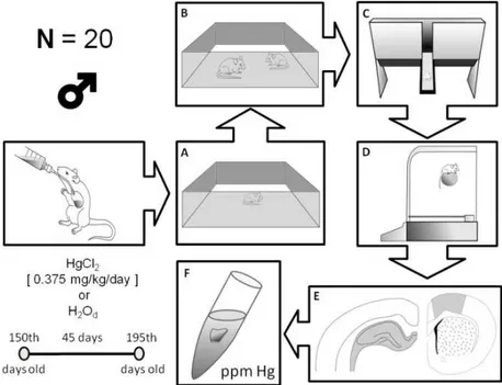
All analyses were conducted in duplicates of the group tissue samples, and the values obtained ranged from a confidence interval of ±10% (r: 0.9992). The methodology is summarized in Figure 1.
Figure 1. Schematic representation of the experimental design utilized in the present study. (A) Open field; (B) social recognition apparatus; (C) elevated T maze; (D) rotarod; (E) hippocampus on the left and motor cortex on the right; (F) sample to measure mercury.
2.9. Statistical Analysis
All values are expressed as the means ± SEM (n = 10 animals per group) for the behavioral assays.
Statistical comparisons between groups were performed using the Student’s t-test for behavioral
analyses and one-way ANOVA followed by Tukey’s test for mercury measurements. Values of
p≤ 0.05 were considered statistically significant. GraphPad Prism 5.0 (San Diego, CA, USA) software
was used for all analyses.
3. Results
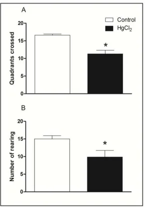
3.1. Chronic HgCl2 Exposure during Late Adulthood Induces Deficits on Spontaneous Locomotor Activity in Rats
Figure 2 illustrates spontaneous locomotor activity evaluated in the open field arena by chronic HgCl2 exposure. The Student’s t-tests revealed that HgCl2-treated animals displayed a reduced
locomotor activity in both horizontal and vertical exploration in the open field. The total number of squares crossed by HgCl2 group was lower than that of the control group (p < 0.05, Figure 2A).
Chronic HgCl2 exposure also decreased the number of rearing in rats in the unfamiliar arena (p < 0.01,
Figure 2. Effects of HgCl2 administration (0.375 mg/kg/day) for 45 days on the locomotor
activity of male Wistar rats evaluated in the open field (5 min). The results are expressed as the mean ± SEM of the: (A) total quadrants crossed; (B) number of rearing. * p < 0.05 compared to control group (Student’s t-test).
3.2. Learning, Short- and Long-Term Memory Impairments Induced by Chronic HgCl2 Exposure during Late Adulthood in Rats
The effects of chronic HgCl2administration during senescence on the male rats’ social recognition memory evaluated in the social recognition task are illustrated in Figure 3. The Student’s t-test
revealed that chronic HgCl2 exposure during adulthood did not alter implicit social recognition ability
(p > 0.05), observed in RID when the same juvenile was re-exposed 30 min after the first encounter.
Figure 3. Effects of HgCl2 administration (0.375 mg/kg/day) for 45 days on the social
recognition memory of male Wistar rats. The results are expressed as mean ± SEM of RIDs (ratio of investigation duration; i.e., the ratio of the second exposure to the first exposure)
The effects of chronic HgCl2 administration during late adulthood on the learning, short- and
long-term memory evaluated in the elevated T maze task are illustrated in Figure 4.
Figure 4. Effects HgCl2 administration (0.375 mg/kg/day) for 45 days on the learning,
short- and long-term memory of male Wistar rats evaluated in the elevated T maze (ETM) test. The results are expressed as the mean ± SEM of the: (A) number of re-expositions (learning function); (B) time in seconds in the enclosed arms for the first time test of long-term memory; and (C) time in seconds in the enclosed arms for the first time test of short-term memory. *p < 0.05 compared to control group (Student’s t-test).
Statistical comparisons revealed that HgCl2 administration during senescence induced a significant
increase in the number of re-expositions to achieve inhibitory avoidance, indicating significant impairments to learning compared with the control group (p < 0.05, Figure 4A). Panel 4B represents
the test and 4C the retest conducted 24 h after the exposures. In the test session related to long-term memory and the retest session related to short-term memory (p < 0.05), the mercury-intoxicated group
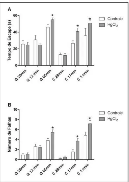
3.3. Chronic HgCl2 Exposure during Late Adulthood Promotes Alterations in Motor Function
In order to evaluate balance and coordination, the animals were tested in the rotarod apparatus. During test sessions, animals were subjected five times to the gyratory cylinder at increasing speeds (16, 20, 25, 28 and 37 RPM) with 60-s intervals between the sessions. The parameters observed were the latency and number until the first fall [16].
Figure 5A shows that the HgCl2 group reduced the latency until the first fall at speeds of 16, 20, 25
and 28 RPM when compared to the control group (p < 0.05). The latency of the intoxicated animals
group was only restored in the last test session (p > 0.05). The HgCl2 group had an increased number
of falls across the rotation increment in the two first sessions (p < 0.05) when compared to the control
group, which was recovered in the next rotation sessions (p > 0.05) (Figure 5B).
Figure 5. Effects of HgCl2 administration (0.375 mg/kg/day) for 45 days on the motor
function of male Wistar rats evaluated in the rotarod apparatus. The results are expressed as mean ± SEM of the: (A) latency in seconds to the first fall; and (B) number of falls.
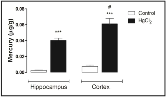
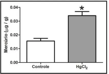
3.4. Mercury Deposition Is Higher in the Cortex than Hippocampus after Chronic Intoxication during Late Life in Rats
Figure 6 displays the mercury concentrations in the hippocampus and cortex of rats (after 45 days of intoxication). Note that the mercury concentration in the hippocampus and cortex is more than
that in the control group. Indeed, ANOVA followed by Tukey’s test indicates that the mercury concentration is higher in the cortex than in the hippocampus region (p < 0.001).
Figure 6. Effects of HgCl2 administration (0.375 mg/kg/day) for 45 days on mercury
(µg/g) deposition in the cortex and hippocampus of male Wistar rats. The results are expressed as the mean ± SEM. *** p < 0.001 compared to the control group; # compared
cortex to hippocampus intoxicated groups (one-way ANOVA followed by Tukey’s test).
4. Discussion
This study demonstrates, for the first time, that long-term chronic HgCl2 intoxication in rats during
senescence promotes functional damage. Chronic HgCl2 exposure induced anxiety-related responses,
short- and long-term memory impairments and motor deficits, as evaluated by different behavioral tests (open field, social recognition, elevated T maze and rotarod). Additionally, it was observed that HgCl2 accumulates in both the hippocampus and cortex regions, but has a higher affinity for
the cortex.
During the last few years, our group has extensively studied the long-lasting consequences of mercury intoxication during the prenatal period [15,18,19]. In this work, our hypothesis was that inorganic mercury exposure during adulthood also promotes functional impairment.
Mercury is able to induce distinct neurotoxic effects that depend on its chemical form (organic compounds, elemental mercury vapor or inorganic salts) [19,20]. It is well documented that organic mercury easily crosses the blood-brain barrier (BBB), and inorganic mercury salts (i.e., HgC12) that are
mercury in both the hippocampus (medium 0.04 µg/g) and cortex (medium 0.06 µg/g) of rat brains. Interestingly, our results highlight that mercury has a higher affinity for the cortex than the hippocampus. The possible mechanism involved in HgCl2 transport through the BBB implies an indirect effect
resulting from interference with the activities of cerebrovascular enzymes involved in BBB transport. In fact, Szumafiska et al. [6] showed the effect of acute doses of HgC12 (6 mg/kg) on Na+/K+ ATPase
activity in the neuropil of all of the cerebral cortical layers, which impairs BBB ion movement across the membrane even after a single dose of inorganic mercury. In addition, Moller-Madsen [21] demonstrated that after i.p. administration of HgCl2, mercury was detected in the cortical layer, but not
after oral administration [23]. However, Pamphlett and Hum [24] detected mercury deposits in lower motor neurons, but not in corticomotor neurons after inorganic mercury intoxication in rodents. The uptake of mercury seems to be through striated muscle and neuromuscular junctions, and finally, it is retrogradely transported to lower motor neuron cell bodies by their axons [25].
Despite claims that HgCl2 concentrations under 1 µg/g are not toxic to in vitro cultured CNS
tissues [26,27], our results affirm that lower concentrations of HgCl2 induce behavioral and
cognitive impairment.
In this sense, our current findings suggest that chronic HgCl2-treated animals reduced locomotor
exploratory activity in both horizontal and vertical exploration in the open field related to 0.06 µg/g of mercury concentration in cortex tissue. In addition, in the forced motor task (rotarod apparatus), inorganic mercury induced motor learning and coordinating impairment, reducing the latency until the first fall during four sessions, which was restored only in the last phase of the test. In accordance with our results, neurodevelopmental studies demonstrated that exposure to HgCl2 in the early postnatal
days induced impairments in motor function and muscular strength, as well as reductions of locomotor and exploratory activities [28,29]. On the contrary, Yasutake et al. [30] demonstrated that after three
weeks of intracerebroventricular injection of inorganic mercury, rodents increased spontaneous locomotor activity in the open field apparatus, indicating that Hg2+-induced hyperactivity was recovered three months after mercury exposure. These contradictory results may be explained by the administration protocol and age of the animals, since we adopted animals at 150 days old that were intoxicated until 195 days of life, which is mimetic of late adulthood and aged periods. It is well documented that ageing reduces motor performance and cognitive functions [31,32], which could be exacerbated by mercury poisoning.
The motor cortex has long been viewed to play an important role in fine motor control and fractionation of movement [33,34], sensorimotor integration and higher order cognitive-motor movements [35]. There are also other studies showing the role of motor cortex in the performance of behavioral tasks utilized in the current study [36–38], as well as showing that alterations on motor cortex can be associated with impaired spontaneous locomotion and incoordination in rodents [39–41].
Cognitive dysfunction was also observed in our work. Learning, short- and long-term memory were reduced in social recognition and T maze tests related to 0.04 µg/g content of mercury in the hippocampus. Previous works have elucidated that distinct forms of memory are mediated by different CNS regions, such as the primary cortex (i.e., prefrontal cortex-PFC) and limbic structures
(i.e., hippocampus). These forms can be classified as declarative or explicit, defined by the ability to
dependent on the striatum and cerebellum [42,43]. Of high importance, PFC plays a pivotal role, since it receives projections from both motor and sensory areas that are crucial for learning and is an intricate neuroanatomical correlation [44]. On the other hand, the hippocampus is involved in anxiety-like behaviors, as well as in memory and learning processes, as a result of its connections with other limbic areas involved in emotional behaviors [45,46]. Our results are in accordance with Yasutake and colleagues [30], which infer that acute doses of inorganic mercury induced cognitive damage in mice.
In the current chronic inorganic mercury intoxication protocol, we suggest that observed behavioral disabilities were related, at least in part, by the cortical and hippocampal mercury content that may interfere with local homeostasis. Considering the high bonding affinity between mercury and sulfur compounds (i.e., thiol groups of proteins, peptides and amino acids), interactions of mercury
compounds with proteins in the CNS may explain some of their effects on neurotransmission. Mercury micromolar concentrations inhibited cholinergic, glutamatergic, GABAergic and dopaminergic systems [47–51], affecting both behavior and cognition. It is well established that cognitive and motivational processes depend on the connections between PFC and limbic structures, which are dependent on the neurotransmitters cited above, and the hippocampus plays a key role in the functioning of these pathways [43].
5. Conclusions
In conclusion, our results provide new evidence that exposure to inorganic mercury during late adulthood induces motor and cognitive impairments associated with low mercury content in cortex and hippocampus structures. Of significance are the current findings indicating that even under in vitro
cytotoxic effects, CNS micromolar concentrations of mercury induce behavioral and motor dysfunction after chronic exposure in late adulthood. The mechanisms involved in the observed neurobehavioral consequences include disruption to the blood brain barrier, mainly the damage activity of Na/K ATPase found in brain microvessels; however, the completed mechanism should be investigated in future research, but the present data provide promising evidence that inorganic mercury can also promote neurotoxic effects, even in the mature brain.
Acknowledgments
The authors thank the Núcleo de Medicina Tropical, Federal University of Pará for allowing the use of their infrastructure for the quantification of mercury. Francisco Bruno Teixeira was supported by a Brazilian Government/Conselho Nacional de Desenvolvimento Científico e Tecnológico (CNPq) fellowship.
Author Contributions
Marcia C. F. Silva, Cristiane S. F. Maia, Rafael R. Lima. Contributed reagents/materials/analysis tools: Francisco B. Teixeira, Marcia C. F. Silva, Cristiane S. F. Maia, Rafael R. Lima. Wrote the paper: Francisco B. Teixeira, Marcia C. F. Silva, Cristiane S. F. Maia, Rafael R. Lima, Rafael M. Fernandes, Luana N. S. Santana, Luanna M. P. Fernandes, Natacha M. M. Costa, Paulo M. A. Farias-Junior, Ademir F. Silva-Junior.
Conflicts of Interest
The authors declare no conflict of interest.
References
1. Bernhoft, R.A. Mercury toxicity and treatment: A review of the literature. J. Environ. Public Health 2012, 2012, doi:10.1155/2012/460508.
2. Goldman, L.R.; Shannon, M.W. American Academy of Pediatrics: Committee on Environmental Health Technical report: Mercury in the environment: Implications for pediatricians. Pediatrics 2001, 108, 197–205.
3. Dyall-Smith, D.J.; Scurry, J.P. Mercury pigmentation and high mercury levels from the use of a cosmetic cream. Med. J. Aust.1990, 153, 409–415.
4. Weldon, M.M.; Smolinski, M.S.; Maroufi, A.; Hasty, B.W.; Gilliss, D.L.; Boulanger, L.L.; Dutton, R.J. Mercury poisoning associated with a Mexican beauty cream. West. J. Med. 2000, 173, 15–18.
5. Clarkson, T.W.; Magos, L. The toxicology of mercury and its chemical compounds.
Crit. Rev. Toxicol.2006, 36, 609–662.
6. Szumafiska, G.; Gadamski, R.; Albrecht, J. Changes of the Na/K ATPase activity in the cerebral cortical microvessels of rat after single intraperitoneal administration of mercuric chloride: Histochemical demonstration with light and electron microscopy. Acta Neuropathol. 1993, 86,
65–70.
7. Rice, K.M.; Walker, E.M., Jr.; Gillette, M.W.C.; Blough, E.R. Environmental mercury and its toxic effects. J. Prev. Med. Public Health2014, 47, 74–83.
8. Smith, J.C.; Allen, P.V.; Turner, M.D.; Most, B.; Fisher, H.L.; Hall, L.L. The kinetics of intravenously administered methylmercury in man. Toxicol. Appl. Pharmacol.1994, 128, 251–256.
9. Szasz, A.; Barna, B.; Gajda, Z.; Galbacs, G.; Kirsch-Volders, M.; Szente, M. Effects of continuous low-dose exposure to organic and inorganic mercury during development on epileptogenicity in rats.
Neurotoxicology2002, 23, 197–206.
10. Huang, C.; Liu, S.; Hsu, C.; Lin-Shiau, S. Neurotoxicological effects of low-dose methylmercury and mercury chloride in developing offspring mice. Toxicol. Lett.2009, 201, 196–204.
11. Cai, L.; Yan, X.B.; Chen, X.N.; Meng, Q.Y.; Zhou, J.N. Chronic all-trans retinoic acid administration induced hyperactivity of HPA axis and behavioral changes in young rats.
Eur. Neuropsychopharmacol. 2010, 20, 839–847.
13. Prediger, R.D.; Fernandes, D.; Takahashi, R.N. Blockade of adenosine A2A receptors reverses short-term social memory impairments in spontaneously hypertensive rats. Behav. Brain Res. 2005, 159, 197–205.
14. Takahashi, R.N.; Pamplona, F.A.; Fernandes, M.S. The cannabinoid antagonist SR141716A facilitates memory acquisition and consolidation in the mouse elevated T-maze. Neurosci. Lett. 2005, 380, 270–275.
15. Maia, C.S.F.; Ferreira, V.M.; Diniz, J.S.; Carneiro, F.P.; de Sousa, J.B.; Costa, E.D.; Tomaz, C. Inhibitory avoidance acquisition in adult rats exposed to a combination of ethanol and methylmercury during central nervous system development. Behav. Brain Res.2010, 211, 191–197.
16. Sharma, D.J.; Sunkaria, A.; Bal, A.; Bhutia, Y.D.; Vijayaraghavan, R.; Flora, S.J.S.; Gill, K.D. Neurobehavioral impairments, generation of oxidative stress and release of pro-apoptotic factors after chronic exposure to sulphur mustard in mouse brain. Toxicol. Appl. Pharmacol. 2009, 240,
208–218.
17. Akagi, H. Analysis of methylmercury in fish and shellfish by dithizone extraction-gas chromatography. Jpn. J. Hyg.1985, 40, 293.
18. Maia, C.S.F.; Ferreira, V.M.; Kahwage, R.L.; do Amaral, M.N.; Serra, R.B.; Santos, S.N.; Nascimento, J.L.M.; Rodrigues, L.G.; Trévia, N.; Picanço-Diniz, C.W. Adult brain nitrergic activity after concomitant prenatal exposure to ethanol and methyl mercury. Acta Histochem. 2010, 112, 583–591.
19. Maia, C.S.F.; Lucena, G.M.; Correa, P.B.; Serra, R.B.; Matos, R.W.; Menezes, F.C.; Santos, S.N.; Sousa, J.B.; Costa, E.T.; Ferreira, V.M.M. Interference of ethanol and methylmercury in the developing central nervous system. Neurotoxicology2009, 30, 23–30.
20. Gallagher, P.J.; Mitchell, J.; Wheal, H.V. Identity of ultrastructural effects of mercuric chloride and methyl mercury after intracerebral injection. Toxicology1982, 23, 261–266.
21. Maier, W.E.; Costa, L.G. Na+/K+ ATPase as a and marker, respectively, for neurotoxicity: Studies with chlordecone, organotins and mercury compounds. Toxicol. Lett.1990, 51, 175–188.
22. Moller-Madsen, B. Localization of mercury in CNS of the rat. II. Intraperitoneal injection of methylmercuric chloride (CH3HgCl) and mercuric chloride (HgC12). Toxicol. Appl. Pharmacol. 1990, 103, 303–323.
23. Moller-Madsen, B.; Danscher, G. Localization of mercury in CNS of the rat I. Mercuric Chloride (HgCl2) per os. Environ. Res.1986, 41, 29–43.
24. Pamphlett, R.; Jew, S.K. Uptake of inorganic mercury by human locus ceruleus and corticomotor neurons: Implications for amyotrophic lateral sclerosis. Acta Neuropathol. Commun. 2013, 1,
doi:10.1186/2051-5960-1-13.
25. Arvidson, B. A review of axonal transport of metals. Toxicology1994, 88, 1–14.
26. Brookes, N.; Kristt, D.A. Inhibition of amino acid transport and protein synthesis by HgC12 and
methylmercury in astrocytes: Selectivity and reversibility. J. Neurochem. 1989, 53, 1228–1237.
27. Choi, B.H.; Kim, R.C. The comparative effects of methylmercuric chloride and mercuric chloride upon DNA synthesis in mouse fetal astrocytes in vitro. Exp. Mol. Pathol.1984, 41, 371–376.
28. Franciscato, C.; Goulart, F.R.; Lovatto, N.M.; Duarte, F.A.; Flores, E.M.; Dressler, V.L.; Peixoto, N.C.; Pereira, M.E. ZnCl2 exposure protects against behavioral and acetylcholinesterase
29. Franco, J.L.; Braga, H.C.; Nunes, A.K.; Ribas, C.M.; Stringari, J.; Silva, A.P.; Garcia Pomblum, S.C.; Moro, A.M.; Bohrer, D.; Santos, A.R.; et al. Lactational exposure to inorganic mercury:
Evidence of neurotoxic effects. Neurotoxicol. Teratol. 2007, 29, 360–367.
30. Yasutake, A.; Marumoto, M.; Yoshida, M. Neurotoxic action of inorganic mercury injected in the intraventricular space of mouse cerebrum. J. Toxicol. Sci. 2010, 35, 767–771.
31. Skinner, H.B.; Barrack, R.L.; Cook, S.D. Age-related decline in proprioception. Clin. Orthop. Relat. Res.1984, 184, 208–211.
32. Sturnieks, D.L.; St George, R.; Lord, S.R. Balance disorders in the elderly. Neurophysiol. Clin. 2008, 38, 467–478.
33. Asanuma, H. Cerebral cortical control of movement. Physiologist1973, 16, 143–166.
34. Evarts, E.V.; Fromm, C.; Kroller, J.; Jennings, V.A. Motor cortex control of finely graded forces.
J. Neurophysiol.1983, 49, 1199–1215.
35. Sanes, J.N.; Donoghue, J.P. Plasticity and primary motor cortex. Annu. Rev. Neurosci. 2000, 23,
393–415.
36. Stigger, F.; Lovatel, G.; Marques, M.; Bertoldi, K.; Moysés, F.; Elsner, V.; Siqueira, I.R.; Achaval, R.; Marcuzzo, S. Inflammatory response and oxidative stress in developing rat brain and its consequences on motor behavior following maternal administration of LPS and perinatal anoxia.
Int. J. Dev. Neurosci.2013, 31, 820–827.
37. Carmel, J.B.; Martin, J.H. Motor cortex electrical stimulation augmentes sprouting of the corticospinal tract and promotes recovery of motor function. Front. Integr. Neurosci. 2014, 8,
doi:10.3389/fnint.2014.00051.
38. Karl, T.; Pabst, R.; Von Hörsten, S. Behavioral phenotyping of mice in pharmacological and toxicological research. Exp. Toxicol. Pathol.2003, 55, 69–83.
39. Helfer, J.L.; Calizo, L.H.; Dong, W.K.; Goodlett, C.R.; Greenough, W.T.; Klintsova, A.Y. Binge-like postnatal alcohol exposure triggers cortical gliogenesis in adolescent rats. J. Comp. Neurol. 2009, 514, 259–271.
40. Oliveira, G.B.; Fontes, E.D.; de Carvalho, S.; da Silva, J.B.; Fernandes, L.M.; Oliveira, M.C.; Prediger, R.D.; Gomes-Leal, W.; Lima, R.R.; Maia, C.S. Mynocycline mitigates motor impairments and cortical neuronal loss induced by focal ischemia in rats chronically exposed to ethanol during adolescence. Brain Res.2014, 1561, 23–34.
41. Teixeira, F.B.; Santana, L.N.; Bezerra, F.R.; de Carvalho, S.; Fontes-Júnior, E.A.; Prediger, R.D.; Crespo-López, M.E.; Maia, C.S.; Lima, R.R. Chronic ethanol exposure during adolescence in rats induces motor impairments and cerebral cortex damage associated with oxidative stress.
PLoS One2014, 26, doi:10.1371/journal.pone.0101074.
42. Packard, M.G.; McGaugh, J.L. Double dissociation of fornix and caudate nucleus lesions on acquisition of two water maze tasks: Further evidence for multiple memory systems.
Behav. Neurosci.1992, 106, 439–446.
43. McDonald, R.J.; White, N.M. A triple dissociation of memory systems: Hippocampus, amygdala, and dorsal striatum. Behav. Neurosci.1993, 107, 3–22.
44. Middleton, F.A.; Strick, P.L. Basal-ganglia “projections” to the prefrontal cortex of the primate.
45. Moser, E.; Moser, M.B.; Andersen, P. Spatial learning impairment parallels the magnitude of dorsal hippocampal lesions, but is hardly present following ventral lesions. J. Neurosci.1993, 13,
3916–3925.
46. Bannerman, D.M.; Rawlins, J.N.; McHugh, S.B.;Deacon, R.M.; Yee, B.K.; Bast, T.; Zhang, W.N.; Pothuizen, H.H.; Feldon, J. Regional dissociations within the hippocampus: Memory and anxiety. Neurosci Biobehav Rev.2004, 28, 273–283.
47. Bondy, S.C.; Agrawal, A.K. The inhibition of cerebral high affinity receptor sites by lead and mercury compounds. Arch. Toxicol.1980, 46, 249–256.
48. Castoldi, A.F.; Candura, S.M.; Costa, P.; Manzo, L.; Costa, L.G. Interaction of mercury compounds with muscarinic receptor subtypes in the rat brain. Neurotoxicology1996, 17, 735–742.
49. Arakawa, O.; Nakahiro, M.; Narahashi, T. Mercury modulation of GABA-activated chloride channels and non-specific cation channels in rat dorsal root ganglion neurons. Brain Res. 1991, 551, 58–63.
50. Rajanna, B.; Rajanna, S.; Hall, E.; Yallapragada, P.R. In vitro metal inhibition of
N-methyl-d-aspartate specific glutamate receptor binding in neonatal and adult rat brain.
Drug Chem. Toxicol.1997, 20, 21–29.
51. Scheuhammer, A.M.; Cherian, M.G. Effects of heavy metal cations sulfhydryl reagents and other chemical agents on striatal D2 dopamine receptors. Biochem. Pharmacol.1985, 34, 3405–3413.
5. APÊNDICE 2
A EXPOSIÇÃO CRÔNICA AO MERCÚRIO INORGÂNICO PROMOVE DÉFICITS MOTORES ASSOCIADOS À MORTE CELULAR E ESTRESSE OXDATIVO NO
CÓRTEX MOTOR DE RATOS ADULTOS
Francisco Bruno Teixeira1, Luana Ketlen Reis Leão1, Nathália Carolina Fernandes
Fagundes1, Luana de Nazaré Silva da Santana1, Rafael Monteiro Fernandes1, Márcia
Cristina Freitas da Silva1, Lilian Lund Amado2, Fernanda Espírito Santo Sagica3,
Edivaldo Herculano Correa Oliveira3, Maria Elena Crespo-Lopez4, Cristiane Socorro
Ferraz Maia5, Rafael Rodrigues Lima1
1 Laboratório de Biologia Estrutural e Funcional, Instituto de Ciências Biológicas,
Universidade Federal do Pará, Belém, Pará, Brasil;
2 Laboratório de Ecotoxicologia, Instituto de Ciências Biológicas, Universidade Federal
do Pará, Belém, Pará, Brasil;
3 Laboratório de Cultura Celular e Citogenética, Instituto Evandro Chagas, Ananindeua,
Pará, Brasil.
4 Laboratório de Farmacologia Molecular, Instituto de Ciências Biológicas,
Universidade Federal do Pará, Belém, Pará, Brasil;
5 Laboratório de Farmacologia da Inflamação e Comportamento, Instituto de Ciências
da Saúde, Universidade Federal do Pará, Belém, Pará, Brasil.
*Autor de Correspondência: Rafael R. Lima, PhD
rafalima@ufpa.br