RESUMO.- [Avaliação da concentração de alérgenos provenientes de ácaros na pelagem e na poeira de do-micílios de cães com dermatite atópica.] O presente es-tudo avaliou a concentração de Der p 1, Der f 1 e Blo t 5, na pelagem e no ambiente domiciliar de cães com dermatite atópica (DA). Para tal, foram selecionados 20 cães com DA
e 20 cães saudáveis, e seus domicílios. O diagnóstico de DA foi baseado nos critérios de Favrot. As amostras de poeira foram colhidas com um aspirador de pó doméstico. Para cada local de colheita, foi aspirado 1m2 por dois minutos. As amostras foram recolhidas em filtros separadamente, transferidas para envelopes plásticos, seladas e mantidas congeladas até serem analisadas pelo método de ELISA. Na pelagem dos cães com DA, a concentração média de Der p 1 foi de 0,25µg/g de poeira e nos cães saudáveis foi de 0,03µg/g. No ambiente de cães com DA, o Der p 1 foi encon-trado em maior concentração média no tapete (2,18 µg/g), seguido pelo sofá (1,53g/g), cama (1,14µg/g) e roupa de cama dos cães (0,64µg/g) e chão (0,14µg/g). As concentra-ções de Der p 1 no tapete, sofá e cama dos cães foram signi-ficativamente maiores que na pelagem de cães com derma -tite atópica (p<0,05). Enquanto que, não houve diferença estatística quando comparadas as concentrações de Der f 1 e Blo t 5 nos diferentes ambientes avaliados (p>0,05). As concentrações de alérgenos Der p 1, Der f 1 e Blo t 5 se equivaleram em ambientes de cães com DA e saudáveis. En
-Evaluation of the concentration of allergens from mites in fur
and households dust of dogs with atopic dermatitis
1Dévaki L. de Assunção2*, Marconi R. de Farias3, Michelle C.R. Barbosa4
and Luiz H. de Araújo Machado5
ABSTRACT.- Assunção D.L., Farias M.R., Barbosa M.C.R. & Machado L.H.A. 2017. Evalu-ation of the concentrEvalu-ation of allergens from mites in fur and households dust of dogs with atopic dermatitis.Pesquisa Veterinária Brasileira37(1):41-46. Mestrado em Ciência Animal, Escola de Ciências Agrárias e Medicina Veterinária, Pontifícia Universida -de Católica do Paraná, BR-376 Km 14, São José dos Pinhais, PR 83010-500, Brazil. E-mail: [email protected]
This study evaluated the concentration of Der p 1, Der f 1 and Blo t 5 in the fur and households of 20 dogs with atopic dermatitis (AD) and 20 healthy dogs. The diagnosis of AD was clinical based on Favrot’s criteria. Dust samples were collected with a domestic va-cuum cleaner. For each site, 1m2 was vacuumed for 2 min. The samples were collected in se-parate filters, transferred into plastic containers, sealed and kept frozen until ELISA analy -sis. In the fur of atopic dogs the average concentration of Der p 1 was 0.25µg/g compared to 0.03µg/g in healthy dogs. In households with atopic dogs the highest concentrations of Der p 1 were found in carpets (2.18µg/g), followed by couches (1.53µg/g), beds (1.14µg/g), dogs’ bed linen (0.64µg/g) and floors (0.14µg/g). The concentrations of Der p 1 on carpets, couches and beds were significantly higher than in atopic dogs’ fur (p<0.05). There was no statistical difference when comparing the concentrations of Der f 1 and Blo t 5 in different environments of atopic dogs (p>0.05). The concentrations of Der p 1, Der f 1 and Blo t 5 were equivalent in atopic and non-atopic dog’s households. Among the allergens studied, Der p 1 was the most commonly found, predominantly in carpets and couches.
INDEX TERMS: Allergens, mites, fur, household dust, dogs, atopic dermatitis.
1 Received on October 27, 2015.
Accepted for publication on May 19, 2016.
2 Mestrado em Ciência Animal, Escola de Ciência Agrárias e Medicina
Ve-terinária, Pontifícia Universidade Católica do Paraná (PUCPR), BR-376 Km 14, São José dos Pinhais, PR 83010-500, Brazil. *Corresponding author: [email protected]
3 Departamento de Clínica Médica de Animais de Companhia, Escola de
Ciência Agrárias e Medicina Veterinária, PUCPR, BR-376 Km 14, São José dos Pinhais, PR 83010-500, Brazil.
4 Faculdade de Medicina de Ribeirão Preto, Universidade de São Paulo
(USP), Av. Bandeirantes 3900, Monte Alegre, Ribeirão Preto, SP 14049-900, Brazil.
5 Departamento de Clínica Veterinária, Faculdade de Medicina
tre os alérgenos estudados, o Der p 1 foi o mais comumente encontrado, prevalecendo no tapete e no sofá.
TERMOS DE INDEXAÇÃO: Alérgenos, ácaros, pelagem, poeira, cães, dermatite atópica.
InTROdUCTIOn
Atopic dermatitis (AD) is an inflammatory and pruritic der -mopathy which is chronic, recurrent, and highly incident amongst dogs (Olivry et al. 2010). Despite its multifactorial origins, the physiopathology of this disease is associated with genetic mutations that lead to alterations in the final keratinization of the epidermis and skin barrier dysfunc-tion, and these changes facilitate the transepidermal pe-netration of environmental allergens, microbial antigens, and primary irritants and exacerbate innate and adaptive immune responses (Hillier 2002, Akdis et al. 2006, Cork et al. 2006, Olivry et al. 2010).
House dust mite allergens (HDMA) are the main fac-tors responsible for the sensitization and development of symptoms of allergic disease in humans (Zavadniak 2000). The most common are the allergens from the mi-tes Dermatophagoides pteronyssinus (D. pteronyssinus) and
Dermatophagoides farinae (D. farinae); these allergens can have epithelial origin, but they can also be enzymes (Arlian & Platts-Mills 2001). The major allergens of mites D. pte-ronyssinus and D. farinae belong to Group 1 (Der p 1 and Der f 1) and Group 2 (Der p 2 and Der f 2) respectively (Ma-dhurantakam et al. 2010).
Exposure to Der p 1 in concentrations higher than 2µg/g of dust is considered a risk factor for sensitization (Eggleston & Arruda 2003) and concentrations above 10 µg/g can cause clinical manifestations of allergies in hu-mans (Upham & Holt 2005). Moreover, allergens from the mite Blomia tropicalis (B. tropicalis), have been implicated in the sensitization of susceptible individuals in regions with tropical or subtropical climates(Cunha et al. 2012).
The majority of dogs with AD have IgE immunoglobu-lins against environmental allergens that can be detected by using intradermal testing (IDT) and/or allergen-specific IgE serology (ASIgES) (Halliwell 2006, Farmaki et al. 2012). Based on the high frequency of positive results in the IDT and ASIgES tests for house dust mites, such as D. farinae
and D. pteronyssinus in dogs, these mites have been consi-dered important sources of environmental allergens asso-ciated with atopic eczema worldwide (Hill & DeBoer 2001, Farmaki et al. 2012).
The presence of the aforementioned mite species and their allergens, including Der f 1, Der p 1, and Group 2 aller-gens, has been previously confirmed in the bedding, skin, hair coat, and dry food of clinically healthy dogs (Eaton et al. 1985, Randall et al. 2003, Brazis et al. 2008). Meanwhile, only few studies have attempted to determine the concen-tration of mite allergens in the fur and household microen-vironment of dogs with AD.
The objective of this study was to evaluate the concen-tration of allergens from the mites D. pteronyssinus (Der p 1), D. farinae (Der f 1), and B. tropicalis (Blo t 5) in the fur and households of dogs with AD.
MATERIAlS And METhOdS
Sample population. The sample was composed of 20 dogs with AD, 20 healthy dogs, and the dogs’ respective households in the city of Curitiba, state of Paraná (PR), Brazil.
The AD diagnosis was based on the medical history and clini-cal examination, and the dogs included in the study met at least six of the eight criteria established by Favrot (Favrot et al. 2010).
Further, the dogs’ symptoms had not significantly improved (as quantified by using an itch scale for measuring pruritus) after up
to eight weeks of exclusion diet with a novel food source of pro-tein and carbohydrate. Thus, the possibility of AD induced by food
(latu sensu) was excluded.
Sample collection. Samples were collected in the househol-ds of the animals during the fall and winter months. The owners were contacted in advance and were instructed not to replace the beddings and to avoid the use of sprays, vacuum cleaners, and cleaning products on the day of the sample collection. Before collection, a digital humidity and temperature monitor (Incoterm 7663.02.0.00, Incoterm Inc., Porto Alegre, Brazil) was used for measuring the temperature and relative humidity of the air in the location.
Samples were collected with a portable domestic vacuum
cle-aner (ELECTROLUX-INGE9, Electrolux Brazil Inc., Curitiba, Brazil)
containing an adaptor close to the siphon for suctioning
(Dustre-am collection, INDOOR Biotechnologies Inc., Charlottesville, USA) so that the aspired material could be retained by a specific filter (Dustream filters, INDOOR Biotechnologies Inc., Charlottesville, USA). Between each vacuuming, the siphon was sanitized by using
a 70% ethanol solution to remove allergen residues completely. For each household, samples were collected from the places where the dogs spent the most time (dog bed and bedding, couch,
floor, and carpets); each 1m2 area was vacuumed for 2 min.
A similar protocol was used to collect samples from the fur of the animals. The owners were contacted in advance and were instructed not to bathe or brush the dogs for at least seven days before sample collection. The animals were restrained and vacuu-ming was performed all over the body for 2 min.
Following sample collection, the dust-containing filters were sealed in a plastic bag, identified, and frozen at -6°C to maintain
the physical integrity of the mites, avoid allergen denaturation, and inhibit mite metabolism and proliferation.
Quantification of HDMA. All dust samples collected from
the households and dogs’ fur were removed from the filters and were filtered through a 0.3 mm sieve mesh (Standard Sieve Series
ASTM, Dual Manufacturing Co. Inc.,Illinois, USA) to remove big
particles and leave only fine dust in the Petri dishes.
For extract preparation, 100 mg of fine dust was transferred
into a test tube, and 2 mL of phosphate buffered saline contai-ning 0.05% Tween 20 (PBS-T), pH 7.4, was added. For samples that were less than 50 mg, 1 mL was added; for samples between 50 and 100 mg, an adjusted proportional volume of PBS-T was added. For resuspension, a shaker was used (VDRL Shaker
TS--2000A, Biotechnologies Inc., Delhi, India) for 24h at 4°C. Samples
were then centrifuged at 2500rpm for 20 min at 4oC. Supernatants
(approximately 1.5mL) were collected with a Pasteur pipette into labeled Eppendorf tubes (Eppendorf Safe-Lock Tubes, Eppendorf Inc., Hamburg, Germany) and were stored at -20oC.
The levels of allergens Der p 1, Der f 1, and Blot 5 were deter-mined through the validated ELISA test (Luczynska et al. 1989) using the monoclonal antibodies anti-Der p 1, anti-Der f 1, and anti-Blo t 5 (EL-DP1, EL-DF1, and EL-BT5, respectively; Indoor
Biothecnologies Inc., Charlottesville, USA).
diffe-rent sites, the Kruskal Wallis test was used, followed by the Dunn’s test for median comparison. The Spearman test was used for
cor-relation analysis. Results were considered significant at p<0.05.
All statistical analyses were performed by using GraphPad Prism®
version 5 software (GraphPad Software Inc., San Diego, Califor-nia).
RESUlTS
For the analysis, 147 samples from 40 households were used. Epidemiological data for both atopic dogs and heal-thy dogs, as well as the physical characteristics and the han-dling procedures at each household, are listed in Table 1.
During this study, the average environmental tempe-rature was 18.8°C and the average relative humidity was 72%.
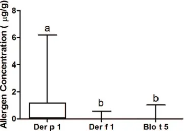
Of the 147 samples collected, Der p 1 was detected in 100 samples (68%), Der f 1 in 14 samples (9.5%), and Blo t 5 in 9 samples (6.1%); Der p 1 was detected at the highest concentration in the households (0.8735µg/g ± 0.1196µg/g) (p<0.00001) (Fig.1).
The average concentrations of the allergens Der p 1, Der f 1, and Blo t 5 in the fur of atopic dogs and healthy dogs, as well as in the household dust are shown in Table 2A and 2B.
In the group of dogs with AD, the concentrations of Der p 1 in the carpets, couches, and dog’s bed linen were signi-ficantly higher than those detected in the fur of these dogs (p<0.05) (Fig.2). No difference was found in the concentra-tions of Der f 1 and Blo t 5 between the different environ-ments tested (p>0.05).
In the group of healthy dogs, the Der p 1 concentrations
in the couches and beds and the Der f 1 concentrations in the beds were significantly higher than the concentrations detected in the fur of these animals (p<0.05). No differen-ces were found in the Blo t 5 concentrations between the different environments tested (p>0.05).
When the concentrations of Der p 1 in the fur and in the microenvironment of dogs with AD were correlated, it was observed that the higher the concentration in the dog’s bed and couch, the higher was the concentration in the dog’s fur (p<0.05).
It was also observed that the concentrations of the mite allergens Der p 1, Der f 1, and Blo t 5 were similar between the microenvironments of dogs with AD and healthy dogs (p>0.05).
dISCUSSIOn
Beginning in the twentieth century, the usual behavior of families changed, and families now spend most of their time in indoor environments. This new lifestyle has incre-ased exposure to allergens, leading to the development of
Fig.1. Concentration of Der p 1, Der f 1, and Blo t 5 (µg/g) in the evaluated samples. The letters are associated with different p values for p<0.00001.
Table 1. Epidemiological data of the dogs with atopic dermatitis and healthy dogs, and the physical characteristics
and handling procedures at the respective households
Dogs with AD Healthy dogs
Gender - Male 05 (25%) 06 (30%)
Female 15 (75%) 14 (70%)
Age (median) 03 years 5.5 years
Age at onset of symptoms (median) 01 year
Castrated (♂♀) 18 (90%) 14 (70%)
Fur length - Short 08 (40%) 08 (40%)
Long 12 (60%) 12 (60%)
Bath frequency - Weekly 20 (100%) 20 (100%)
Shampoo used - Neutral 03 (15%) 20 (100%)
Therapeutic 17 (85%) 00
Regular use of anti-flea/anti-mite agents 16 (80%) 14 (70%)
Regular brushing 10 (50%) 05 (25%)
Habitat - Indoor 20 (100%) 20 (100%)
Number of inhabitants (median) 03 (02-05) 04 (01-05) Number of rooms (median) 11 (10-14) 09 (07-12) Flooring - Floating wood tile 12 (60%) 06 (30%)
Cold 06 (30%) 11 (55%)
Paviflex tiles 00 02 (10%)
Wood parquet 01 (05%) 00
Carpet 01 (05%) 01 (05%)
Cleaning frequency- Daily 01 (05%) 01 (05%)
2x a week 08 (40%) 02 (10%)
3x a week 04 (20%) 00
4x a week 01 (05%) 00
Weekly 06 (30%) 14 (70%)
Every 10 days 00 01 (05%)
Fortnightly 00 02 (10%)
Weekly bed linen changes 20 (100%) 20 (100%)
Dogs bed cleaning 00 00
Table 2 A and B. Average concentration of the allergens der p 1, der f 1, and Blo t 5 (µg/g) detected in household dust
and in the fur of dogs with atopic dermatitis and healthy dogs. The letters correspond to different p values for p<0.05
A Dogs with atopic dermatitis
Der p 1 Der f 1 Blo t 5
Dog´s bed 1.14a 0.00a 0.00a
Fur 0.25b 0.00a 0.01a
Floor 0.14abc 0.00a 0.03a
Dog´s bed linen 0.64abc 0.00a 0.05a
Couch 1.53ac 0.02a 0.00a
Carpet 2.18ac 0.07a 0.06a
B Healthy dogs
Der p 1 Der f 1 Blo t 5
Dog´s bed 1.82a 0.05a 0.07a
Fur 0.03b 0.00b 0.01a
Floor 0.72ab 0.00ab 0.00a
Dog´s bed linen 0.00ab 0.00ab 0.23a
Couch 1.24ac 0.04ab 0.05a
allergic diseases (Platts-Mills 2009). Dogs have also expe-rienced these changes because they have adapted to their owner’s lifestyle; thus, these animals now live more fre-quently in apartments or indoors, and spend most of their time in the proximity of their owners.
Mite allergens are ubiquitous and are capable of causing al -lergic reactions in humans, dogs, and several other animal species (Randall et al. 2003, Gehring et al. 2005, Raffan et al. 2005, Allan & Novak 2006).
D. pteronyssinus is abundant in Southern and Central America (Thomas 2010). Previous studies performed in various Brazilian cities (Geller 1996, Medeiros et al. 2002, Rego et al. 2003, Rullo et al. 2005) showed that Dermato-phagoides pteronyssinus and Blomia tropicalis are impor-tant sensitizing agents in human patients with rhinitis and allergic asthma.
D. pteronyssinus populations grow preferably at tem-peratures of 25°C and a relative air humidity above 60% (Arlian & Platts-Mills2001, Jackson et al. 2005). Notably, in this study, sample collection was performed during the fall and winter months, when the average temperatures were low and the relative air humidity was approximately 72%; this fact may have contributed to the maintenance of
D. pteronyssinus populations and extended the viability of the mite’s allergens.
The allergens from D. pteronyssinus are expected to play an important role in sensitizing dogs with AD in Brazil be-cause they are present in house dust.
The Der p 1 allergen is a cysteine protease with a mo-lecular weight of approximately 24 kDa; this protease is active at an acidic pH and is similar to the one found on the surface of mammalian skin. This allergen facilitates epithelial permeability through several mechanisms: the degradation of occludin, claudin, and junction proteins; the induction of IgE-independent inflammation mediated by
basophile and mastocyte activation; the induction of IL-4 secretion; the inhibition of the development of Th1 res-ponses, both by a direct effect on T cells and by modula-ting IL-12 secretion by dendritic cells; and the activation of a variety of cell types following PAR-2 receptor cleavage. These pro-inflammatory functions can contribute to aller -gic sensitization (Bizikova et al. 2015).
D. farinae and its allergens peak at temperatures betwe-en 25 and 30°C and a relative air humidity of 50 to 60% (Jackson et al. 2005), which may explain the lower concen-trations of Der f 1 in the microenvironments studied when compared with the concentrations detected in other Brazi-lian states and in countries with temperate climates.
In the northern hemisphere, the sensitization and in-crease of AD symptoms in dogs have been associated with exposure to D. farinae allergens (Nuttall et al. 2006).
Wes-tern blot protein studies showed that 80 to 90% of AD dogs sensitized to D. farinae extracts have specific IgE antibodies
against D. farinae proteins with a molecular weight betwe-en 60 and 98 kDa, suggesting that high molecular weight
D. farinae allergens, such as Der f 15 and Der f 18, are the most important factors contributing to the exacerbated cli-nical manifestations of AD (McCall et al. 2001, Weber et al. 2003). Hou et al. (2005), characterized the IgG response to
D. farinae antigens in healthy dogs and in AD dogs by using immunoblotting and electrophoresis techniques, and the two major bands detected corresponded to proteins with molecular weights of 98 kDa (Der f 15) and 44 kDa. Un -fortunately, no tests are available for the quantification of these allergens intradermally or in the environment, and information is not available concerning their concentration in domestic dust samples.
B. tropicalis (Blo t 5) allergens have been suggested as a frequent cause of sensitization in children with rhinitis and allergic asthma(Farias 2007). B. tropicalis can be found in tropical climates, and sensitization to these allergens is common in dogs with AD living in these regions (Cunha et al. 2012).
Der p 1, Der f 1, and Blo t 5 concentrations were low in the households evaluated in our study when compared with previously reported results (Fariaset al. 2015). This can be explained by a combination of factors such as pro-per cleaning of the environment, frequent changings of the animal’s bed linen, presence of floors that facilitated the cleaning process, presence of few rugs and carpets in the environment, and low number of inhabitants.
Carpets and couches were the sites with the highest concentrations of the Der p 1 allergen in the households of atopic dogs, whereas Der p 1 accumulated mostly in the beds and couches in the households with healthy dogs. This could be because of difficulties in sanitizing these sites or because the owners reported that vacuuming or washing the beds is not customary practice.
In the remaining microenvironments studied, the al-lergen concentrations were similar between the environ-ments associated with either atopic dogs or healthy dogs, indicating that both groups of dogs were equally exposed to allergens. Therefore, atopic dogs must develop clinical symptoms because of genetic defects in the epidermal
rier and in junction proteins (Cork et al. 2006, Marsella & Samuelson 2009). Epidermal barrier dysfunction facilita-tes contact between the environmental and microbial aller-gens and the defense cells in the epidermis, which stimu-lates the inflammatory response in the skin (Olivry et al. 2010, Kubo et al. 2012). Changes in the microstructure of the epidermis in atopic dogs were shown to lead to pruri-tus and to trigger activation of the inflammatory response (Homey et al. 2006).
In this study, the low concentrations of Der p 1, Der f 1, and Blo t 5 detected in the fur of atopic dogs tested may re-flect the handling of these animals, which involved weekly baths, regular brushing, and maintenance in clean environ-ments.
When the Der p 1 allergen concentration in the fur was correlated with the microenvironment of the dogs with AD, it was observed that the higher the concentration of this allergen in the dog’s bed and couch, the higher was its con-centration in the fur. Thus, the environment may have been a source of mite allergens to the dog, and these allergens are found in their fur after frequenting their ecological ni -ches.
The mite allergen Der p 1 in the microenvironment of dogs with AD had a concentration higher than 2 µg/g of dust in the carpet. It is not currently known if Der p 1 is ca-pable of sensitizing and triggering symptoms in dogs with AD at these concentrations.
Allergens from group 3, 6, and 9 are serine proteases that, like Group 1 allergens, can compromise epithelial integrity by interfering with cell junctions (Bizikova et al. 2015). Although these proteases comprise 80% of the to-tal of proteases detected in house mite extracts, the con-centration of these allergens was not assayed in this study. Further studies are required to establish the importance of these allergens in the development of AD in dogs.
COnClUSIOnS
Der p 1 was the main allergen found in the dog microen-vironment, especially on the couch, the dog’s bed, and the carpet.
This result did not differ between the microenviron-ments of dogs with AD and healthy dogs.
Dogs can carry mite allergens on the fur after frequen -ting their ecological niches.
REfEREnCES
Akdis C.A., Akdis M., Bieber T., Bindslev-Jensen C., Boquniewicz M., Eigen -mann P., Hamid Q., Kapp A., Leung D.Y., Lipozencic J., Luger T.A., Muraro A., Novak N., Platts-Mills T.A., Rosenwasser L., Scheynius A., Simons F.E., Spergel J., Turjanmaa K., Wahn U., Weidinger S., Werfel T. & Zuberbier T. 2006. Diagnosis and treatment of atopic dermatitis in children and adults: European Academy of Allergology and Clinical Immunology/ American Academy of Allergy, Asthma and Immunology/Practall Con-sensus Report. J. Allergy. Clin. Immunol. 61:969-987.
Allan J.P. & Novak N. 2006. The pathophysiology of atopic eczema. Clin. Exp. Dermatol. 31:89-93.
Arlian L.G. & Platts-Mills T.A.E. 2001. The biology of dust mites and the re-mediation of mite allergens in allergic disease. J. Allergy Clin. Immunol. 107:406-413.
Bizikova P., Pucheu-Haston C.M., Eisenschenk M.N.C., Marsella R., Nuttall
T. & Santoro D. 2015. Review: Role of genetics and the environment in the pathogenesis of canine atopic dermatitis. Vet. Dermatol. 26:95-e26. Brazis P., Serra M., Selle´s A., Dethioux F., Biourge V. & Puigdemont A. 2008.
Evaluation of storage mite contamination of commercial dry dog food. Vet. Dermatol. 19:209-214.
Cork M.J., Robinson D.A. & Vasilopoulos Y. 2006. New perspectives on epi-dermal barrier dysfunction in atopic dermatitis: gene-environment in-teractions. J. Allergy Clin. Immunol. 118:3-21.
Cunha V.E.S., Silva M.H. & Faccini J.L.H. 2012. Serological identification of house dust mite allergens in dogs with atopic dermatitis. Pesq. Vet. Bras. 32:917-921.
Eaton K.K., Downing F.S., Griffiths D.A., Hockland S. & Lynch S. 1985. Hou -sedust mites (D. pteronyssinus) in pets’ beds and their relation to dust allergy. Clin. Allergy 15:151-154.
Eggleston P.A. & Arruda L.K. 2003. Ecology and elimination of cockroaches and allergens in the home. J. Allergy Clin. Immunol. 107:422-429. Farias M.R. 2007. Dermatite atópica canina: da fisiopatologia ao
tratamen-to. Clin. Vet. 69:48-62.
Farias M.R., Barbosa M., Arruda L.K. & Rosario Filho N. 2015. Evaluation of the concentration of the coat of dogs aeroallergens (Canis lupus fami-liaris) and the dust from families of children with asthma and or allergic rhinitis. World Allergy Organ. J. 8(Suppl.1):A171.
Farmaki R., Saridomichelakis M.N., Leontides L., Papazahariadou M.G., Gioulekas D. & Koutinas A.F. 2012. Dust mite species in the households of mite-sensitive dogs with atopic dermatitis. Vet. Dermatol. 23:222-e45.
Favrot C., Steffan J., Seewald W. & Picco F.A. 2010. A prospective study on the clinical features of chronic canine atopic dermatitis and its diagno-sis. Vet. Dermatol. 21:23-31.
Gehring U., Brunekreef B., Fahlbusch B., Wichmann H.E. & Heinrich J. 2005. Are house dust mite allergen levels influenced by cold winter weather? Allergy 60:1079-1082.
Geller M. 1996. Alergia aos ácaros no Rio de Janeiro. J. Bras. Med. 71:164-169.
Halliwell R. 2006. Revised nomenclature for veterinary allergy. Vet. Immu-nol. Immunopathol. 114:207-208.
Hill P.B. & DeBoer D.J. 2001. The ACVD task force on canine atopic der-matitis (IV): environmental allergens. Vet. Immunol. Immunopathol. 81:169-186.
Hillier A. 2002. Definitively diagnosing atopic dermatitis in dogs. Vet. Med. 97:198-208.
Homey B., Steinhoff M., Ruzicka T. & Leung D.Y. 2006. Cytokines and che-mokines orchestrate atopic skin inflammation. J. Allergy Clin. Immunol. 118:178-189.
Hou C.C., Pemberton A., Nuttal T. & Hill P.B. 2005. IgG responses to anti-gens from Dermatophagoides farinae in healthy and atopic dogs. Vet. Immunol. Immunopathol. 106:121-128.
Jackson P.A., Foster P.A., Hart J.B., Helps R.C. & Shaw E.S. 2005. Prevalence of house dust mites and dermatophagoides group 1 antigens collected from bedding, skin and hair coat of dogs in south-west England. Vet. Dermatol. 16:32-38.
Kubo A., Nagao K. & Amagai M. 2012. Epidermal barrier dysfunction and cutaneous sensitization in atopic diseases. J. Clin. Invest. 122:440-447. Luczynska C.M., Arruda L.K., Platts-Mills T.A., Miller J.D., Lopez M. &
Chap-man M.D. 1989. A two-site monoclonal antibody ELISA for the quantifi -cation of the major Dermatophagoides spp. allergens, Der p 1 and Der f 1. J. Immunol. Methods. 118:227-35.
Madhurantakam C., Nilsson O.B., Konradsen J., Saarne T. & Hogbom E. 2010. Crystal structure of the Dog Lipocalin Allergen Can f 2: Implica-tions for Cross-reactivity to the Cat Allergen Fel d 4. J. Mol. Biol. 401:68-83.
Marsella R. & Samuelson D. 2009. Unravelling the skin barrier: a new para -digm for atopic dermatitis and house dust mites. Vet. Dermatol. 20:533-538.
D.A., Terra S.A., Amorim W.W., Pinho R.S., Araujo M.I. & Carvalho E.M. 2002. Association between mite allergen (Derp 1, Der f 1, Blo t 5) levels and microscopic identification or skin prick test in asthmatic subjects. Int. Arch. Allergy Immunol. 129:237-241.
McCall C., Hunter S., Stedman K., Weber E., Hillier A., Bozic C., Rivoire B. & Olivry T. 2001. Characterization and cloning of a major high molecular weight house dust mite allergen (Der f 15) for dogs. Vet. Immunology. Immunopathol. 78:231-247.
Nuttall T., Hill P.B., Bensignor E. & Willemse T. 2006. House dust and forage mite allergens and their role in human and canine atopic dermatitis. Vet. Dermatol. 17:223-235.
Olivry T., Deboer D.J., Favrot C., Jackson H.A., Mueller R.S., Nuttall T. & Pré-laud P. 2010. Treatment of canine atopic dermatitis: clinical practice gui-delines from the International Task Force on canine atopic dermatitis. Vet. Dermatol. 21:233-248.
Platts-Mills T.A.E. 2009. Indoor allergens, p.539-555. In: Ibid. (Eds), Middleton’s Allergy: principles and practice. 7th ed. Mosby Elsevier, Phi-ladelphia.
Raffan E., Lawrence H., Henderson T., Nelson S., Isherwood D., McArdle C. & Nuttall T. 2005. Prevalence of the group I Dermatophagoides al-lergens Der p 1 and Der f 1 in homes with no dogs, healthy dogs and Dermatophagoides-sensitized atopic dogs in Liverpool. Vet. Dermatol. 16:253-260.
Randall A., Hillier A., Cole L.K., Kwochka K.W., Needham G. & Wassom D.L. 2003. Quantitation of house dust mites and house dust mite allergens in the microenvironment of dogs. Am. J. Vet. Res. 64:1580-1588.
Rego F.X., Barros M.T., Matos G., Kalil J., Bruim P.F.C., Tobias K.R., Chap-man M.D. & Arruda L.K. 2003. Hammocks as important source of ex-posure to mite allergen in northeast Brazil. J. Allergy Clin. Immunol. 111(2):S239.
Rullo V.E., Solé D., Arruda L.K., Nakamura C., Valente V., Nóbrega F.J. & Nas-pitz C.K. 2005. Exposição a alérgenos e a endotoxina, sensibilização e ex-pressão clínica da doença alérgica: estudo de coorte. Revta Bras. Alerg. Imunopatol. 28:292-297.
Thomas W.R. 2010. Geography of house dust mite allergens: review arti-cle. Asian Pac. J. Allergy Immunol. 28:211-224.
Upham J.W. & Holt P.G. 2005. Environment and development of atopy. Curr. Opin. Allergy Clin. Immunol. 5:167-172.
Zavadniak A.F. 2000. Verificação da potência de extratos alergênicos e da exposição à alérgenos domiciliares: contribuição ao tratamento de doenças alérgicas. Dissertação de Mestrado, Universidade Federal do Paraná, Curitiba.