2019
UNIVERSIDADE DE LISBOA
FACULDADE DE CIÊNCIAS
DEPARTAMENTO DE BIOLOGIA VEGETAL
Micro and macroevolution of multidrug resistant
Klebsiella pneumoniae plasmids
Ana Margarida Pires Lavado de Sousa Modesto
Mestrado em Biologia Molecular e Genética
Dissertação orientada por:
João Perdigão, PhD;
ii
Micro and macroevolution of multidrug resistant
Klebsiella pneumoniae plasmids
Ana Margarida Pires Lavado de Sousa Modesto
The results herein presented were partially included on two posters at the 29th European Congress of Clinical Microbiology & Infectious Diseases - Amsterdam, Netherlands, 13-16 April 2019 “Emergence of arr2 ribosyl transferase in Klebsiella pneumoniae clinical isolates in Portugal and additional rifampicin resistance mechanisms in Acinetobacter baumannii, Escherichia coli and Pseudomonas aeruginosa mutants” “blaCTX-M-15 dissemination across multiple plasmidic scaffolds among multidrug resistant Klebsiella pneumoniae clinical isolates
iv AGRADECIMENTOS
Nunca teria conseguido realizar este trabalho e ter tido força para escrever esta tese sem o apoio de muitas pessoas, pelo que não poderia deixar de lhes agradecer formalmente.
Ao meu orientador João Perdigão, por ter sido impecável a guiar-me no laboratório, pela paciência que teve a ensinar-me o que precisei de aprender para este trabalho, pela paciência que teve com o tempo que demoro a almoçar, pela experiência no ECCMID em Amesterdão que não teria sido possível sem ele... a lista é muito longa, mas resume-se a uma profunda gratidão por me ter ajudado a crescer nos 2 anos que passei sob a sua orientação!
À Prof.ª Dr.ª Aida Duarte, não só pela disponibilização de material e das estirpes biológicas que estudei, mas por ter sido minha co-orientadora não oficialmente no papel, pois sem toda a sua ajuda e conhecimento valioso nesta área o meu trabalho simplesmente não se realizaria.
Aos membros do grupo MM140, pela boa disposição e energia que trazem para o laboratório e gabinete, algo que me ajudou a manter motivada, e às restantes pessoas da Faculdade de Farmácia da Universidade de Lisboa que se disponibilizaram para me ajudar e oferecer material quando necessário. Aos professores das cadeiras que frequentei no primeiro ano do mestrado, destacando-se os pertencentes à antiga coordenação do mBMG (Professores Manuel Gomes, Rita Zilhão e Jorge Marques Silva) por terem construido um mestrado que me dotou de conhecimentos diversos na área da biologia molecular e que me deu as ferramentas necessárias para entrar neste mundo da investigação.
Ao meu professor de canto António Ramos, que me acompanha há 19 (!) anos e que se tornou uma pessoa muito importante na minha vida cuja amizade estimo com carinho.
A todos os meus amigos do Colégio Moderno que terão sempre um lugar especial no meu coração (Bernardo Pernadas, Carla Belo, Filipe Rocha, Sara Cabral e Tiago Fernandes), sempre dispostos a criar “grupos de apoio” para nós todos que sofremos com as teses, trabalhos e exames da faculdade, e ao meu amigo da faculdade Tomás Ochôa May, por ser uma uma fonte de inspiração e motivação diária, alguém com quem posso sempre contar.
Às minhas leais e preciosas confissoras, e eternas companheiras de “fangirl”, Catarina Costa e Patrícia Alexandra, por estarem aqui sempre para mim, pela força e amor que me dão todos os dias e por aceitarem os lados mais estranhos da minha personalidade. Penso que expresso o quanto vos adoro e o quanto vocês significam para mim com regularidade suficiente para não me ter que alongar. Estou muito grata por vos ter na minha vida!
Finalmente, aos meus pais, pelo amor incondicional, por todo o apoio que me dão diariamente que vai desde a pequenas coisas como darem-me boleia até à faculdade até pagarem-me as propinas, por me darem uma casa onde cresci feliz, por todos os valores que me ensinaram, por sempre me encorajarem a ser feliz e a lutar por ter sucesso na vida, e sobretudo por acreditarem em mim e nas minhas escolhas. Não poderia pedir pais melhores!
vi ABSTRACT
Klebsiella pneumoniae is a Gram-negative bacillus from the Enterobacteriaceae family responsible for a significant number of community-acquired infections worldwide. K. pneumoniae strains are frequently multidrug resistant (MDR), that is, resistant to three or more classes of antibiotics. The increasing emergence of K. pneumoniae strains producing extended-spectrum β-lactamases (ESBLs) and plasmid-encoded carbapenemases has become a major cause of concern worldwide due to the lack of alternative therapeutic options and the high morbidity and mortality rates associated with infections by these strains in hospital settings. Antibiotic resistance is often acquired by the horizontal transfer of resistance genes on mobile elements like plasmids. Plasmids are a key vehicle for bacterial evolution and they promote the lateral gene transfer between different genera of bacteria via conjugation and transformation. Their replication is regulated by the basic replicon. In the past it was believed that it was not possible for plasmids with the same replicon type to coexist in the same cell. Based on this concept of incompatibility, plasmids were sorted into different Inc groups. Additionally, antimicrobial resistance genes can be mobilized through the acquisition of mobile elements in plasmids, such as transposable elements, conjugative plasmids and integrons.
To characterize plasmid diversity among K. pneumoniae, 183 K. pneumoniae clinical isolates from Portugal were subjected to whole genome sequencing by a second generation platform (Illumina), and their plasmidome was characterized. Herein, IncFIB is the most prevalent replicon from the Inc family, with its prevalence showing a slight increasing trend over the last 20 years. Seven of these isolates, five of which classified as MDR, were further subjected to whole genome sequencing by a third generation platform (PacBio), producing long-read sequence data. In total, the complete sequence of 20 plasmids was obtained. The majority of these plasmids carry genes coding for β-lactamases and aminoglycoside resistance. CTX-M-15 was the only ESBL enconded on a plasmid and it always found on a similar Tn3 transposon. Moreover, this study allowed the identification of blaLAP-1 on a K.
pneumoniae isolate for the first time. To investigate the structural diversification of the plasmids herein characterized, we then compared their scaffold with the scaffold of a global datset of plasmids available at the pAtlas database. Our results show that IncFIB(K) plasmids are the most conserved Inc-type plasmids while simultaneously identifying the same plasmidic scaffolds across distinct genetic backgrounds and geographically distributed isolates.
The mobilization of these drug resistance plasmids to other Gram-negative bacteria responsible for hospital-associated infections through conjugation and transformation was also assessed. IncF plasmids were successfully transferred by conjugation to Escherichia coli but not to Acinetobacter baumannii or Pseudomonas aeruginosa.. Additionally, the transfer of a ColRNAI plasmid to E. coli was possible by transformation.
This study comprises the largest ongoing sequencing effort of K. pneumoniae clinical isolates from Portugal. We also demonstrate the horizontal transfer of ESBLs to other Enterobacteriaceae via efficient conjugation by megaplasmids harbouring a diverse genetic content. Moreover, we showed that the gene content of these plasmids, which is not limited to antibiotic resistance, can potentially contribute to high rates of persistence in its host strain even in the absence of the selective pressure of an antibiotic.
viii RESUMO
Klebsiella pneumoniae é uma bacteria Gram-negativa pertencente à familia Enterobacteriaceae. Esta é responsável por um número significativo de infecções hospitalares em todo o mundo, afectando essencialmente grupos que compreendem, entre outros, indivíduos imunocomprometidos ou com certas outras infecções e doenças. Apesar de os antibióticos constituirem uma arma essencial para o combate deste tipo de infecções, a notável plasticidade genética destas bactérias leva à rápida emergência de estirpes resistentes a antibióticos. K. pneumoniae é uma bactéria frequentemente multi-resistente (MDR), isto é, resistente a três ou mais classes de antibióticos. O aumento da incidência de casos com infecções hospitalares envolvendo estirpes MDR produtoras de β-lactamases de espectro alargado (ESBLs) e carpabenemases constitui um motivo de preocupação crescente, já que estas se encontram associadas a elevadas taxas de morbilidade e mortalidade.
A resistência a antibióticos pode estar associada a mecanismos intrínsecos, ou pode ser adquirida por mutações em genes no cromossoma associados ao mecanismo de acção de antibioticos ou pela transferencia horizontal de genes de resistência. Este último mecanismo é mediado por elementos móveis, dos quais se destacam os plasmídeos. Os plasmídeos podem ser transmitidos entre bactérias por conjugação (transferência de DNA mediada por canais que unem a célula dadora, com o plasmídeo, e a célula receptora, que recebe o plasmídeo) ou transformação (entrada de DNA livre no exterior da bactéria). A replicação dos plasmídeos é regulada pelo seu replicão, uma curta sequência de DNA com a origem de replicação e genes que codificam para proteinas envolvidas no início da replicação. No passado, pensava-se que as bactérias não podiam conter plasmídeos com o mesmo tipo de replicão, um fenómeno designado “incompatibilidade plasmídica” usado para a classificação de plasmídeos em vários grupos Inc; todavia, alguns estudos têm vindo a questionar este conceito. Certos grupos Inc são restrictos a espécies ou géneros de bactérias específicos, enquanto que outros apresentam uma maior amplitude quanto ao seu hospedeiro, podendo disseminar-se por várias famílias de bactérias. A sequência e estrutura do replicão, bem como a presença de vários replicões no mesmo plasmídeo (designados plasmídeos multireplicão) são os factores mais importantes na determinação do leque de hospedeiros capazes de albergar um plasmídeo. A diversificação e evolução dos plasmídeos é mediada por outros elementos móveis: transposões (sequências de DNA capazes de saltar de um ponto para outro do cromossoma, do cromossoma para o plasmídeo ou entre plasmídeos), transposões conjugativos (com característias estruturais e funcionais mistas dos plasmídeos e dos transposões) e integrões (responsáveis pela captura e integração de cassetes génicaspor eventos de recombinação).
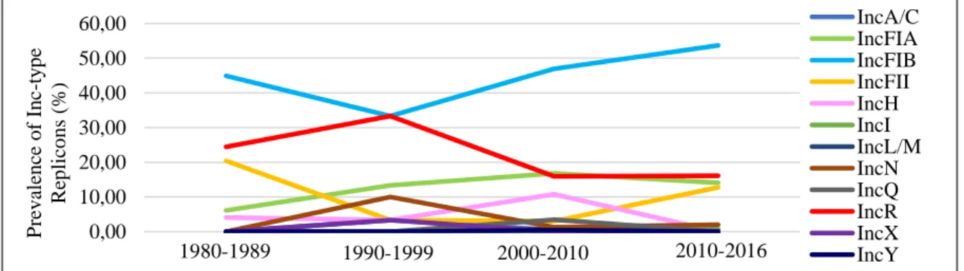
Por serem elementos importantes na evolução microbiana e na transferência de genes de resistência a antibióticos, o principal objectivo deste trabalho foi a análise e caracterização molecular de plasmídeos. Este estudo consiste no maior projecto de sequenciação de isolados clínicos de K. pneumoniae em Portugal. No total, 183 isolados de K. pneumoniae obtidos em diferentes hospitais de Lisboa ao longo de 36 anos foram sequenciados por uma plataforma de segunda geração (Illumina). Nesta fase inicial, averiguámos quais os replicões mais prevalentes, a sua distribuiçao temporal e a sua associação com certos STs. Os resultados obtidos mostram que o replicão IncFIB é o mais frequente na maioria dos STs destes isolados portugueses e reportam uma tendência para o aumento da sua prevalência nos últimos anos. Adicionalmente, estes resultados apontam para a forte disseminação temporal deste replicão em Portugal e em diversos STs. Por outro lado, outros replicões como IncR ou IncFII demonstram uma diminuição da sua prevalência ou valores estáveis ao longo do tempo.
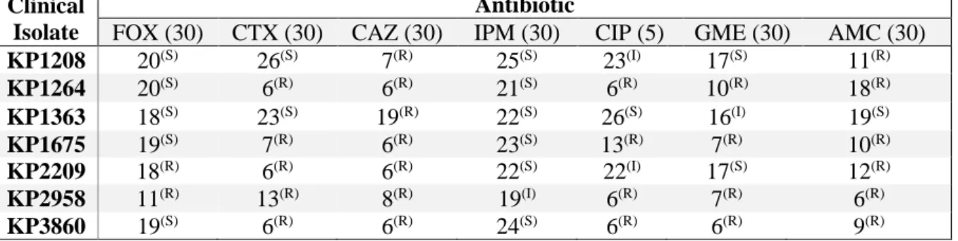
Posteriormente, sete isolados de STs distintos foram escolhidos para uma análise mais detalhada dos seus plasmídeos e do respectivo conteúdo génico através de sequenciação genómica completa por uma plataforma de terceira geração (PacBio). Cinco destes isolados são MDR e apresentam resistência concomitante à cefotaxima, mas nenhum mostrou ser resistente ao carbapenemo imipenemo. A assemblagem dos contigs foi realizada de duas formas: uma usando somente as reads obtidas por PacBio, e uma abordagem híbrida em que se combinaram as reads obtidas pelas duas plataformas de
ix
sequenciação. Foram obtidos um total de 20 plasmídeos, maioritariamente comprendendo replicões do tipo IncFII(K) e IncFIB(K). Dois dos isolados contêm megaplasmídeos do mesmo grupo de incompatibilidade; tratam-se, porém, de plasmídeos que são multireplicão, e em ambos os casos estes replicões encontram-se associados a divergência nucleotídica e terão acumulado polimorfismos potencialmente envolvidos na alteração da especificidade dos mecanismos de incompatibilidade, ou simplesmente à sua inactivação.
A maioria dos plasmídeos aqui estudados contém genes que codificam para proteínas membranares, sobretudo transportadores ABC e proteínas involvidas na mediação da conjugação. Para além destes, outros foram detectados, nomeadamente genes que codificam para proteínas fágicas, mobilização de ferro e resistência ao mercúrio. Quanto aos genes de resistência a antibióticos, as classes mais frequentemente encontradas são β-lactamases e proteinas de resistência aos aminoglicosídeos. Notavelmente, neste estudo foi descrita pela primeira vez a β-lactamase LAP-1 num isolado de K. pneumoniae; esta foi ainda a primeira detecção da enzima LAP em Portugal. O contexto genético deste gene foi descrito neste trabalho, juntamente com o contexto genético dos genes arr-2, conferindo resistência ao antibiótico rifampicina a um dos isolados, e blaCTX-M-15, a única ESBL presente nestes
plasmídeos e associada a um transposão Tn3 com uma estrutura semelhantte em todos eles.
De seguida, a diversificação estrutural dos megaplasmídeos deste estudo foi estudada por comparação com plasmídeos da base de dados pAtlas, obtidos de isolados da família Enterobacteriaceae e de regiões geográficas diversas. Os nossos resultados mostram que os plasmídeos IncFIB são os mais conservados não só em Portugal mas noutras regiões do globo, o que nos diz que a vasta disseminação de resistência a antibióticos se encontrará intimamente ligada a plasmídeos com este replicão. Destaca-se ainda a detecção de plasmídeos estruturalmente idênticos aos descritos neste estudo em diversas regiões geográficas e backgrounds genéticos, atestando assim o seu enorme potencial para a disseminação global de genes de resistência a antibióticos.
Por forma a corroborar a capacidade destes megaplasmideos de se disseminarem em multiplos hospedeiros, foram realizados ensaios de conjugação entre dois dos sete isolados clínicos de K. pneumoniae e as seguintes bactérias de Gram-negativo: Acinetobacter baumannii, Escherichia coli e Pseudomonas aeruginosa. O único receptor que foi possível conjugar foi E. coli. Usando o primeiro dador, obtivemos três conjugantes, os quais ganharam resisência às cefalosporinas de terceira geração cefotaxima, ceftazidima, e ao β-lactâmico amoxicilina na presença no inibidor ácido clavulânico. Usando o segundo dador, obtivemos dois conjugantes, os quais ganharam resistência aos antibioticos cefotaxima, ceftazidima, ciprofloxacina, gentamicina, tetracilina, sulfametoxazol-trimetoprim e ao β-lactâmico amoxicilina na presença do inibidor ácido clavulânico. No entanto, os níveis de resistência a estes antibióticos difere entre estes ultimos dois transconjugantes. Por forma a esclarecer estas diferenças, realizaram-se ensaios usando o inibidor de bombas de efluxo reserpina. Estes ensaios demostraram que as diferenças observadas não se devem exclusivamente a mecanismos de efluxo, uma vez que as reduções observadas ao nível das concentrações mínimas inibitórias de determinados antibióticos não foram significativas.
Na fase final, procurámos estudar a estabilidade de alguns dos megaplasmídeos aqui caracterizados na ausência da pressão selectiva de um antibiótico. Os resultados obtidos demonstram que, apesar do seu tamanho e da ausência desta pressão, todos os isolados à excepção de um apresentam uma elevada estabilidade plasmídica. Estes resultados sugerem que o extenso conteúdo génico presente nestes megaplasmídeos poderá acarretar outras vantagens selectivas para o hospedeiro, contribuindo desta forma para a presença e persistência de genes de resistência a antibióticos na ausência de uma pressão selectiva e no meio ambiente.
Palavras-chave: Klebsiella pneumoniae; Resistência a antibióticos; Plasmídeos; Transferência horizontal de genes.
xi TABLE OF CONTENTS Agradecimentos ... iv Abstract ... vi Resumo ... viii Table of Contents ... xi
List of Figures ... xiii
List of Tables ... xiv
List of Abbreviations ...xv
1. Introduction ...1
1.1 Klebsiella pneumoniae: an Emerging Healthcare-associated Pathogen ...1
1.2 The Antibiotic Resistance Crisis ...1
1.2.1Antibiotic Action Mechanisms ...1
1.2.2 Antibiotic Resistance Mechanisms ...1
1.2.3 K. pneumoniae: Antimicrobial Resistance in Europe...2
1.2.4 Emergence of ESBL and Carbapenemase-producing K. pneumoniae and Enterobacteriaceae ...3
1.3 Plasmids in Enterobacteriaceae ...5
1.3.1 Plasmids are Important Vehicles for the Spread of Antimicrobial Resistance ...5
1.3.2 Plasmid Incompatibility Groups ...5
1.3.3 Plasmid Diversity in Enterobacteriaceae ...6
1.3.3.1 Plasmid Diversity in K. pneumoniae ...7
1.3.4 Plasmid Cross-species Compatibility ...8
1.3.5 Mobile Genetic Elements as Drivers of Plasmid Diversification ...8
1.4 Objectives ...9
2. Materials and Methods ...10
2.1 Clinical Isolates and Gram-Negative Strains Used ...10
2.2 Culture Media and Solutions ...10
2.3 Isolation of Genomic DNA from K. pneumoniae and Whole genome Sequencing ...10
2.4 Plasmid DNA Extraction and Visualization by Agarose Gel Electrophoresis ...11
2.5 Bioinformatic Analysis of NGS Data ...11
2.6 Drug Susceptibility Testing and Minimum Inhibitory Concentration (MIC) Assays ...12
2.7 Conjugation Assays ...12
2.8 Transformatio Assays ...12
xii
2.8.2 Transformation by Electroporation ...13
2.9 Amplification of a Conserved Region of blaCTX-M-15 ...13
2.10 Plasmid Stability Assays Assays ...13
3. Results and Discussion ...14
3.1 The plasmidome of K. pneumoniae in Portugal is Diverse but Dominated by IncFIB-Type and ColRNAI Replicons ...14
3.2 Long-read Sequencing Enables Plasmid Structural Analysis ...15
3.3 Gene Content and Structural Analysis of Plasmidic Scaffolds ...18
3.4 Genetic Context of Antibiotic Resistance Genes ...18
3.4.1 blaCTX-M-15 is Located on a Truncated Tn3 Transposon ...21
3.4.2 First blaLAP-1 in a K. pneumoniae isolate ...22
3.4.2 arr-2 is Located on a Novel Class 1 Integron ...22
3.5 Phylogenetic Analysis of K. pneumoniae Megaplasmids ...22
3.6 Horizontal Transfer of K. pneumoniae Plasmids ...25
3.7 Plasmid Stability Assays Assays ...28
3.8 Concluding Remarks and Future Perspectives ...29
4. References ...30
xiii LIST OF FIGURES
Figure 1.1. Distribution of K. pneumoniae isolates susceptible and resistant to fluoroquinolones,
third-generation cephalosporins, aminoglycosides and carbapenems in EU/EEA countries, 2017 ...3
Figure 1.2. Percentage of K. pneumoniae invasive isolates with resistance to third-generation cephalosporins in EU/EEA countries, 2017 ...4
Figure 1.3. Epidemiological situation of carbapenemase-producing Enterobacteriaceae in 2018 ...5
Figure 1.4. Distribution of different Inc groups isolated from human, animal and environment in Europe, Asia and the Americas ...7
Figure 1.5. Distribution of genes encoding resistance to different antimicrobial classes carried by the most frequent Inc types: IncF, IncI, IncA/C and IncH ...7
Figure 1.6. Schematic representation of a class 1 integron ...9
Figure 3.1. Prevalence (%) of (A) Inc and (B) Col-Like Replicons in 183 K. pneumoniae clinical isolates from Portugal ...14
Figure 3.2. Distribution of replicos from 12 Inc groups throughout the years ...14
Figure 3.3. Agarose gel electrophoresis gel of plasmid extraction from seven K. pneumoniae clinical isolates ...14
Figure 3.4: Physical maps of the Inc megaplasmids from this study ...19
Figure 3.5: Scaffold comparison of plasmid-associated contigs ...20
Figure 3.6. Comparison of blaCTX-M-15 genetic context in three K. pneumoniae clinical isolates ...21
Figure 3.7. Genetic context of blaLAP-1 on the plasmid 1264HYB7 ...22
Figure 3.8. Genetic context of arr-2 on on In1264, a class I integron on the plasmid 1264HYB7...22
Figure 3.9. Phylogenetic analysis of IncFIA, IncFIB and IncFII plasmids. ...23
Figure 3.10. Agarose gel electrophoresis of blaCTX-M-15 PCR products from the donor, recipient and transconjugants obtained in the conjugation assays between Kp2209 and E. coli 1.1. ...27
Figure 3.11. Plasmid stability assay for Kp1264 ...22
Supplemetary Figure 5.1. Scaffold comparison of the plasmids obtained by the hybrid assemblies and HGAP with the same replicons and similar sizes ...41
Supplemetary Figure 5.2. Scaffold comparison of the pAtlas plasmids with a stronger phylogenetic relatioship with plasmids from this study ...43
xiv LIST OF TABLES
Table 2.1. K. pneumoniae isolates used in this study. ...10
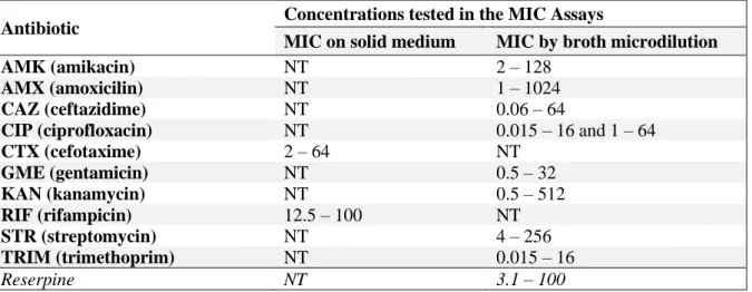
Table 2.2. Range of concentrations tested in the MIC Assays ...12
Table 3.1. Distribution of Inc-type replicons on 183 K. pneumoniae isolates from 37 STs ...15
Table 3.2 Kirby-bauer disk diffusion susceptibility tests for seven K. Pneumoniae clinical isolates from this study ...15
Table 3.3. Size, replicons and antibiotic resistance genes present on the plasmids used in this study, belonging to seven K. pneumoniae clinical isolates. ...17
Table 3.4. pAtlas plasmids with a stronger phylogenetic relationship with the Inc plasmids from this study. ...25
Table 3.5. Kirby-Bauer disk diffusion susceptibility tests of three transconjugants: EC2209_A, EC2209_B and EC2209_C ...26
Table 3.6. Kirby-Bauer disk diffusion susceptibility tests of two transconjugants: 3860EC_A and 3860EC_B ...26
Table 3.7. MIC assays for EC3860_A, EC3860_B, E. coli 1.1 and KP3860, with and without reserpine ...26
Table 3.8. MIC assays for STR and TRIM ...27
Table 3.9. Kirby-Bauer disk diffusion susceptibility tests for isolates A, B and C, obtained from plasmid stability assays ...28
Supplemetary Table 5.1. Culture Media used in this study ...39
Supplemetary Table 5.2. Composition of the solutions used for genomic DNA extraction ...39
Supplemetary Table 5.3. Composition of the solutions used for plasmid extraction ...39
Supplemetary Table 5.4. Antibiotics used in this study and their stock concentration ...39
Supplemetary Table 5.5. HGAP and hybrid assembly of seven K. pneumoniae clinical isolates. ...40
Supplemetary Table 5.6. Size of plasmid assemblies and therein identified replicons by the HGAP assembly ...40
Supplemetary Table 5.7. Size of plasmid assemblies and therein identified replicons by the hybrid assembly ...40
Supplemetary Table 5.8. Gene content of 14 out of the 20 plasmids from study. ...42
Supplementary Table 5.9. Antibiotic resistance genes present on the chromosome of seven K. pneumoniae isolates ...44
xv LIST OF ABBREVIATIONS
AMC: Amoxicillin-Clavulanate Acid AMK: Amikacin AMX: Amoxicillin Bp: Base Pairs CAZ: Ceftazidime CIP: Ciprofloxacin CHL: Chloramphenicol CTX: Cefotaxime DDW: Double-distilled Water DMSO: Dimethyl Sulfoxide
DNTP: Deoxyribonucleotide Triphosphate EDTA: Ethylenediamine Tetraacetic Acid EEA: European Enconomic Area
ESBL: Extended-Spectrum β-Lactamase EU: European Union
FOX: Cefoxitin GME: Gentamicin
HGAP: Hierarchical Genome Assembly Process
Inc: Incompatibility IPM: Imipenem IS: Insertion Sequence LB: Luria-Bertani
MDR: Multidrug Resistant MHA: Müeller-Hinton Agar MHB: Müeller-Hinton Broth
MIC: Minium Inhibitory Concentration NGS: Next Generation Sequencing OD: Optical Density
ORF: Open Reading Frame PCR: Polymerase Chain Reaction
RIF: Rifampicin
SDS: Sodium Dodecyl Sulphate SOB: Super Optimal Broth
SOC: Super Optimal Broth with Catabolite Repression ST: Sequence Type STR: Streptomycin SXT: Sulfamethoxazole-Trimethoprim TE: Tris-EDTA TET: Tetracycline TRIM: Trimethoprim
1
1.
INTRODUCTION
1.1 Klebsiella pneumoniae: an Emerging Healthcare-associated Pathogen
Klebsiella pneumoniae is a Gram-negative, rod-shaped, non-motile bacillus that belongs to the Enterobacteriaceae family. This bacterium is facultatively anaerobic, oxidase-negative, catalase-positive, lactose-fermenting and non-spore-forming1. It is usually encapsulated, this being one of the
most important factors for virulence2,3, since it grants protection against phage predation, increases
resistance to serum complement and it allows the bacteria to escapse the host’s inflammatory system (phagocytosis and macrophages/neutrophils intracellular killing)4–6. Aside the clinical settings, which
represent a more recent ecological niche, K. pneumoniae can be found on the soil, sewage, surface waters, plants and animals7–10. In humans, the primary reservoir for this bacterium is mostly the gastrointestinal
tract, although it can also be found in the nasopharynx, it being infrequent on the skin8.
Although K. pneumoniae typically colonises humans’ mucosal surfaces without causing pathology11, it is responsible for a significant number of community-acquired infections worldwide12.
Furthermore, Klebsiella-associated infections are strongly linked with hospitalization and elevated morbidity and mortality rates in hospital settings9,13, namely by causing nosocomial outbreaks and
triggering a wide range of infections such as urinary infections, pneumonia, septic shock and meningitis6,14. The groups with higher risk of being infected by this opportunistic pathogen are mainly
the neonates, the elderly, and immunocompromised individuals often with other severe underlying infections, certain comorbidities (like diabetes) or life-threatening diseases9,15,16. Additional risk factors
that lead to the increase of K. pneumoniae infection rates include: previous antibiotic therapy, invasive procedures (tracheal intubation, central venous catheterization, surgery), blood transfusion and organ transplantation, the size of the hospital, the duration of hospitalization and patient readmission6,16–18.
1.2 The Antibiotic Resistance Crisis 1.2.1 Antibiotic Action Mechanisms
In nature, bacteria and fungi produce chemicals to attack other microbes that compete for the same resources. These act by blocking or destabilizing processes or structures essential for microbial growth and survival. Antibiotics can be either bacteriocidal (if they kill bacteria) or bacteriostatic (if they interrupt bacterial growth)19. These drugs have three main targets: (1) cell wall synthesis, (2) nucleic
acid synthesis and (3) protein synthesis20. The main class of antibiotics from the first group are β-lactams
(penicillins, cephalosporins, monobactams, and carbapenems), which bind to cell wall synthesis enzymes, causing the disruption of the peptidoglycan coat and consequent bacterial lysis21,22. In the
second group, fluoroquinolones inhibit the bacterial DNA gyrase, needed for bacterial replication to prevent excessive DNA positive supercoiling23. Other examples of antibiotics from this second group
are sulfonamides and trimethoprim, which inhibit DNA synthesis by disrupting the cell’s folic acid metabolism24, and rifampicin, which inhibits the bacterial RNA polymerase, responsible for
transcription25. In the third group, aminoglycosides and tetracyclines inhibit protein synthesis by
blocking the ribosome’s 30S subunit22, while chloramphenicol interfers with the 50s subunit26; these
three classes lead to premature termination of mRNA translation22.
1.2.2 Antibiotic Resistance Mechanisms
The notable genetic plasticity of bacteria allows them to respond to a wide array of environmental pressures; therefore, it only takes months to years after the start of the routine administration of a new antibiotic for resistant bacterial strains to rise27. For example, penicillin
resistance was first detectef in the mid 1940s, only two years of its introduction28. Alarmingly, some
2
is shortened29,30. There are multiple forms of antibiotic resistance, falling either into the intrinsic or the
acquired resistance categories19.
Intrinsic resistance is based on the bacteria’s structural or functional characteristics and is a result of phenotypic changes without the uptake of additional genetic information; it can be a consequence of a reduced membrane permeability30. Gram-negative bacteria are naturally insusceptible
to many antibiotics due to the presence of an outer-membrane; this factor, together with the saturated nature of the fatty acid chains within their lipopolysaccharide layer, contributes to a decrease of membrane fluidity and permeability in these bacteria31,32. Intrinsic resistance can also be granted by
porins, nonspecific diffusion channels present on the outer-membrane that not only allow the entry of specific key nutrients but also block the influx of a number of antibiotics33.
Antibiotics can also be expeled by efflux pumps, which keep the intrabacterial concentrations of certain drugs at low and not toxic levels by expelling them at a faster rate than they can enter the cell; for example, TET-resistant bacteria frequently overproduce efflux pumps34,35. These membranal
structures can be specific for one drug or export a variety of molecules34. However, unlike what happens
with TET, the action of efflux pumps in some antibiotics usually leads to low-levels of resistance, so, for higher resistance levels, bacteria typically depend on acquired resistance mechanisms, although this is not always the case34,35.
Antibiotic resistance can be acquired either by the horizontal transfer of resistance genes or by spontaneous chromosomal mutations in genes associated with the antibiotic’s mechanism of action20,27.
An example of the latter is quinolone resistance in Enterobacteriaceae23. Acquired resistance has many
possible origins, such as the modification or degradation of the antibiotic’s active site. Enterobacteriaceae most commonly gain resistance to β-lactams by the acquisition of genes coding for β-lactamases, enzymes that neutralize the β-lactam ring before these antibiotics can reach the peptidoglycan coat22. In the 1980s, the increased prevalence of β-lactamases like TEM-1 e SHV-1 lead
to the introduction of 3rd generation cephalosporins (like ceftazidime an cefotaxime) in hospital settings;
this new selective pressure lead to the rapid rise of novel lactamases: Extended-Spectrum β-Lactamases (ESBLs)36. These grant resistance to all β-lactams but carbapenems and cephamycins36,37.
Modification or overproduction of the target enzyme can also lead to acquired resistance; for example, RIF resistant strains usually emerge due to specific mutations which are expected to decrease binding of the RNA polymerase to this drug38. Last but not the least, the acquisition of alternative metabolic
pathways to those inhibited by the drug can lead to acquired antibiotic resistance20.
1.2.3 K. pneumoniae: Antimicrobial Resistance in Europe
K. pneumoniae strains are frequently multidrug resistant (MDR), that is, resistant to three or more classes of antibiotics39. The increasing emergence of MDR K. pneumoniae strains producing
ESBLs and plasmid-encoded carbapenemases has become a major cause of concern worldwide due to the lack of alternative therapeutic options and the high morbidity and mortality rates associated with infections by these strains40-42. Antimicrobial inappropriate and irrational prescribing and overuse in
clinical and food-producing settings, together with the absence of monetary incentives and the challenging regulatory requirements for the production of new antibiotics, has been stipulated to be the root of this antibiotic resistance crisis43–46.
Data from the 2017 “Surveillance of antimicrobial resistance in Europe” report47 show that, in
2017, 34.1% of the K. pneumoniae clinical isolates in Europe were resistant to at least one of the following four antimicrobial groups: fluoroquinolones, third-generation cephalosporins, aminoglycosides and carbapenems. The most common resistance phenotype in this study was the combination of resistance to fluoroquinolones, third-generation cephalosporins and aminoglycosides. Fluoroquinolones was revealed to be the class with the highest population-weighted mean resistance percentage (31.5%) and it was followed by third-generation cephalosporins (31.2%), aminoglycosides
3
(24.1%) and carbapenems (7.2%). Cases of clinical isolates resistant to at least two of these four groups were more comon than cases of single resistance. Figure 1.1 shows a substantial inter-country variability: countries on the south and east of Europe (Bulgaria, Greece, Poland, Lithuania, Romania, Slovakia and Italy) reported a higher resistance percentage to the four antimicrobial groups mentioned above when compared to northern european countries (Finland, Sweden, Norway and Denmark).
Figure 1.1. Distribution of K. pneumoniae isolates susceptible and resistant to fluoroquinolones, third-generation cephalosporins, aminoglycosides and carbapenems in EU/EEA countries, 2017. Adapted from: ECDC (2018)47.
1.2.4 Emergence of ESBL and Carbapenemase-producing K. pneumoniae and Enterobacteriaceae
ESBLs have become an important focus of attention over the last years. It is believed that the ESBL-spread phenomenon had its origins in Europe, most likely due to the selective driving forces of third-generation cephalosporins consumption, which began here36. Notably, in the ECDC report
mentioned above47, 87.8% of the K. pneumoniae isolates resistant to third-generation cephalosporins
were found to be positive for the presence of an ESBL. Once again, important geographical differences are evident: eight countries reported a statistically significant increase of K. pneumoniae clinical isolates resistant to third-generation cephalosporins from 2014 to 2017 (Finland, Netherlands, United Kingdom, Germany, Spain, Hungary, Portugal and Lithuania), while only three reported a decrease (Latvia, Romania and Slovakia). The distribution of K. pneumoniae isolates resistant to third-generation cephalosporins in Europe in 2017 is evident in Figure 1.2. Countries on the north report up to 99% of isolates susceptile to this class of antibiotics, while in the south-east regions of Europe the percentange of resistance can be higher than 50% .
One of the most epidemiologic relevant ESBL in K. pneumoniae is the CTX-M family. In fact, in Europe there has been a significant rise of these β-lactamases and a decrease of the TEM and SHV variants in the past few years48. Theses β-lactamases are inhibited by clavulanate and tazobactam and
grant high levels of resistance to CTX, while also conferring resistance to ceftriaxone and aztreonam49,50.
Although Enterobacteriaceae carrying CTX-M β-lactamases have spread all over the world, eastern Europe, South America, and Japan are hot areas for their emergence51. During the 1990s, the same
4
CTX-M-like enzymes were isolated and identified for the first time in distant countries and continents – for example, CTX-M-1 was first detected in an Escherichia coli isolate from Japan in 1986 (this enzyme was originally named FEC-1)52 and in a K. pneumoniae isolate from California in 198753. The
rapid and strong dissemination of these enzymes is probably dued to international travel51.
A variety of ESBLs have been reported in K. pneumoniae isolates from Europe. On northern countries, the most commom ESBLs are from the CTX-M and SHV families48. In Spain, there is a
notable higher prevalence of the enzymes CTX-M-9, CTX-M-14, CTX-M-10 and TEM-4; in Portugal, studies of individual hospitals report TEM, SHV and CTX-M-like ESBLs, and a predominant spread of CTX-M-14 and TEM-52, alongside with TEM-24, CTX-M-15, CTX-M-32 and SHV-12 which are commonly detected in both countries54–57. In Italy, TEM β-lactamases are the ESBLs most commonly
reported, as well as CTX-M-1, -15 and -32; these enzymes are less predominant in K. pneumoniae than they are in E. coli58,59. ESBL-producing K. pneumonia in eastern Europe most commonly carry
CTX-M-3, but also SHV-2 and SHV-560–62.
The CTX-M-15 enzyme seems to be wide-spread in almost all european countries51. This
lactamase grants resistance to ceftazidime, which is not the case for the majority of CTX-M-type β-lactamases50,63. CTX-M-15 has been increasingly associated with community and hospital-acquired
infections48,64–67. This enzyme was first isolated in India, in the year 200063. In Portugal, the first K.
pneumoniae strain producing CTX-M-15 was isolated in 200368. The current CTX-M-15 pandemic
dissemination is partially atributed to the E. coli strain O25:H4-ST131, which has been detected in the majority of european countries and has been found to harbour different plasmids, all of them carrying this enzyme69,70.
Regarding carbapenem resistance driven by carbapenemase-producing bacteria, it is perceptible that resistance levels in K. pneumoniae have been increasing throughout the years. To better study this rise, the European Antimicrobial Resistance Genes Surveillance Network (EURGen-Net) developed the project “European Survey of Carbapenemase-Producing Enterobacteriaceae”71. Here, seven
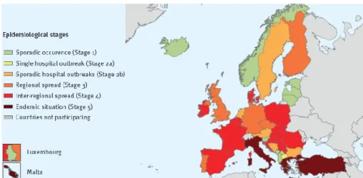
epideomological stages were established, ranging from Stage 0 (no cases reported) to Stage 5 (endemic situation). While in 2010 five of the participating countries were classified as stage 0 (Austria, Bulgaria, Estonia, Iceland and Slovenia), in 2018 all 37 countries from this study reported cases of infections by a carbapenemase-producing Enterobacteriaceae (Stage 1 or higher). From 2015 to 2018, 11 countries reported a worsening of the epidemiological stage (Bosnia and Herzegovinaa, Croatia, Cyprus, Czech Republic, Finland, Iceland, Ireland, Kosovo, North Macedonia, Portugal and Serbia), while only one country reported a decrease (Slovenia). No changes were described for the remaining 25 countries. Greece, Italy, Malta and Turkey reported an endemic situation both in 2015 and in 2018. The epidemiology of carbapenemase-producing Enterobacteriaceae in 2018 is described in Figure 1.3. K.
penumoniae is the Enterobacteriacea where carbapenem-resistance is more predominant.47
Figure 1.2. Percentage of K. pneumoniae invasive isolates with resistance to third-generation cephalosporins in EU/EEA countries, 2017. Adapted from: ECDC (2018)47.
5
In the countries that provided genotypic data to this study as to understand the underlying molecular basis of resistance, the most frequently detected carbapenemase genes were blaVIM, blaKPC,
blaNDM and blaOXA-48, followed by blaIMP, blaGES, blaFRI, blaIMI and blaSME71.
Figure 1.3. Epidemiological situation of carbapenemase-producing Enterobacteriaceae in 2018. Brolund et al. (2019)71.
1.3 Plasmids in Enterobacteriaceae
1.3.1 Plasmids are Important Vehicles for the Spread of Antimicrobial Resistance
Plasmids are bacterial, circular DNA molecules capable of self-replication. Although they carry no genes indispensable for host cell’s growth when under non-stressed conditions, resistance genes located on plasmids grant a selective advantage to the bacteria under antimicrobial pressure72. Plasmids
carrying antimicrobial resistance genes are a key vehicle for bacterial evolution since these promote the horizontal gene transfer between bacteria from different genera. This is particularly true when they carry multiple genetic markers that confer resistance to distinct classes of antibiotics, since this offers a selective advantage to the host when several antimicrobial drugs are simultaneously administered73.
There are two possible ways for a plasmid to be taken up by bacteria, one being transformation – the stable and functional uptake, integration and expression of extracellular DNA by bacterial cells that previously reached a physiological state of competence under specific growth conditions74,75. Many
genera of pathogenic bacteria are naturally transformable, competence being triggered by specific environmental changes like lack of nutrients or celular density, and sometimes being dependent on the growth-stage76,77. In lab settings, competence can be achieved with treatment with high concentrations
of Ca2+ and heat shock74,78. Bacteria that are difficult to transform (due to the presence of thick cell walls
or capsules, for example) or that are not naturally transformable can also be subjected to electroporation, a technique where cells are mixed with the DNA and then exposed to short high-voltage electrical pulses to permeabilize the cell envelope79,80. These procedures allow the cell to take up double-stranded DNA74.
Another process for plasmid mobilization is conjugation. Plasmid conjugative transfer is mediated by cell-to-cell channels (pili) through which DNA can be transfered from the donor cell (containing the conjugative plasmid) to the recipient cell72.
1.3.2 Plasmid Incompatibility Groups
Plasmid replication is regulated by its basic replicon – a small sequence of DNA (typically not larger than 1.8 Kbp) that comprehends the ori (origin of replication) and other sequences coding for specific replication initiation proteins81-83. Bacteria can carry multiple plasmids; however, in the past it
was belived that plasmids with the same replicon type could never coexist in the same cell, due to a phenomenon called plasmid incompatibility83. This is caused by a competition for factors involved in
replication control and/or DNA equipartitioning during cell division, which inevitably gives a selective advantage to the plasmids that, for example, can replicate faster due to their smaller size or are less
6
toxic82,83. Plasmid incompatibility is also strongly related to another phenomenon, plasmid exclusion –
the presence of certain membrane proteins preventing conjugation with a donor cell carrying the same plasmid. pTraT and pTraS are partially responsible for this, the first protein reducing the cell’s ability to form stable mating aggregates (surface exclusion) and the latter reducing DNA transfer within stable mating aggregates (entry exclusion)84. With the first formal system of plasmid classification, plasmids
were sorted into distinct incompatibility (Inc) groups81. This classification method consisted on the
transfer of a plasmid of an unknown Inc group into a bacterial strain carrying a plasmid of a previously identified Inc group. The plasmid that was being tested would be assigned to the same Inc group as the plasmid from the recipient strain if one of these molecules was eliminated in the population85.
Recent studies question this concept of incompatibility by denoting the stable maintenance of plasmids harbouring the same origin of replication in a celular line86. During cell division, plasmids
replicate and segregate within minutes to oposite polls of the cell; here, they may replicate once again87.
The partitioning of plasmids from different Inc groups is independent and even happens at different times of the cell cycle, which is crucial for the cell to maintain both plasmids86,87. If replicons acumulate
mutations in the genes responsible for this stochastic process of plasmid partitioning, the plasmid incompatibility specificity might be altered88. It has been theorized that this frequently happens in
multireplicon plasmids, where one replicon is strongly conserved while the other tends to diverge. Mutations in one of the replication initiation genes can even lead to the emergence of a novel replicon and, consequently, changes in replicon incompatibility88.
In 1988, a new plasmid classification method was developed, based on the recognition of the basic replicons frequently found on Enterobacteriaceae using hybridization tecniques82. Since those old
plasmid identification systems were time-consuming and non-pratical for large scale analysis, Caratolli and collegues developed a PCRbased replicon typing (PBRT) technique, a more efficient, fast and inclusive method of plasmid classification. 18 pairs of primers were designed to perform five multiplex and three simplex-PCRs89, recognizing the most representative Inc groups circulating among
Enterobacteriaceae: IncFIA, FIB, FIC, HI1, HI2, I1-Ig, L/M, N, P, W, T, A/C, K, B/O, X, Y, F, and FIIA82. PBRT was later improved with the implementation of real-time PCR, wich made this tecnique
faster and more sensitive to the detection of low-copy plasmids90.
Considering how these classification methods focus solely on the classic and most common replicons, it is clear that they fail to detect divergent or novel replicons. In order to complete the typing and classification of all the plasmids circulating in Enterobacteriaceae, other replicons should be cloned and sequenced89. Additional big challenges for plasmid replicon typing are multireplicon plasmids and
the fact that some plasmids can cointegrate, originating, by recombination events, a new multireplicon that might not be easily detected91–93. Therefore, the most accurate methodology to characterize a
plasmid is by subjecting its full-length DNA sequence to new-generation sequencing and then annotating its resistance and virulence genes, as well as its replicon(s) using PlasmidFinder. PlasmidFinder is an online plataform with a database built on plasmid replicon sequences obtained from a total of 559 fully sequenced Enterobacteriaceae plasmids available on the NCBI database94.
1.3.3 Plasmid Diversity in Enterobacteriaceae
IncF, IncI, IncA/C and IncH are some of the most common replicons in Enterobacteriaceae in Europe, Asia and the Americas95. Their distribution is summarized in Figure 1.4, and the antibiotic
classes most frequently found on these plasmids is summarized in Figure 1.5.
IncF are large (>100 Kbp) low-copy-number conjugative plasmids that can harbour more than one replicon – usually FII together with IncFIA or FIB, or other replicons with a broader host range (a concept discussed in 1.3.5)92,95. The antibiotic resistance genes most frequently found on IncF plasmids
code for ESBLs and other β-lactamases (in particular blaTEM, blaSHV, blaCTX-M and carbapenemases),
7
Figure 1.4. Distribution of different Inc groups isolated from human, animal and environment in Europe, Asia and the Americas. “Other”: ColE, IncB/O, IncK, IncL/M, IncN, IncP, IncR, IncT, IncU, IncW, IncX, IncY and IncZ. Adapted from:
Rozwandowicz et al. (2018)95.
Figure 1.5. Distribution of genes encoding resistance to different antimicrobial classes carried by the most frequent Inc types: IncF, IncI, IncA/C and IncH. The “Other” antibiotics group includes genes encoding resistance to: trimethoprim,
chloramphenicol, florfenicol, colistin, fosfomycin. Adapted from: Rozwandowicz et al. (2018)95.
CTX-M-15 spread is highly associated with IncF plasmids, mainly the subtype IncFII95,96. The
blaCTX-M-1 gene is commonly carried both on IncN and IncI1 plasmids73,98. In Spain the United Kingdom,
IncK plasmids have been linked with CTX-M-14 dissemination, while in Asia the gene coding this enzyme is typically found on IncF plasmids99–101. CTX-M-3disseminated in Europe and China in a
conjugative plasmid from the IncL/M family95. Genes coding for carbapenemases are mostly found on
the following families: IncL/M, IncA/C, IncF, IncHI1, IncX3 and novel IncN and IncHI1 variations, as well as the rare IncT family73. oqxA and oqxB, coding for efflux pumps, have been identified on an
IncX1 plasmid, as well as on IncF plasmids in E. coli isolates from China and Eastern Europe102,103. Regardless of these trends, it is clear that most of these antibiotic resistance genes have a
notable potential for the spread among a large repertoire of plasmids95.
Although the high prelavence of these plasmids is strongly linked to an antibiotic selective pressure, their distribution is ubiquitous95,104. The presence of virulence factors in association with
resistance genes in plasmids provides a strong selective advantage73,105. For example, the existence of
virulence clusters on the scaffold of some IncF plasmids might explain their spread among Enterobacteriaceae in the ausence of an antibiotic pressure106,107; IncI1 plasmids have also been found
to carry genes coding for type IV pili, contributing to adhesion and bacterial invasion107.
1.3.3.1 Plasmid Diversity in K. pneumoniae
The PCR-based replicon typing method mentioned above did not allow the typing of most K. pneumoniae plasmids89. However, with the advances in genomics and whole-genome sequencing,
8
the complete sequencing of K. pneumoniae plasmids is now possible. The plasmids carried by this bacterium are generally from the IncF family, in particular IncFII. FIIk, the most frequent replicon identified in K. pneumoniae, is often linked with replicons of the FIB-type. FIBpKPQIL and FIBpKPN
are the FIB-type replicons more recurrently reported in K. pneumoniae plasmids109. Plasmids harbouring the pKPN-CZ variant have been found to carry blaCTX-M-15 and type 3 fimbriae110. However,
blaCTX-Mgenes on Klebsiella isolates are typically located on plasmids that are very similar to E. coli
plasmids and that differ from the ones carrying blaKPC. Multireplicon plasmids carrying IncFIIk and IncR/IncN are als common in K. pneumoniae, these being linked with the spread of blaKPC-2 in Italy,
USA, China, Russia and Taiwan111. The blaKPC-2 and blaKPC-3 genes have also been linked with
IncFIIkpKpQIL plasmids112,113.
Another class highly represented in K. pneumoniae is Col-Like plasmids, in particular ColRNAI73,114. Bacteria usually carry around 20 copies of these small plasmids (around 6–40 Kbp) per
cell95. These highly mobilizable vectors have mostly, but not exclusively, been associated with
carbapenem and quinolone resistance in K. pneumoniae and other Enterobacteriaceae114-116.
1.3.4 Plasmid Cross-species Compatibility
Certain plasmids can only replicate and be stably maintained under specific conditions tailored to a few species (narrow-host range plasmids), while others are promiscuous and have the capability to replicate in a medium or broad range of hosts117. The IncF family is restricted to Enterobacteriaceae,
while IncA/C, IncL/M, IncP, IncQ, IncR, IncU and IncW plasmids have a broad range of bacterial hosts95. Plasmid transfer between bacteria from different species, genera or families can be limited by
factors such as interactions at the cell surface blocking conjugative contact, DNA degradation by the new host’s restriction systems or the absence of the machinary needed for plasmid replication in the new host118. Other factors incluce G+C content, oligonucleotide frequencies and codon usage119.
The determination of the plasmid’s host range is frequently dictated by the structure of its origin of replication117. The precise coordenation of replicon recognition and the initiation of replication is
depended on the origin’s nucleotide sequence, on the presence of specific proteins recognizing specific sites at the ori and modifying its DNA structure, and on the presence of DNA tertiary structures at the origin118. Some plasmids, like the ones from the broad-host range family IncQ, seem to be independent
from the host for replication, and this is a key factor for their maintenance in many baterial hosts. These types of plasmids often proceed with the first priming step by carrying genes coding for their own primases (RepA, RepB and RepC)117,120. On the other hand, ColEl-type plasmids can only partially
replicate in Pseudomonas spp. when the medium is supplemented with the purified E. coli enzymes gyrase and DNA polymerase I118.
Overreplication or runaway (uncontroled) replication of low-copy-number plasmids has been proven to lead to the death of the bacterial host121,122. Therefore, failure in keeping the replication
frequency in a low level is another explanation for the inability of narrow-host range plasmids to replicate in certain hosts118. The existence of several replication origins in multireplicon plasmids is also
a factor that can play in favour of their broad-host range, if the different replicons are functional in different hosts116. For example, Da SilvaTatley and Steyn identified a plasmid that harboured a
pUC1-like and an IncF-pUC1-like replicon, both active and functional in distinct bacterial hosts123.
1.3.5 Mobile Genetic Elements as Drivers of Plasmid Diversification
Antimicrobial resistance genes can be mobilized between bacteria through the acquisition of mobile elements in conjugative plasmids72,124.
Transposable elements, one type of mobile elements, are considered to be "jumping genes", since they are able to move (“jump”) from one location to another in the genome, either within a plasmid
9
or the chromossome, or between these two molecules124. Insertion sequence elements (IS), short
sequences of DNA (0.5–2 Kbp) coding for a transposase, are the simplest example of a transposable element125. Similar to ISs, ISCR elements are small mobile elements exhibiting a rolling circle
mechanism for transposition and recombination126. Another type of transposable elements are
transposons, which typically consist of a pair of an IS element and a central DNA feature which expression changes the cell phenotype124. Transposition requires the ends of the transposon to be flanked
by the same IS element (usually short inverted repeats)and the presence of a transposase, an enzyme at the end of a transposon that catalyzes this event of transposition125. Some well-studied transposons
found in Enterobacteriaceae are the Tn3 transposon (associated with β-lactam resistance), the Tn5 transposon (encoding resistance to the aminoglycosides kanamycin and neomycin, among others), the Tn10 transposon (encoding tetracycline resistance) and the Tn21 transposon (linked with resistance to streptomycin, spectinomycin, sulphonamides and often containing the mercury resistance operon)124,125.
Conjugative transposons are transposon-like elements, but these have a distinct method of excision and integration. These also combine features of plasmids, since after excision they form a closed circular structure; however, unlinke plasmids, these do not replicate. After excision, conjugative transposons will either integrate in another part of the chromosome or br transfered to other cells by conjugation to then integrate into the recipient's genome127.
Finally, integrons are another type of mobile elements that, unlike transposons, are based on site-specific recombination by an enzyme called integrase124. The amino acid sequences of the gene
coding for these enzymes is what allows integron classification – class 1 Integrons carry intI1, class 2
intI2, class 3 carry intI3, and class 4 carry intI4128. The first class, whose structure is represented in
Figure 1.6, is the most commom. Integrons usually carry gene cassettes, small DNA sequences with a single gene (that can code for antibiotic resistance, for example) and a recombination site129.
Figure 1.6. Schematic representation of a class 1 integron. The integron conserved region comprises the integrase gene intl1
(coding for the enzyme responsible for integration), as well as an integration site (attI1) and promotors (P1 and P2) for the transcription of the genes on the variable region of the integron (“Resistance gene 1” and “Resistance gene 2”). The arrows represent the direction of transcription. The attC regions are the recombination sites. qacE∆1 and sulI confer resistance to quaternary ammonium compounds and sulfonamides. Adapted from: Carattoli (2001)128.
1.4. Objectives
The primary goal of this study relies in the molecular characterization of plasmids, particularly megaplasmids (>100 Kbp) associated with MDR K. pneumoniae isolates. This study encompasses clinical isolates obtained from different hospitals in Portugal, in the Lisbon region, over a time span of 36 years. Second and third generation NGS data was obtained for these isolates with the aim of understanding plasmid diversity and their overall genetic plasticity under a global context. The specific objectives were:
✓ To characterize the gene content and genetic environment of plasmid-associated drug resistance genes, as well as their contribution to horizontal transfer of drug resistance;
✓ To study the mobilization of Klebsiella pneumoniae drug resistance plasmids to other Gram-negative bacteria responsible for hospital-associated infections through conjugation and transformation.
✓ To investigate the stability of studied plasmids in its host bacterial isolates in the absence of antimicrobial selective pressure;
10
2.
MATERIALS AND METHODS
2.1. Clinical Isolates and Gram-Negative Strains Used
The initial part of this study includes a total of 183 K. pneumoniae clinical isolates collected in four different hospitals and laboratories in Lisbon over a period of 36 years, between 1980-2016. Then, from these, seven representing different sequence types (listed on Table 2.1) were studied more in depth.
Table 2.1. K. pneumoniae isolates used in this study.
Clinical Isolate Origin Biological Sample Year Genome length (bp) ST
KP1208 Hospital A Urine 2006 5631566 35 KP1264 Hospital A Blood 2007 5495084 147 KP1363 Hospital B Blood 2005 5580655 16 KP1675 Hospital B Blood 2008 5787724 48 KP2209 Hospital A Urine 2008 5937559 133 KP2958 Hospital A Blood 2010 5727377 14 KP3860 Hospital A Blood 2013 5764368 397
The E. coli isolates CF604 (harbouring a pCFF14 plasmid) and CF804 (harbouring a pC804 plasmid) were used together with the seven isolates from Table 2.1 for plasmid extraction.
RIF-resistant spontanous mutants named 1.1 (previously obtained from other studies from Acinetobacter baumanni ATCC19606 (S531F), E. coli ATCC25922 (S540Y) and Pseudomonas aeruginosa ATCC9997 (D521G)) were chosen as recipients in conjugation assays. E. coli BL21DE3
(F– e14– (McrA–) hsdR (rK– mK–) glnV44 thr-1 leuB6 thi-1 lacY1 fhuA21 mcrB hflA150::Tn10 (TetR))
was used for transformation assays. 2.2. Culture Media and Solutions
The culture media, solutions and antibiotics used are listed on Supplementary Tables 5.1 to 5.4. 2.3. Isolation of Genomic DNA from K. pneumoniae and Whole Genome Sequencing
The DNA of 183 K. pneumoniae clinical isolates was extracted using a protocol from Soolingen et al. (1999)130.
The bacteria were incubated overnight in LB Agar at 37°C, and at least one loopful of culture was transfer into 400 µL of TE (1×). The bacterial suspensions were heated at 90°C for 20 minutes and cooled down at room temperature. Then, 5 µL of lysozyme (10 mg/mL) were added and mixed by vortexing before an incubation step a 37°C for at least 2 hours, and 75 µL of Solution A were added and mixed by slowly vortexing before another incubation step for 10 minutes at 65°C. Afterwards, 100 µL of a 5M NaCl solution and 100 µL of a prewarmed (65°C) CTAB/NaCl solution were added sequentially. The mixtures were vortexed until they became white and incubated for 10 minutes at 65°C before adding 750 µL of Solution B. After vortexing it again for a few seconds, this mixture was centrifuged (20 minutes, 16000g, room temperature). The supernatants were carefully transferred to sterile eppendorfs. Then, 450µL of isopropanol were added before storing the extracted DNAs at -20°C overnight. The next day, the DNAswere centrifuged (15 minutes, 16000g, 4°C). Most of the supernatant was removed and 1 mL of ethanol (70%) was added and mixed by turning the eppendorfs a few times upside down. After a final centrifugation step (5 minutes, 16000g, 4°C), most the supernatant was discarded and the remaining ethanol was removed with a pipette. To make sure there was no residual ethanol left on the DNA pellets, the eppendorfs were left with the lid opened for 5 to 10 minutes. The pellets were diluted in 100 μL of double-distilled water.
The genomic DNA obtained was sequenced in an Illumina HiSeq 4000 platform and a PacBio RSII platform in the Genome Institute of Singapore as part of a collaboration between Faculty of
11
Pharmacy of the University of Lisbon, London School of Hygiene and Tropical Medicine and Genome Institute of Singapore.
2.4. Plasmid DNA Extraction and Visualization by Agarose Gel Electrophoresis
Plasmid DNA was extracted from the seven K. pneumoniae isolates from Table 2.1 and the E.
coli isolates CF604 and CF804 using a protocol adapted from Birnboim and Dolly (1979)131.
The bacteria were were grown overnight in LB Agar and then 1.5 mL of culture were centrifuged (13000g, 4 minutes, room temperature). The pellets were gently ressuspended in 100 μL of Solution I, placed on ice for 30 minutes, after which 200 μL of Solution II was added and gently mixed. After a second ice incubation for five minutes, 300 μL of Solution III was added and gently mixed, followed by a centrifugation step (15000g, 5 minutes, 4°C) and transfer of the supernatants to sterile eppendorfs. Afterwards, 1 mL of ethanol (97%) was added to the collected supernatants and the mixtures were incubated at -20 ºC over-night, after which DNA pellets were collected by centrifugation (15000g, 10 minutes, 4°C) and washed in cold ethanol (70%). After another centrifugation step (15000g, 3 minutes, 4°C), ethanol was carefully removed and the pellets dilluted in 100 μL of DDW.
Plasmid DNA was visualized by agarose gel electrophoresis of 45 μL of each DNA samples, which ran for approximately 17 hours in a 0.7% agarose gel (routine grade agarose, Nzytech) made with TAE 1× at 60V and 4°C. The gel was stained after the run, by immersing it on 200 mL of TAE 1x containing 0.5 µg/ml of Ethidium Bromite (BioRad).
2.5. Bioinformatic Analysis of NGS Data
PacBio sequencing reads were assembled using the Pacific Biosciences SMRT Analysis Server portal and the therein implemented Hierarchical Genome Assembly Process (HGAP). Illumina sequencing data was de novo assembled using SPAdes assembler implemented on the Unicycler pipeline for optimization of best k-mer values, contig correction and rotation (https://github.com/rrwick/Unicycler). Short and long read data obtained by Illumina and PacBio were also assembled using Unicycler using a hybrid assembly approach by bringing short read based contigs using long read sequence data. The sequence types were determined using the pubMLST portal (https://pubmlst.org/kpneumoniae/).
Plasmid replicons and drug-resistance genes were initially detected using PlasmidFinder and ResFinder, available at “Center for Genomic Epidemiology” (http://www.genomicepidemiology.org/). Only the plasmids which replicons had a coverage equal or bigger than 60% and a percentage of identidy of at least 95% were considered (except for Col-Like plasmids, for which the cutt-of for the percentage of identidy was 80%)94. Screening for drug resistance genes was complemented with AMRFinder
(https://ncbi.nlm.nih.gov/pathogens/antimicrobial-resistance/AMRFinder/) and the NCBI Bacterial Antimicrobial Resistance Reference Gene Database (Accession PRJNA313047). The circularized contigs were annotated using RAST (http://rast.nmpdr.org/). BRIGG software (BRIG-0.95-dist) and Mauve (v. 20150226 10 (c) 2003-2015) were used for plasmid homology comparison. Easyfig (v. 2.2.3) was used for creating comparative physical maps.
A global replicon-based phylogenetic analysis was carried out by comparing plasmid sequences herein obtained against 199 IncFIA, 891 IncFIB and 562 IncFII plasmid sequence downloaded from the pATLAS database (http://www.patlas.site). Downloaded sequences were mapped against reference plasmid sequences obtained in this study using Snippy with the -ctgs option. SNPs were extracted from VCF files, coverage validated and concatenated into a pseudo-molecule alignment where its position represent a homologous position across the entire dataset. jModeltest (2.1.10 v20160303) was used for statistical selection of best-fit models of nucleotide substitution and Seaview (v 4.6) was used for the construction of the phylogenetic trees.