BIOPATHOLOGY OF TUMOR HETEROGENEITY
IN SYNOVIAL SARCOMA
PORTO
1994
BIOPATHOLOGY OF TUMOR HETEROGENEITY
IN SYNOVIAL SARCOMA
BIOPATOLOGIA DA HETEROGENEIDADE
TUMORAL NO SARCOMA SINOVIAL
JOSÉ MANUEL PEDROSA BAPTISTA LOPES
Dissertação de candidatura ao grau de Doutor apresentada à Faculdade
de Medicina da Universidade do Porto
PORTO
1994
Artigo 48° , § 3
o- A Vacuidade não responde pelas doutrinas expendidas na
disser-tação (Regulamento da Faculdade de Medicina do Porto - Decreto-Lei n° 19 337,
de 29 de Janeiro de 1931).
Ao abrigo do Art. 8
odo Decreto-Lei n° 388/70 fazem parte integrante desta
Dis-sertação os seguintes trabalhos já publicados, em vias de publicação, ou
envia-dos para publicação:
I. Lopes JM, Bjerkehagen B, Sobrinho-Simões M, Nesland JM: The
ultrastruc-tural spectrum of synovial sarcomas: A study of the epithelial type
differentia-tion of primary tumors, recurrences, and metastases. Ultrastruct Pathol 17:
137-151, 1993.
II. Lopes JM, Bjerkehagen B, Holm R, Bruland 0 , Sobrinho-Simões M, Nesland
JM: Immunohistochemical profile of synovial sarcoma with emphasis on the
epithelial-type differentiation. A study of 49 primary tumours, recurrences and
metastases. Pathol Res Pract 190: 168-177, 1994.
III. Lopes JM, Bjerkehagen B, Holm R, Bruland 0 , Sobrinho-Simões M, Nesland
JM: The proliferative activity of synovial sarcoma. An immunohistochemical
evaluation of Ki-67 labeling indices of 52 primary and recurrent tumors.
Ultras-truct Pathol 19: 101-106, 1995(em publicação).
IV. Lopes JM, Hannisdal H, Bjerkehagen B, Bruland 0 S , Danielsen HE,
Pettersen EO, Sobrinho-Simões M, Nesland JM: Synovial sarcoma. DNA ploidy
and proliferation (PCNA and Ki-67) markers in the evaluation of prognosis
(en-viado para publicação).
V. Lopes JM, Bruland 0 S , Bjerkehagen B, Silva MC, Holm R, Pettersen EO,
Solheim 0 P , Sobrinho-Simões M, Nesland JM: Synovial sarcoma.
Immunohisto-chemical expression of P-glycoprotein and glutathione S transferase-pi and
clini-cal drug resistance (enviado para publicação).
Em cumprimento do disposto no referido Decreto-Lei declara que participou
ac-tivamente na recolha e estudo do material incluído em todos os trabalhos, tendo
redigido os textos com activa colaboração dos outros autores.
NOTA EXPLICATIVA
A presente Dissertação está escrita em Inglês na sua quase totalidade,
ex-ceptuando o Sumário e Conclusões, pelo facto de o Doutor Jahn Nesland ter sido
o seu co-orientador.
ACKNOWLEDGMENTS
I wish to express my gratitude to the following persons and institutions:
Prof. Manuel Sobrinho-Simões, for the privilege of having him as my scientific
supervisor, for invaluable help during my professional and academic career, and
for his superior scientific orientation and constant stimuli during the preparation
and discussion of the present Thesis.
Prof. Jahn Nesland, who was my scientific co-supervisor, for his never failing
interest, help and excellent supervision throughout the experimental work and
preparation of the present Thesis.
Prof. Daniel Serrão, for his interest during the initial steps of m y academic
ca-reer, and for his indulgence during the present project.
I am also grateful to the co-authors of the studies who have made this project
possible.
Also the technical assistance of several persons during the preparation of each
study of the present Thesis is acknowledged.
Prof. Jorge Soares, Prof. Carlos Lopes, and Dra. Manuela Lacerda, Departments
of Pathology of Lisboa, Porto, and Coimbra Oncology Institutes, respectively, for
access to study some cases of this rare tumor.
Department of Pathology - Norwegian Radium Hospital and Institute for
Can-cer Research - Oslo - Norway, for providing financial support and excellent
work-ing conditions to carry out most of the studies included in the present Thesis
during the years 1992-1994.
Medical School of Porto University, Department of Pathology - Hospital S. João,
and IPATIMUP, for providing working facilities and financial support.
CONTENTS
INTRODUCTION 13
PAPERS
I. The ultrastructural spectrum of synovial sarcomas: A study of
the epithelial type differentiation of primary tumors,
recur-rences, and metastases 25
II. Immunohistochemical profile of synovial sarcoma with
empha-sis on the epithelial-type differentiation. A study of 49 primary
tumours, recurrences and metastases 43
III. The proliferative activity of synovial sarcoma. An
immunohis-tochemical evaluation of Ki-67 labeling indices of 52 primary
and recurrent tumors 55
IV. Synovial sarcoma. DNA ploidy and proliferation (PCNA and
Ki-67) markers in the evaluation of prognosis 63
V. Synovial sarcoma. Immunohistochemical expression of
P-glycoprotein and glutathione S transferase-pi and clinical drug
resistance 81
CONCLUSIONS 98
[Tumors are "heterogeneous" in several ways. There is the heterogeneity among
cancers in different individuals who nominally have the same type of disease. It
is this heterogeneity which fuels the search for prognostic indicators and for
methods to individualize therapy. A second type of heterogeneity is that seen
within the same patient over the course of time. The biological, as well as the
clinical, characteristics of an "early", preinvasive tumor are not the same as
ex-hibited by the same cancer when it has disseminated. This type of heterogeneity
is acknowledged by Fould's concept of "progression".
Heterogeneity is also seen within a single tumor at any one time. Histological
examination of tumor samples reveals considerable differences in the
morpho-logy of cancer cells in different areas of the same lesion. Host infiltrating and
connective tissue are not evenly distributed. Areas of necrosis may be present.
Depending upon tumor size, marked disturbances in vasculature can occur,
lea-ding to focal differences in oxygen tension, pH, substrate supply, and waste
drai-nage. Related in part to this structural heterogeneity is heterogeneity in growth
compartments. The cells within a tumor may be cycling or noncycling, quiescent
or reproductively dead. If cycling, they may be at any stage in the cycle. Insofar
as stage of cell cycle may influence cellular properties such as membrane
bio-chemistry, antigen expression, sensitivity to immune killing, drug cytotoxicity, and
ability to metastasize, tumors will be heterogeneous in regard to those
proper-ties.]
INTRODUCTION
The major steps in our understanding of synovial sarcoma (SS) are presented in
the following historical background in a chronological order.
1852 - Chassignac (quoted by Moberger et al
53) describes tumors arising from
joint capsules, tendon sheaths and serous bursae.
1894 - Herdie (quoted by Lejars et al
41) uses the term "sarcoma synovial du
genou" to describe a tumor in an adult female.
1910 - Lejars and Rubens-Duval
41report the first adequately described case of
SS under the name of "synovial endothelioma".
1927 - Smith
71introduces the term "synovioma".
1936 - Knox
35introduces the term " synovial sarcoma".
1944 - Murray et al
54describe in vitro cultures of human SS and demonstrate
that the two apparently morphological distinct cell types of SS are not
generi-cally separate.
1951 - Tillotson et al
72describe "synovial fibrosarcoma" as a distinct type of
SS.
1952 - King
34suggests that SS is derived from connective tissue cells and not
from special synovial cells.
1960 - Vincent
78suggests that SS arises from specialized mesenchymal cells near
or distant to a joint, bursa or tendon sheath and that it rarely enters a joint
cav-ity.
1960 - Luse
43reports the first ultrastructural description of SS and favors a
dis-tinct similarity of tumor cells with normal synovial lining.
1965 - Enzinger
15characterizes 3 histological subtypes of SS: "pseudoglandular",
"fibrosarcoma", and "endothelioid".
1966 - Ghadially and Roy
22report experimentally produced SS.
1968 - Moberger et al
53describe 3 different types of SS: "synovioblastic",
"syno-viocytic", and "fibrosarcoma".
1971 - Gabbiani et al
20report an electron microscopic study of a typical SS and
show little resemblance of tumor cells with synovial membrane.
1976 - Fernandez and Hernandez
18describe the "poorly differentiated" SS.
1977 - Mackenzie u questions the existence of "monophasic" SS as an histological
entity.
1981 - Krall et al
36describe the ultrastructural features supporting the
recogni-tion of "monophasic" SS.
1981 - Hajdu et al
25report SS strains that are transplantable into nude mice.
1982 - Miettinen et al
4 7describe the presence of keratin proteins in SS.
1982 - Farris and Reed
17report examples of "monophasic glandular" SS.
1987 - Turc-Carel et al
7 5report a characteristic chromosomal translocation, t (X;18)
(pll.2;qll.2) in SS.
Present situation
SS is a rare malignant soft tissue tumor that represents 5 to 10% of all soft
tis-sue sarcomas
14. SS is most prevalent in adolescents and young adults
14.
How-ever, it can occur in any age, from newborns to elderly patients up to the ninth
decade «w-
14^.
1».
67.
72.
80. There is a tendency for male predominance
14. SS occurs
pre-dominantly in the extremities being especially frequent in the thigh and knee
regions
14. Unusual locations include head and neck region
14-
26-
65W( soft palate
4S,
tongue
30-
52, tonsil
13, maxillofacial region
55-
6y, scapular region
42, esophagus
u,
pharyngeal
32, laryngeal
S1, and nasopharynx n region, and orbit
62), abdominal
wall
2'
6-
19, mediastinum
79, retroperitoneum
16-
70, heart
68, large vessels
23-
48, vagina
58, intra-osseous
64, bursa
10, and intra-articular
46-
63.
At present, there is general consensus regarding the existence of 3
morphologi-cal subtypes of SS
H<
17'
18-
36: "biphasic", "monophasic" ("fibrous" and "epithelial"
variants), and "poorly differentiated". However, the pathogenic relationship
be-tween these subtypes of SS remains unclear
n-
12-
21-
44. In addition, the relationship
between cell proliferation and cell differentiation in different subtypes of SS is
still unsettled . This applies particularly to progression of SS.
Three main reasons raised our interest in SS. Firstly, the characteristic epithelial
differentiation present in many SS is rather curious in a well established
malig-nant soft tissue tumor s^tm^so.sv^ Secondly, the vivid controversy on the
histo-genesis of "synovial sarcoma", since the synovial origin has been ruled out
20-
21-
39-
49.
Thirdly, persistent guarded prognosis of SS in spite of some therapy
improve-ments reported in the last years 4.5,7,19,24,27,28,31,37,38,40,56,57,61,66,73,74,76,80,81^
Despite the huge amount of information available in the literature, several
ques-tions on the biopathology of this peculiar neoplastic entity remain to be
clari-fied.
Aims
1. To investigate the relationship between biphasic (BSS) and monophasic (MSS)
variants of synovial sarcoma (SS).
Questions:
a) Does this subdivision (BSS and MSS) of synovial sarcoma reflect a different
histogenesis ?
b) Is MSS (of the fibrous type) the exact counterpart of the spindle cell
compo-nent of BSS or a poorly differentiated variant of BSS ?
2. To investigate the relationship between cells in solid/glandular component and
the surrounding spindle cells of BSS.
Questions:
a) Are the solid/glandular and spindle cell components of BSS the result of a
divergent (epithelial and mesenchymal) differentiation or do they reflect the
re-sult of an evolution and transformation (transition) between them?
b) What is the relationship between cell differentiation and cell proliferation in
both components of BSS ?
3. To investigate the clinicopathologic features that influence prognosis in SS.
Questions:
a) What is the role of the treatment regimes in the control of local and distant
disease in SS ?
b) Are there any clinical or morphological (tumor a n d / o r host) features, apart
from TNM staging, that can be used to predict the relapse-free survival a n d / o r
overall survival of the patients ?
c) Does the ploidy status a n d / o r the growth fraction of the tumors influence the
prognosis of the patients ?
4. To investigate the relationship between the immunohistochemical expression
of drug resistance markers (P-glycoprotein and glutathione S transferase-pi) and
clinical drug resistance of SS.
Questions:
a) Is there any relationship between the expression of both drug resistance
mark-ers and proliferation and/or differentiation of SS ?
b) What is the relationship between drug resistance and clinical progression of
SS (and survival of the patients harboring SS)?
c) Does the expression of both drug resistance markers predict the response to
chemotherapy of untreated primary SS and/or is this expression influenced by
chemotherapy ?
The five studies that are reproduced as papers I to V of the present Thesis
sub-stantiate our attempts to find answers to the aforementioned questions.
Since each paper has its own Introduction, Material and Methods, Discussion/
Conclusions, and References, we decided to simplify as much as possible the
structure of this Introduction as well as those of References (pp. 16-24; 102-106)
and Conclusions (pp. 98-102).
References
1. Amr SS, Shibaki NK, Hajj HA: Synovial sarcoma of the esophagus. Am J
Oto-laryngol 5: 266-269, 1984.
2. Berkheiser SW: Synovioma-like tumor (synovial sarcoma) of the abdominal
wall: Report of a case. Ann Surg 135: 114-117, 1952.
3. Bloch MJ, Iozzo RV, Edmundo H, Brooks JJ: Polypoid synovial sarcoma of the
esophagus. Gastroenterology 92: 229-233, 1987.
4. Brodsky AT, Burt ME, Hajdu SI, Casper E, Brennan MF: Tendosynovial
sar-coma. Clinicopathological features, treatment and prognosis. Cancer 72: 484-489,
1992.
5. Buck P, Mickelson R, Bonfoglio M: Synovial sarcoma: A review of 33 cases.
Clin Orthop 156: 211-215, 1981.
6. Cadman NL, Soule EH, Kelly DJ: Synovial sarcoma: An analysis of 134
tu-mors. Cancer 18: 613-627, 1965.
7. Collin C, Godbold J, Hajdu S, Brennan M: Localized extremity soft-tissue
sar-coma: An analysis of factors affecting survival. J Clin Oncol 5: 601-612,1987.
8. Corson JM, Weiss L, Banks-Schlegel SP, Pinkus GS: Keratin proteins in
syno-vial sarcoma. Am J Surg Pathol 7: 107-109, 1983.
9. Crocker DW, Stout AP: Synovial sarcoma in children. Cancer 12: 1123-1133,
1959.
10. Dardick I, O'Brien PK, Jeans MT, Massiah KA: Synovial sarcoma arising in
an anatomical bursa. Virchows Arch Pathol Anat 397: 93-101, 1982.
11. Dardick I, Ramjohn S, Thomas MJ, Jeans D, Hammer SP: Synovial sarcoma.
Inter-relationship of biphasic and monophasic subtypes. Pathol Res Pract 187:
871-885, 1991.
12. Dickersin GR: Synovial sarcoma. A review and update, with emphasis on the
ultrastructural characterization of the nonglandular component. Ultrastruct Pathol
15: 379-402, 1991.
13. Engelhardt J, Leafstedt SW: Synovial sarcoma of tonsil and tongue base. South
Med J 76: 243-244, 1983.
14. Enzinger FM, Weiss SW: Synovial sarcoma. In Stamathis (ed): Soft-Tissue
tumors (ed 2). St. Louis: CV Mosby, 1988, pp 659-688.
15. Enzinger FM: Recent trends in soft tissue pathology. In: Tumors of bone and
soft tissue. Chicago: Year Book Medical Publishers, 1965, pp 315-332.
16. Eusebi V, Russomanno E: Case of synovial sarcoma localized in the
retrope-ritoneum. Arch Ital Anat Istol Patol 43: 260-270, 1970.
17. Farris KB, Reed RJ: Monophasic, glandular, synovial sarcomas and
carcino-mas of the soft tissues. Arch Pathol Lab Med 106: 129-132, 1982.
18. Fernandez BB, Hernandez FJ: Poorly differentiated synovial sarcoma. A light
and electron microscopic study. Arch Pathol Lab Med 100: 221-223, 1976.
19. Fetsch JF, Meis JM: Synovial sarcoma of the abdominal wall. Cancer 72:
469-477, 1993.
20. Gabbiani G, Kaye GI, Lattes R, Manjo G: Synovial sarcoma. Electron
micros-copic study of a typical case. Cancer 28: 1031-1039, 1971.
21. Ghadially FN: Is synovial sarcoma a carcinosarcoma of connective tissue?
Ultrastruct Pathol 11: 147-151, 1987.
22. Ghadialy FN, Roy S: Experimentally produced synovial sarcoma. Cancer 19:
1901-1908, 1966.
23. Golomb HM, Gorny J, Powell W, Graff P, Ultmann JE: Cervical synovial
sar-coma at the bifurcation of the carotid artery. Cancer 35: 483-489,1975.
24. Golouh R, Vuzevski V, Bracko M, Heul RO, Cervek J: Synovial sarcoma. A
clinicopathological study of 36 cases. J Surg Oncol 45: 20-28, 1990.
25. Hajdu SI, Lemos LB, Kozakewich H, Helson L, Beattie EJ: Growth pattern
and differentiation of human soft tissue in nude mice. Cancer 17: 90-98, 1981.
26. Harrison E, Black BM, Denine KD: Synovial sarcoma primary in the neck.
Arch Pathol 71: 137-141, 1961.
27. Hashimoto H, Daimaru Y, Takeshita S, Tsuneyoshi M, Enjoji M: Prognostic
significance of histologic parameters of soft tissue sarcomas. Cancer 70: 2816-2822,
1992.
28. Henderson SA, Davis R, Nixon JR: Synovial sarcoma: A clinicopathological
review. Int Orthop 15: 251-255, 1991.
29. Heppner GH, Miller BE: Tumor heterogeneity: Biological implications and
therapeutic consequences. Cancer Met Rev 2: 5-23, 1983.
30. Holtz F, Magielski JE: Synovial sarcomas of the tongue base. Arch
Otolaryn-gol 111: 271-272, 1985.
31. Ivanov-Dutescu R, Tomeno B, Mallet F, Laugier A, Forest M: Synovialsarcome.
Etude d'une serie de 46 cas. Rev Chir Orthop 78: 1-7,1992.
32. Jernstrom P: Synovial sarcoma of the pharynx: Report of a case. Am J Clin
Pathol 24: 957-961, 1954.
33. Jorgensen LJ, Lyon H, Myhre-Jensen O, Nordentoft A, Sneppen O: Synovial
sarcoma. An immunohistochemical study of the epithelial component. APMIS 102:
191-196, 1994.
34. King ESJ: Tissue differentiation in malignant synovial tumors. J Bone Joint
Surg 34: 97-115, 1952.
35. Knox LC: Synovial sarcoma. Am J Cancer 28: 461-480, 1936.
36. Krall RA, Kostianovsky M, Patchefsky AS: Synovial sarcoma. A clinical,
pa-thological, and ultrastructural study of 26 cases supporting the recognition of a
monophasic variant. Am J Surg Pathol 5: 137-151, 1981.
37. Lack EE, Steinberg SM, White DE, Kinsella T, Glatstein E, Chang AE,
Rosen-berg SA: Extremity soft tissue sarcomas: Analysis of prognostic variables in 300
cases and evaluation of tumor necrosis as a factor in stratifying higher-grade
sarcomas. J Surg Oncol 41: 263-73, 1989.
38. Ladenstein R, Treuner J, Koscielniak E, d'Oleire F, Keim M, Gadner HJurgens
H, Niethammer D, Ritter J, Schmidt D: Synovial sarcoma of childhood and
ado-lescence. Report of the German CWS-81 study. Cancer 71: 3647-3655, 1993.
39. Leader M, Patel J, Collins M, Kristin H: Synovial sarcomas. True
carcinosar-comas? Cancer 59: 2096-2098, 1987.
40. Lee S-Y, Jeon D-G, Kim S-S: Synovial sarcoma of the extremities. Int Orthop
17: 293-296,1993.
41. Lejars MM, Rubens-Duval: Les sarcomes primitifs des synoviales articulaires.
Rev Chir 41: 751-783, 1910.
42. Letts H, Singh I: Synovial sarcoma in the scapular region of a 12-year-old
child: A case report. Pediatrics 41: 1004-1007, 1968.
43. Luse SA: A synovial sarcoma studied by electron microscopy. Cancer 13:
312-322, 1960.
44. Mackenzie DH: Monophasic synovial sarcoma - A histological entity?
Histo-pathology 1: 151-157, 1977.
45. Massarelli G, Tanda F, Salis B: Synovial sarcoma of the soft palate: Report of
a case. Hum Pathol 9: 341-345, 1978.
46. McKinney CD, Mills SE, Fechner RE: Intraarticular synovial sarcoma. Am J
Surg Pathol 16: 1017-1020, 1992.
47. Miettinen M, Lehto VP, Virtanen I: Keratin in epithelial-like cells of classical
biphasic synovial sarcoma. Virchows Arch ( Cell Pathol) 40: 157-161, 1982.
48. Miettinen M, Santavirta S, Slatis P: Intravascular synovial sarcoma. H u m
Pa-thol 18: 1075-1077, 1987.
49. Miettinen M, Virtanen I: Synovial sarcoma - A misnomer. Am J Pathol 117;
18-25, 1984.
50. Miettinen M: Keratin subsets in spindle cell sarcomas. Keratin subsets are
widespread but synovial sarcoma contains a distinctive keratin polypeptide
pat-tern and desmoplakins. Am J Pathol 138; 505-513, 1991.
51. Miller LH, Santaella LL, Miller T: Synovial sarcoma of the larynx. Trans Am
Acad Ophthalmol Otolaryngol 80: 448-451, 1975.
52. Mir-Abedey M: Considerations of the base of the tongue and its tumors. Ann
d'Otolaryngol Chirurg Cervico-Fac 79: 547-561, 1962.
53. Moberger G, Nilsonne U, Friberg S Jr: Synovial sarcoma: Histological features
and prognosis. Acta Orthop 111 (suppl): 3-38, 1968.
54. Murray MR, Stout AP, Pogogeff IA: Synovial sarcoma and normal synovial
tissue cultivated in vitro. Ann Surg 120: 483-851, 1944.
55. Nunez-Alonso C, Gashti EM, Christ ML: Maxillofacial synovial sarcoma:
Light-and electron-microscopic study of two cases. Am J Surg Pathol 3: 23-30, 1978.
56. Oda Y, Hashimoto H, Takeshita S, Tsuneyoshi M: The prognostic value of
immunohistochemical staining for proliferating cell nuclear antigen in synovial
sarcoma. Cancer 72: 478-485, 1993.
57. Oda Y, Hashimoto H, Tsuneyoshi M, Takeshita S: Survival in synovial
sar-coma. A multivariate study of prognostic factors with special emphasis on the
comparison between early death and long-term survival. Am J Surg Pathol 17:
35-44, 1993.
58. Okagaki T, Ishida T, Higers RD: A malignant tumor of the vagina
resem-bling synovial sarcoma: a light and electron microscopic study. Cancer 37:
2306-2320, 1976.
59. Ordonez NG, Mahfouz SM, Mackay B: Synovial sarcoma: An
immunohisto-chemical and ultrastructural study. Hum Pathol 21: 733-749, 1990.
60. Pack GT, Ariel IM: Synovial sarcoma (malignant synovioma): A report of 60
cases. Surgery 28: 1047-1084, 1950.
61. Rapjal S, Moore RH, Karakousis CP: Synovial sarcoma. A review of
treat-ment and survival in 52 patients. NY State J Med 84: 17-19, 1984.
62. Ratnatunga N, Goodlad JR, Sankarakumaran N, Seimon R, Nagendran S,
Fletcher CDM: Primary biphasic synovial sarcoma of the orbit. J Clin Pathol 45:
265-267, 1992.
63. Raynal L, Fievez M, Collard M, Cornil Y, El BS: Intra-articular primary
syno-vial sarcoma. Acta Orthop Belg 42: 318-328, 1976.
64. Rose AG, Uys CJ, Brawn E: Intra-osseous synovial sarcoma. S Afr Med J 61:
673-674, 1982.
65. Roth JA, Enzinger FM, Tannenbaum M: Synovial sarcoma of the neck:
Fol-low-up study of 24 cases. Cancer 35: 1243-1253, 1975.
66. Santavirta S: Synovial sarcoma. A clinicopathological study of 31 cases. Arch
Orthop Trauma Surg 11: 155-159, 1992.
67. Schmidt D, Thum P, Harms D, Treuner J: Synovial sarcoma in children and
adolescents. Cancer 67: 1667-1672, 1991.
68. Sheffield EA, Corrin B, Addis BJ, Gelder C: Synovial sarcoma of the heart
arising from a so-called mesothelioma of the atrio-ventricular node.
Histopatholo-gy 12: 191-202, 1988.
69. Shmookler BM, Enzinger FM, Brannon RB: Orofacial synovial sarcoma. A
clinicopathologic study of 11 new cases and review of the literature. Cancer 50:
269-276, 1982.
70. Shmookler BM: Retroperitoneal synovial sarcoma: A report of four cases. Am
J Clin Pathol 77: 686-691, 1982.
71. Smith LW: Synovioma ta. Am J Pathol 3: 355-364, 1927.
72. Tillotson JF, McDonald JR, Janes JM: Synovial sarcomata. J Bone Joint Surg
33: 459-473, 1951.
73. Tsujimoto M, Aozasa K, Ueda T, Morimura Y, Komatsubara Y, Doi T:
Multi-variate analysis for histologic prognostic factors in soft-tissue sarcomas. Cancer
62: 994-998, 1988.
74. Tsuneyoshi M, Yokoyama K, Enjoji M: Synovial sarcoma. A
clinicopathologi-cal and ultrastructural study of 42 cases. Acta Pathol Jpn 33: 23-36, 1983.
75. Turc-Carel C, Dal Cin P, Limon J, Rao U, Li FP, Corson JM, Zimmerman R,
Parry DM, Cowan JM, Sandberg AA: Involvement of chromosome X in primary
cytogenetic change in human neoplasm: Nonrandom translocation in synovial
sarcoma. Proc Natl Acad Sci USA 84: 1981-1985, 1987.
76. Varela-Duran J, Enzinger FM: Calcifying synovial sarcoma. Cancer 50:
345-352, 1982.
77. Verma A, Mehta S, Mann SB, Radotra BD: Synovial sarcoma of the
naso-pharynx. Ear Nose Throat J 69: 347-349, 1990.
78. Vincent RG: Malignant synovioma. Ann Surg 152: 777-788, 1960.
79. Witkin GB, Miettinen M, Rosai J: A biphasic tumor of the mediastinum with
features of synovial sarcoma: A report of four cases. Am J Surg Pathol 13:
490-499, 1989.
80. Wright PH, Sim FH, Soule EH, Taylor WF: Synovial sarcoma. J Bone Joint
Surg 64-A: 112-122, 1982.
81. Zito RA: Synovial sarcoma: An Australian series of 48 cases. Pathology 16:
45-52, 1984.
Paper I
The Ultrastructural Spectrum of Synovial
Sarcomas: A Study of the Epithelial Type
Differentiation of Primary Tumors,
Recurrences, and Metastases
José M. Lopes, MD
Department of Pathology, Medical School of
Porto, Hospital S João, 4200 Porto, Portugal
Bodil Bjerkehagen, MD
Department of Pathology, The Norwegian
Radium Hospital, Montebello, 0310 Oslo,
Norway
Manuel Sobrinho-Simoes, MD, PhD
Department of Pathology, Medical School of
Porto, Hospital S João, 4200 Porto, Portugal
Jahn M. Nesland, MD, PhD
Department of Pathology, The Norwegian
Radium Hospital, Montebello, 0310 Oslo,
Norway
Synovial sarcomas (SS) are malignant soft tissue tumors of unknown origin. Their classification as carcinomas (monophasic synovial sarcomas, MSS) or carcinosarcomas (Diphasic synovial sarcomas, BSS) still raises controversy. In an attempt to settle this controversy, an ultrastructural study was under-taken of 25 primary SS (12 BSS and 13 MSS), 5 recurrences (3 BSS and 2 MSS), and 2 metastases (2 BSS) based upon precise selection of different aspects of BSS and MSS on numerous semithin sec-tions from each case. Ultrastructural markers of epi-thelial type differentiation of neoplastic cells were found in every type of cellular component of SS re-gardless of the tumoral pattern (biphasic or not). No major differences were found between MSS and the nonglandular areas of BSS (not even regarding the presence of abortive glandular luminal. Cytoarchitec-tural transitions were frequently observed; these included spindle to epithelioid cell types and fascicu-lar to solid (MSS and BSS) or fascicufascicu-lar to glandufascicu-lar (BSS) patterns. These findings support the assump-tion that spindle cells of SS are neoplastic and may evolve to glandular cells in SS. Based on the cytoge-netic data pointing to a common pathogenesis of both phenotypes (BSS and MSS), SS may represent true carcinomas of soft tissues with a biphasic and/ or monophasic pattern depending on the degree of differentiation.
KEY WORDS: carcinoma of soft tissue, carcinosar-coma of soft tissue, synovial sarcarcinosar-coma, ultrastruc-ture.
INTRODUCTION
Synovial sarcomas (SS) are soft tissue
tu-mors of unknown origin
1that, based on
ul-trastructural and immunohistochemical
The authors express their sincere thanks to Inga Fin-seth, Liv Inger HãFin-seth, Ellen Hellesylt, Elisabeth Moisted, Mette Myre, and Paula Silva for technical assistance and to Jo Fátima Magalhães and Kristin Funder for typing the manuscript.
Address correspondence to J. M. Lopes.
evidence, most investigators consider
car-cinosarcomas or carcinomas of soft
tis-sues.
2 7It is the presence of biphasia
(clear-cut alternating areas of epithelial and
spindle cells) that is used for the diagnosis
of biphasic synovial sarcoma (BSS).
1Monophasic synovial sarcoma (MSS) lacks
distinct biphasia and consists only of
epi-thelial or spindle/epithelioid cells.
1"
5,8"
16Although many investigators have
de-scribed immunohistochemical and/or
ultra-Ultrastructural Pathology, 1 7 : 1 3 7 - 1 5 1 , 1993 Copyright © 1 993 Taylor & Francis 0191-3123/93 $10.00 + .00
27
138
J. M. Lopes et al
structural features supporting epithelial
type differentiation of spindle cells of BSS
and s p i n d l e / e p i t h e l i o i d cells of
M S S2 -6.8 -10.1216 t h e r e
|
a t i o n s hj p between
both subtypes is still unsettled. Some
be-lieve that MSS is the phenotypic
counter-part of the spindle (monophasic fibrous
variant) and epithelial (monophasic
epithe-lial variant) components of B S S .
8 9 1 1Oth-ers claim that fibrous MSS represents a
poorly differentiated variant of BSS and
not the spindle cell component of BSS.5
The use of terms such as epithelial and
epithelioid (ie, epithelial like) to describe
cells of SS still raises controversy. Dardick
et al
5 used the term epithelioid to describethe appearance of MSS cells, whereas
Dickersin1 5 used the same term to describe
cells both in the interglandular zones of
BSS and in MSS. Finally, despite numerous
descriptions of cytoarchitectural
transi-tions in B S S ,
2'6'
8 1 0'1 3 1 5 1 5 no tumor cellstransitional between spindle and epithelial
components have been noted in BSS
ac-cording to Dardick et al.5
To clarify the aforementioned
controver-sies, w e made a specifically oriented
ultra-structural study of the different
compo-nents of SS in a series of 12 cases of BSS
and 13 cases of MSS.
MATERIALS AND METHODS
Twenty-five cases of primary SS were
se-lected from the files of the ultrastructural
units of the Pathology Department of the
Norwegian Radium Hospital, Norway (18
cases) and the Medical School of Porto,
Portugal (7 cases). In 6 cases, material
f r o m recurrences (5 tumors) and
metasta-ses (2 tumors) w a s also available for
ex-amination.
In every case, extensive sampling (4 to
1 5 paraffin blocks per primary tumor and
per recurrence or metastasis) w a s
avail-able for routine histologic evaluation. The
cases were classified, on the basis of light
microscopic and i m m u n o h i s t o c h e m i c a l
features according t o the criteria
estab-lished by Enzinger and Weiss,1 BSS {n =
12) and MSS (n = 13). Clinicopathologic
features of the cases are given in Table 1 .
All the MSS were of the fibrous t y p e .
TABLE 1 Clinicopathologic Features of SS
Patient Location Case age Patient ofnumber (years) sex tumor
Biphasic 1 48 Female Thigh 2„ t 58 Female Trunk 3* 38 Female Arm 4 18 Male Leg 5 81 Male Thigh 6 17 Male Leg 7 55 Female Knee 8* 61 Male Abdominal wall 9 ' 28 Male Thigh 10 20 Female Pharynx 1 1 13 Male Knee 12 26 Female Thigh Monophasic 13 15 Male Leg 14 28 Female Leg 15* 31 Male Mediastinum 16 42 Male Thigh 17 39 Male Retroperitonium 18* 53 Male Retroperitonium 19 48 Female Arm 20 43 Male Thigh 21 32 Male Knee 22 25 Female Thigh 23 19 Female Arm 24 35 Female Thigh 25 17 Male Hand
*Cases with recurrences available for ultrastruc-tural study.
'Cases with metastases available for ultrastruc-tural study.
Ultrastructural study was performed on
material fixed in phosphate-buffered 2 . 5 %
glutaraldehyde (13 primary tumors and 7
recurrences or metastases) or in buffered
1 0 % formaldehyde (7 primary tumors).
The specimens were postfixed in buffered
1 % o s m i u m t e t r o x i d e , d e h y d r a t e d in
graded ethanols, and embedded in an
Epon-Araldite mixture. In the remaining 5
primary tumors ultrastructural study w a s
made of small fragments retrieved f r o m
paraffin blocks according to the method
described by Johannessen.1 7 Ultrathin
sec-tions w e r e m o u n t e d on copper grids,
stained w i t h uranyl acetate and lead
U l t r a s t r u c t u r a l Spectrum of Synovial Sarcomas
trate, and examined under the transmis
sion electron microscope.
Semithin sections (8 to 10 blocks per
tumor), cut with glass knives and stained
with toluidine blue, were thoroughly scruti
nized in every case for light microscopic
orientation of the different areas of the tu
mors that were afterward specifically stud
ied at the ultrastructural level.
For the sake of simplicity, the cases
were divided according to their histologic
classification into BSS, MSS, and recur
rence/metastasis.
BSS (ii = 12)
In 6 tumors we were able to select from
the semithin sections areas showing dis
tinct biphasia (glandular or solid pattern
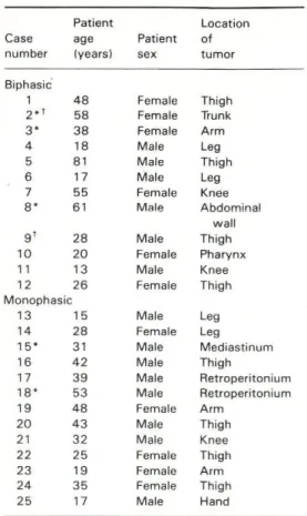
and spindle cell pattern. Fig. 1). In the re
maining 6 tumors only spindle and epithe
lioid cell components, without clearcut
biphasia but showing a solid or fascicular
pattern, were observed (Fig. 2). All the tu
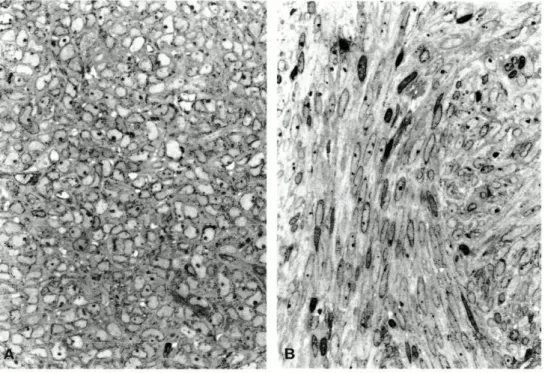
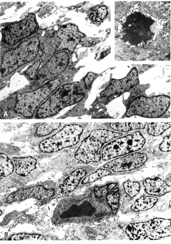
FIG. 1 BSS: Solid (A) and glandular (B) a,
thelioid
(B) cells. Toluidine blue, x
139
mors consisted of epithelioid and spindle
cells. The epithelioid cells were columnar,
polygonal, or round and had more abun
dant cytoplasm than the spindle cells.
Transitions between spindle and epitheli
oid cells could be found in 10 of the tu
mors (Figs. 2B and 3). We also selected
for ultrastructural study areas showing an
architectural transition between a glandu
lar or solid pattern and patternless foci
(Fig. 3).
MSS (ii = 13)
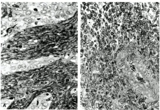
In 7 tumors there was a clear predomi
nance of spindle cells arranged in a fasicu
lar pattern (Fig. 4A). In the remaining 6 tu
mors a haphazard mixture of spindle and
epithelioid cells predominated (Fig. 4B).
Clusters of epithelioid cells (Fig. 5A) and
transitions between spindle and epithelioid
cells could be found in 6 cases (Figs. 4B
and 5B) and were also specifically selected
for ultrastructural study.
■eas alternating with spindle (A) and
epi-560.
V i •
•
i <• ■>V -I
s i : 1 ■ !,v
*vi *^«
• -4-ÎS* 1 4»
''." .»%.
t-
,; y-
-•-V'
■ : \ * , " ' * ' ■ * *•-'*?• ^
i
' ■ " . ' ■* ^ ." *
ffc '
'
J A | . ■ " *'<'
i**
i ' Í v , *t ?
#
I
*im
v
r
•h.
♦i*
"é
t
Q-
1 *
/ >
*5
. .•
>;Y/ ■ > ; t e , '
âs.
m
'ti
si
li? "#'•
Í. w • * ■ >FIG. 2 fiSS; So//tf epithelioid (A) antf fascicular spindle/epithelioid (B) areas without
distinct biphasia. Toluidine blue, x 560.
FIG. 3 fiSS: Cytoarchitectural transitions of glandular (A) antf so/Zd (B) areas fo sty/
rounding spindle/epithelioid cells. Toluidine blue, x 560.
Ultrastructural Spectrum of Synovial Sarcomas
141
m-*
v.
J
Û''
1
m
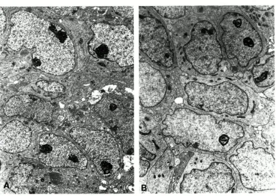
F I G . 4 /WSS; Spindle (A) and spindle/epithelioid (B) ce//s w/f/7 fascicular pattern.
To-luidine blue, x 5 6 0 .
R e c u r r e n c e s (n = 5) and
M e t a s t a s e s (n = 2)
Semithin sections of recurrent and/or
metastatic tumors showed neoplastic cell
shape components and architecture similar
to those of the corresponding primary t u
-mors. Spindle cells were the only cell
com-ponent present in 1 recurrence of MSS; all
the other tumors showed both spindle and
epithelioid cells.
RESULTS
BSS
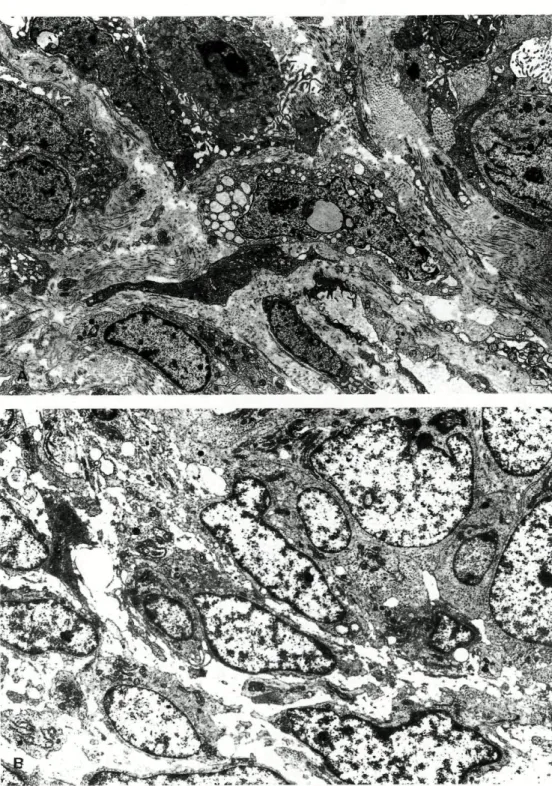
Electron microscopy confirmed a clear-cut
biphasic pattern with prominent glandular
and/or solid foci in 6 cases. The glandular
structures were lined by columnar,
polygo-nal, cuboid, or sometimes flattened cells
that rested on a continuous basal lamina
(Figs. 6A and 7). The neoplastic cells were
well to moderately equipped with
organ-elles. Golgi complexes and aggregates of
intermediate filaments were commonly
present. Cilia and ciliary bodies and
bundles of tonofilaments were found in 3 t u
-mors (Fig. 7B). The luminal surface of the
cells had short to long microvilli projecting
into the lumen, which contained dense and
amorphous material or cellular debris (Figs.
7A and 8A, inset) The nuclei were oval
with prominent nucleoli. The cells were
connected by different types of
well-differentiated junctions (ie, tight junctions
and desmosomes) and intermediate
junc-t i o n s . Injunc-tracellular lumina were ofjunc-ten
present, whereas intracellular lumina were
rarely observed (Fig. 7A).
Nests of epithelioid cells showing minor
degrees of glandular differentiation were
also seen in areas classified by light
mi-croscopy as biphasic. These nests rested
also on a continuous basal lamina.
Intercellular lumina with microvilli were
found in the spindle/epithelioid cell
compo-nents between solid and glandular areas of
1 tumor with clear-cut biphasia (Fig. 9A).
142
J. M. Lopes et al
<f * f ¥ Mrm
•f
~* '/f <
,.'-, «
• ■ > |,«\ •'
/
:•
fes" ."
■■■'fit i <*
".^-■•-,K" ^i •
Afli F I G .
5 MSS: Clusters of epithelioid cells (A) and transitions between spindle and
epithelioid cells (B). Toluidine blue, x 560.
The cells composing the other 6 BSS
(without apparent biphasia) were of spin
dle or epithelioid type (Fig. 10A). Both
types of cells showed less equipped cyto
plasm than the cells lining the glandular lu
mina. Aggregates of intermediate fila
ments, Golgi complexes, and cilia were
usually less conspicuous than those in the
cells of glandular or solid areas of SS with
biphasia. Bundles of tonofilaments were
never observed. Lipid inclusions and lyso
somes were frequently found. Oval nuclei
w i t h prominent nucleoli were present in
epithelioid cells (Fig. 10A). Intercellular
spaces with filopodia and abortivelike mi
crovilli were found in 6 cases (Fig. 11).
M o s t of the tumors s h o w e d intercellu
lar junctions of the paired subplasmalem
mal density (PSD) type1 8 and, rarely, of
the desmosome type connecting spindle
and epithelioid cells. A discontinuous ba
sal lamina was found around some spin
dle and epithelioid cells in most of the
cases. In 1 case, a continuous basal lam
ina was seen around some epithelioid cell
clusters w i t h o u t glandular differentiation
(Fig. 6A).
Stroma between neoplastic cells was al
most virtual in most glandular and solid ar
eas (Figs. 7A and 10A). Moderately abun
dant stroma with bundles of collagen fibrils
of the banded type (Figs. 6A, 7B, and 8A;
long spacing type in 3 tumors) within a
loose or floccular matrix predominated in
spindle/epithelioid (Fascicular) areas w i t h
out biphasia. In 3 cases an electrondense
(basal laminalike) matrix was seen around
some cells.
All sorts of cytoarchitectural transitions
could be found in most BSS; these in
cluded spindle to epithelioid cell types and
fascicular to solid or glandular patterns
(Figs. 6B and 8A). The interface between
some alternating glandular or solid areas
and fascicular areas showed focal absence
of a basal lamina and, thus, a distinct con
tiguity between cell components (Fig. 6B).
Transitions of cytologic features within
32
FIG. 6 BSS: (A) Glandular (upper right) and solid areas surrounded by a continuous
basal lamina. Uranyl acetate and lead citrate, x 3,630. (B) Solid area (upper
right) with transition to surrounding epithelioid cells. No basal lamina is
present. Uranyl acetate and lead citrate, x 3,630.
FIG.
7
1 4 4
BSS: (A) Glandular area with an apparently intracytoplasmic lumen. Uranyl
acetate and lead citrate, x 4,840. (B) Periphery of glandular area surrounded
by a distinct basal lamina. Bundles of tonofilaments are present (arrows).
Note the cilium in the spindle cell (lower left). Uranyl acetate and lead citrate,
x 8,580.
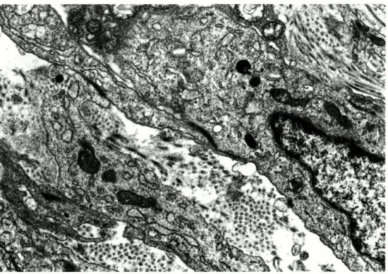
FIG. 8 (A) BSS: Spindle and epithelioid transitional cells. Uranyl acetate and lead
ci-trate, x 3,600. (Inset) The tumor had distinct glandular areas with
intercellu-lar lumens. Uranyl acetate and lead citrate, x 6,600. (B) MSS: Spindle and
epithelioid transitional cells. Scattered filopodia are present. Uranyl acetate
and lead citrate, x 3,630.
35
1 4 6
J. M. Lopes et al
F I G . 9 (A) BSS: Spindle cell component with glandular differentiation. Uranyl
ace-tate and lead citrate, x 6,940. (B) MSS: Intercellular lumen lined with
mi-crovilli from neighboring epithelioid cells. Uranyl acetate and lead citrate,
x 8,840.
each p a t t e r n c o u l d also be o b s e r v e d
whether biphasia was present or not.
MSS
In 6 tumors the cell exhibited a spectrum
of shapes ranging f r o m a long spindle
ap-pearance to a more round or polygonal
epi-thelioid appearance (Figs. 8B and 10B). In
the remaining 7 cases there was a
predom-inance of spindle cells.
Epithelioid cells were better equipped
w i t h organelles (Fig. 10B) than spindle
cells, especially w i t h cilia-related
struc-t u r e s , aggregastruc-tes of instruc-termediastruc-te
fila-m e n t s , and Golgi cofila-mplexes. Nuclei were
fusiform or oval and displayed prominent
nucleoli in epithelioid cells (Fig. 10B).
Inter-cellular junctions of the PSD type were
ob-served in most of the cases, and a
discon-tinuous basal lamina w a s more frequently
found surrounding epithelioid cells (Fig.
12) than spindle cells. In 1 case a
well-developed glandular structure w i t h
mi-crovilli and desmosomes was seen in an
area composed of epithelioid cells (Fig.
9B). A continuous basal lamina
surround-ing clusters of epithelioid cells w a s
ob-served in this case.
Stroma was almost absent, particularly
in predominantly epithelioid cell-rich areas
(Fig. 10B). It was moderately abundant
and contained dense (basal lamina-like)
material or banded collagen fibrils within a
loose or floccular matrix in the spindle
cell-rich areas (Figs. 8B and 12). Collagen
fi-brils of the long spacing type were found
in the matrix of 8 tumors.
Comparison of BSS and MSS
If one excludes the glandular areas that by
definition are pathognomonic of BSS, the
ultrastructural findings of MSS were
Ultrastructural Spectrum of Synovial Sarcomas
147
lar to those of the nonglandular
(interglan-dular areas and large solid or fascicular
ar-eas without biphasia) arar-eas of BSS,
namely regarding the features of epithelial
type differentiation (Table 2) and the
ma-trix. Major differences were the presence
of more numerous cilia-related structures
and less prominence of intermediate
fila-ment aggregates in the cytoplasm and
dis-continuous basal lamina around the
neo-plastic cells of MSS. Long spacing type
collagen fibrils were more frequently found
in the stroma of MSS than in BSS. A tumor
of each group showed glandular
differenti-ation within apparently nonglandular solid
areas in the semithin sections (Fig. 9).
Comparison of Primary and
Recurrent/Metastatic SS
Ultrastructural study of recurrences was
performed in 5 cases (3 BSS and 2 MSS)
and of metastases in 2 cases (BSS; Table
1 ). No major differences were found in the
cellular components and architecture in
comparison to the respective primary
tu-mors, except for the greater prominence of
nucleoli and abundance of polyribosomes
in recurrent and metastatic tumor cells.
DISCUSSION
SS is a relatively frequent soft tissue tumor
of unknown histogenesis that is most
prevalent in adolescents and young
adults.
1The tumor raises particular
inter-est because of its frequent double
pheno-typic histologic pattern, which often
in-cludes glandular foci.
Biphasic and monophasic (fibrous or
ep-ithelial) types are extremes of the
histo-logic spectrum of SS according to most
in-vestigators. Recent data, mainly based on
immunohistochemical and ultrastructural
FIG. 10 (A) BSS: Epithelioid cells without glandular structures. Uranyl acetate and
lead citrate, x 3,510. (B) MSS: Epithelioid cells in a monophasic tumor.
Note the similarity to the cells in (A). Uranyl acetate and lead citrate,
x 3,520.
148
J . M. Lopes et al
■
;:
^t lu* '&£■<.'
FIG. 11 BSS: Abortive glandular structure lined by epithelioid cells. Uranyl acetate
and lead citrate, x 7 1,400.
studies, support the epithelial (monopha
sic) or epitheliomesenchymal (biphasic) na
ture of SS.
2"
7Monophasic subtypes are difficult to di
agnose,
4"
6,8 16and some investigators have
even questioned their true existence.
19Al
though poorly differentiated SS are recog
nized as a distinct subgroup because of
their ominous prognosis,
1'
2 0no well
defined criteria for the histologic grading of
SS have been established so far.
To address the problem of neoplastic
differentiation in SS, we followed in this
study the criteria of Enzinger and Weiss
1as
a basis for the comparison between ultra
structural features of glandular and non
glandular areas of BSS as well as between
nonglandular areas of BSS and MSS. To
achieve this goal and to avoid misrepresen
tations at the ultrastructural level of the
different histologic aspects, we carefully
selected from each case the most repre
sentative semithin sections. These sec
tions were afterward used to precisely ori
ent the sites most appropriate for
ultrastructural study.
Our results clearly show the existence
of a spectrum varying from epithelial type
differentiation (abortive glandular struc
tures) to true epithelial differentiation
(welldeveloped glandular structures) in all
cellular components of SS. In this context,
PSD type junctions
18and discontinuous
basal lamina observed in spindle and epi
thelioid components, both in primary and
in recurrent SS (biphasic or monophasic
patterns), should be considered putative
signs of primitive epithelial type differenti
ation. Although rarely, we have also ob
served desmosomes and discrete glandular
structures in the nonglandular areas of
both tumor subtypes. This further sup
ports the epithelial nature of these tumors,
regardless of the predominant morphologic
appearance of SS.
Another argument in favor of the epithe
38
Ultrastructural Spectrum of Synovial Sarcomas
1 4 9
FIG. 12 MSS: PSD between and discontinuous basal lamina around spindle and
epi-thelioid cells. Uranyl acetate and lead citrate, x 11,400.
TABLE 2 Ultrastructural Features Suggestive of Epithelial Type
Differentiation in Spindle and Epithelioid Cell Components of Fibrous
MSS and in Interglandular and/or Solid Areas without Biphasia of BSS
Number of cases
BSS
MSS excluding glandular areas Characteristic
(/) -
15) (n - 17) Intercellular spaces 7 10 Microvilli 1 1 Intercellular junctions PSD 13 14 Desmosomes 1 3 Tight junctions 1 2 Basal lamina Continuous 1 1 Discontinuous 6 12Intermediate filament aggregates 5 11
150
J. M. Lopes et al
liai differentiation of SS is the frequent
demonstration of epithelial markers in the
neoplastic cells, again regardless of their
histologic appearance. Although epithelial
membrane antigen and cytokeratins of the
simple epithelial type keratins (8, 18, and
19) are being increasingly described in
sev-eral other types of soft tissue tumors,
21,22the same does not hold true for the
strati-fied epithelial type cytokeratins, which,
like others,
6,21we have observed in all the
cellular components of SS.
Transitions between cellular
compo-nents of glandular or solid areas and
non-glandular or solid areas could be found in
our series, as has been reported
previ-ously.
26,8'
10,13,15,16We also found all sorts
of transitions between spindle and
epitheli-oid cell components of SS. Epithelial type
differentiation was more prominent in
epi-thelioid cells than in spindle cells. This
dif-ference was more evident in tumors
dis-playing a biphasic pattern than in those
displaying a monophasic pattern, but we
repeatedly found unequivocal signs of
epi-thelial type differentiation in areas of SS
exclusively or almost exclusively
com-posed of spindle cells.
Taking all this together, we believe that
there is enough evidence to support the
neoplastic nature of spindle cells of both
BSS and MSS and, furthermore, in
con-trast to Dardick et al,
5that spindle cells are
able to transform and evolve into glandular
cells of SS. We concur with Dickersin
15that the coexistence of biphasic and
monophasic patterns in the same tumor
re-flects different degrees of differentiation
(transitions) of SS and not a combination
of tumors with different natures
(carcino-sarcoma and carcinoma), as proposed by
Dardick et al.
5There is growing evidence that
plastic cell shapes and the degree of
neo-plastic differentiation are dependent on
cell-cell and cell-matrix interactions. This
could explain why carcinomas growing in
the superficial mesenchyma (eg, the
so-called pseudosarcomatous carcinomas of
mucosa and skin) display a predominantly
spindle cell architecture. It remains to be
seen whether the mesenchyma where SS
occur plays a role in the acquisition of the
epithelioid (or true epithelial) architecture
of neoplastic cells.
Nomenclature and criteria for the
histo-logic grading of SS still raises
contro-versy. In the most recent series, SS have
been considered carcinomas or
carcino-sarcomas of soft tissues depending on
the type of differentiation.
311,13Cytoge-netic data
23,24point, however, to a
com-mon pathogenesis for both phenotypes
(BSS and MSS). Based on this and on our
own results, we suggest that SS
repre-sent true carcinomas of soft tissues with
biphasic and/or monophasic patterns
de-pending on the degree of differentiation.
REFERENCES
1. Enzinger FM, Weiss S. Synovial sarcoma. In: Stamathis G Soft Tissue Tumors (2nd ed). Phil-adelphia: Mosby; 1988:659-688.
2. Miettinen M, Virtanen I. Synovial sarcoma —a misnomer. Am J Pathol. 1984; 117:18-25. 3. Leader M, Patel J, Collins K, Kristen H.
Syno-vial sarcoma: true carcinosarcomas? Cancer. 1987;59:2096-2098.
4. Miettinen M, Letho V-P, Virtanen I. Monopha-sic synovial sarcoma of spindle-cell type: epi-thelial differentiation as revealed by ultrastruc-tural features, content of prekeratin and binding of peanut agglutinin. Virchows Arch
Cell Pathol. 1983;44:187-199.
5. Dardick I, Ramjohn S, Thomas MJ, Jeans D, Hammar SP Synovial sarcoma. Interrelation-ship of biphasic and monophasic subtypes.
Pathol Res Pract. 1991;187:871-885.
6. Sumitomo M, Hirose T, Kudo E, Sano T, Shino-miya S, Hizawa K. Epithelial differentiation in synovial sarcoma. Correlation with histology and immunophenotypic expression. Acta
Pathol Jpn. 1989;39:381-387.
7. Ghadially FN. Is synovial sarcoma a carcino-sarcoma of connective tissue? Ultrastruct
Pathol. 1 9 8 7 ; 1 1 : 1 4 7 - 1 5 1 .
8. Krall RA, Kostianovsky M, Pathchefsky AS. Synovial sarcoma: a clinical, pathological, and ultrastructural study of 26 cases supporting the recognition of a monophasic variant. Am J
Surg Pathol. 1 9 8 1 ; 5 : 1 3 7 - 1 5 1 .
9. Abenoza P, Manivel JC, Swanson PE, Wick MR. Synovial sarcoma: ultrastructural study and immunohistochemistry analysis by a com-bined peroxidase-antiperoxidase/avidin-peroxidase complex procedure. Hum Pathol. 1986;17:1107-1115.
10. Ordonez NG, Mahfouz SM, MacKay B. Syno-vial sarcoma: an immunohistochemical and ultrastructural study. Hum Pathol. 1990;21: 7 3 3 - 7 4 9 .
1 1. Farris KB, Reed RJ. Monophasic glandular,
Ultrastructural Spectrum of Synovial Sarcomas
151
synovial sarcomas and carcinomas of the softtissues. Arch Pathol Lab Med. 1 982; 106:1 2 9 -132.
12. Golough R, Venzevski V, Bracko M, Van der Heul RD, Cervek J. Synovial sarcoma: a clini-copathological study of 36 cases. J Clin
On-col. 1990;45:20-28.
13. Tsuneyoshi M, Yokoyama K, Enjoji M. Synovial sarcoma: a clinicopathological and ultrastruc-tural study of 42 cases. Acta Pathol Jpn. 1983;33:23-36.
14. Mickelson MR, Brown GA, Maynard JA, Cooper RR, Bonfiglio M. Synovial sarcoma: an electron microscopic study of monophasic and biphasic forms. Cancer. 1980:45:2109-21 18.
15. Dickersin GR. Synovial sarcoma: a review and update, with emphasis on the ultrastructural characterization of the nonglandular compo-nent. Ultrastruct Pathol. 1991;15:379-402. 16. Fisher C. Synovial sarcoma: ultrastructural and
immunohistochemistry features of epithelial differentiation in monophasic and biphasic tu-mors. Hum Pathol. 1986;17:996-1008. 17. Johannessen JV. Use of paraffin material for
electron microscopy. Pathol Annu. 1977; 12: 1 8 9 - 2 2 4 .
18. Quinonez G, Simon GT. Cellular junctions in a
spectrum of human malignant tumors.
Ultrastruct Pathol. 1988; 1 2:389-405.
19. Mackenzie DH. Monophasic synovial sarcoma —a histological entity?
Histopathol-ogy. 1977;1:151-157.
20. Fernandez BB, Hernandez FJ. Poorly differenti-ated synovial sarcoma: a light and electron microscopic study. Arch Pathol Lab Med. 1976;100:221-223.
2 1 . Miettinen M. Keratin subsets in spindle cell sarcomas. Keratins are widespread but syno-vial sarcoma contains a distinctive keratin polypeptide pattern and desmoplakin. Am J
Pathol. 1991;138:505-513.
22. Litzky LA, Brooks J. Cytokeratin immunoreac-tivity in malignant fibrous histocytoma and spindle cell tumors; comparison between fro-zen and paraffin-embedded tissues. Mod
Pathol. 1992;5:30-34.
23. Dal Cm P, Rao U, Jani-Sait S, Karakousis C, Sandberg AA. Chromosomes in the diagnosis of soft tissue tumors. I. Synovial sarcoma.
Mod Pathol. 1992;5:357-362.
24. Knight J, Reeves BR, Smith S, et al. Cytoge-netic and molecular analysis of synovial sar-coma. IntJ Oncol. 1992;1:747-752.
Accepted in revised form December 3, 1992.
Paper II
Path. Res. Pract. 190, 168177 (1994)
Immunohistochemical Profile of Synovial Sarcoma with
Emphasis on the Epithelialtype Differentiation
A Study of 49 Primary Tumours, Recurrences and Metastases
J. M. Lopes
1, B. Bjerkehagen
2, R. Holm
2, Õ. Bruland
3,
M. SobrinhoSimões and J. M. Nesland
21Unit of Molecular Pathology - IPATIMUP, Medical Faculty, Porto, Portugal;
Departments of 2Pathology and Medical Oncology3, The Norwegian Radium Hospital
and The Norwegian Cancer Society, Oslo, Norway
SUMMARY
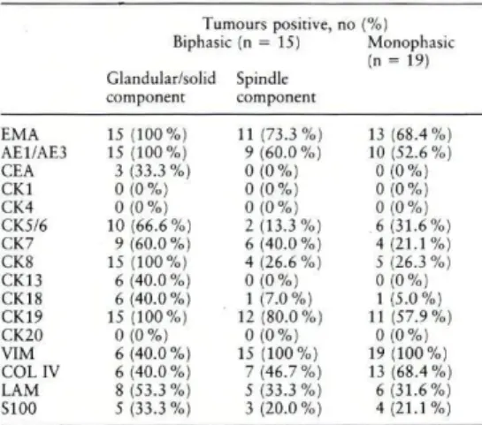
The relationship between btphasic (BSS) and monophasic (MSS) subtypes of synovial sarcoma (SS) as well as the relationship between cells of solidlglandular areas and the spindle cells of BSS remain controversial. In order to further evaluate the immunohisto chemical phenotype ofSS we studied 34 primary tumours (15 BSS; 19 MSS), 7 recurrences (4 from primary BSS; 3 from primary MSS) and 8 metastases (7 BSS; one MSS), using several antibodies (EMA, CEA, keratins I, 4, 5/6, 7, 8, 13, 18,19, 20, vimentin, collagen IV and lammin) that work in paraffinembedded material. Spindle cells outside solidl glandular areas of BSS and m MSS showed immunoreactwity for kera tins Sib, 7,8,18 and 19. The transition of solid/glandular areas to surrounding spindle cells also showed keratin staining and failed to show a distinct separation regarding the immunoreactivity for laminin and collagen IV. Peripheral cells of solid/glandular areas were immunoreactive for vimentin. No major differences were observed between immunophenotypical cell profiles of BSS and MSS, apart from the exclusive immuno staining of solidl glandular areas of BSS for keratin 13 and CEA. Downgrading of keratin and extracellular matrix antigens immunoreactwity was observed when primary tumours were compared to recurrent and/or metastatic tumours of both subtypes (MSS and BSS). We conclude that SS should be regarded as 'carcinomas of soft tissues with an immunohistochemical phenotype depending on the degree of epithelial differentiation: spindle cells (MSS and BSS) predominantly expressing simple keratins, and poorly differentiated (solid/glandular) as well as welldifferentiated (glandular) areas (BSS) expressing, in addition, complex epithelialtype keratins.
Introduction Several immunohistochemical studies of SS have been published in the last ten years'. W ' 1 5. '72 12 3'2 4'2 62 72 9. Synovial sarcomas (SS) are soft tissue tumours of Most of these studies have clearly shown the epithelial unknown origin9. Based on ultrastructural and immuno differentiation of SS. However, individual keratins were histochemical studies, most authors consider them either only evaluated in a few series1 7'2 1'2 7 and in a limited as carcinosarcomas or as carcinomas depending on the number of precisely classified SS. The limited number of presence or absence of biphasia6'1 5'1 9'2 0. ■ cases studied to date contributes to explain why some 03440338/94/01900168$3.50/0 *> 1994 *>y Gustav Fischer Verlag, Stuttgart
controversies still persist regarding the pattern of immu-noreactivity of SS.
The most vivid controversy resides on the relationship between biphasic (BSS) and monophasic (MSS) subtypes of SS and also on the relationship between cells in solid/glan-dular areas and the surrounding spindle cells of BSS6'. Immunohistochemistry studies of BSS on record de-scribe the almost constant exptession of epithelial-type (keratins and/or EMA) and mesenchymal-type (vimentin) markers in solid/glandular and spindle cell components, respectively. MSS show lower and variable degrees of expression of the aforementioned markers1-3-6- i». is, 17-21, 23, 24, 26, 27, 19 Qfcfr jm m u n o r ea c t i v i t y of solid/glandular component is an infrequently reported finding; to the best of our knowledge such immunoreactivity has usually been detected in BSS co-expressing keratins and EMA, as well as in rare MSS also co-expressing keratins and EMA5'2 3'2 7. Although S100 protein reactivity was described in some BSS and MSS cases, they also usually co-expressed keratins and EMA9-11-23. Other markers (e.g., desmin, smooth-muscle actin, Factor VIII) used in the differential diagnosis with othet soft tissue tumours were consistently negative in all reported series5-6-23-24-26-27. Vimentin reactivity is fre-quently described in spindle cell component of BSS and MSS, and also occasionally in cells of solid/glandular component of few cases23-24-26-27. Few studies of collag-en IV and laminin expression depicted continuous staining along solid/glandular component of BSS22-23-27 and also, in one series23, between groups of cells of the spindle cell component of BSS and MSS.
Solid/glandular component of BSS of the largest series showed co-expression of keratins and EMA6-2'. Spindle cell component of BSS is variably described as non-reactive6-17-18 or focally reactive in most cases for bne or both of these markers4-5."-15-23-24.26.2~-2').
Apart from these discrepancies it also remains to be clarified if there is any relationship between the expression of individual keratins and other immunohistochemical markers of SS.
The availability of commercial antibodies for individual keratins and the improvement in the processes of unmask-ing keratin epitopes led us to study the keratin profile together with several other immunohistochemical markers of the neoplastic cells and the matrix of a large series of paraffin-embedded SS, in an attempt to settle at least some of the aforementioned controversies.
Material and Methods
Biphasic (BSS) and monophasic (MSS) synovial sarcomas of 36 patients were selected from the files of the Department of Pathology, The Norwegian Radium Hospital (25 cases) and Porto Medical School (11 cases).
Thirty-four primary tumours (15 BSS; 19 MSS), 7 recurrences (4 from BSS; 3 from MSS) and 8 metastases (7 from primary BSS and one from primary MSS) were available for examination. In 2 patients with primary BSS only recurrence or metastases were available for immunohistochemical study.
In every tumour, several blocks (4—15 paraffin-blocks per primary tumour and per recurrence and/or metastasis) were
Immunohistochemical Profile of Synovial Sarcoma - 169 Table 1. Clinico-pathologic features of synovial sarcomas
Biphasic Age (years) Sex Location
M 48 F Thigh
*2f
58 F Trunk ,3o o 38 F Arm *4° 18 M Leg *5 81 M Thigh 6 32 M Thigh *7 17 M Leg *8 55 F Knee *9+ 6 1 M Abdomina wall *10t 28 M Thigh l l f 33 F Trunk 1 2o o 38 M Tongue 13 20 M Foot 14 45 M Thigh 15 14 M Knee 16 35 M Knee 17 46 F Foot Monophasic MS 15 "19 28 *20 31 • 2 1 f 42 *22 17 23 7 24 40 25 t t 30 ! ,26 39 - 2 7 53 28 40 29° 61 "30 48 *31 43 *32 32 33 2S *34 25 • 3 5 19 • 3 6 35 M F M Leg Leg Mediastinum M M Thigh Hand M Thigh F Pharynx M Foot M Retroperitoneum M F F Retropcritoneum Leg Foot F Arm M M Thigh Knee F F F Thigh Thigh Arm F ThighCases with ultrastructural study* and cases with recurrences f and/or metastases ° available for study. Cases 9 and 12 without primary tumours available for immunohistochemistry. F = fe-male; M = male.
available for routine histological examination. The cases were classified according to the light microscopic, immunohistochem-ical and electron microscopic criteria of Enzinger and Weiss9. The clinico-pathological features of the cases are summarized in Table 1. All MSS were of the fibrous type. One recurrence and one metastasis from BSS showed distinct biphasia, whereas all the other recurrent and metastatic tumours displayed monophasia.
lmmunobistochem istry
Consecutive sections were stained using the avidin-biotin-peroxidase complex (ABC) method12. Deparaffinized sections were redhydrated, washed with phosphat-buffered saline (PBS11 pH 7.4 and treated with enzyme (Table 2). The sections were then treated with 0.3 % hydrogen peroxide (H->0>) in methanol for 30 minutes to block endogenous peroxidase. To unmask the epitopes of keratins 7 and IS, the sections were microwaved in an «antigen retrieval" solution'". The sections were then incubated