DoI: 10.2298/avb1306581D UDK: 636.32/.38+616.995.132:616-08+615.284:612.12
tHe effect of tHe intensity of Parasitic infection WitH Strongyloides papillosus and albendaZole tHeraPy on biocHemical Parameters in
sHeeP blood
DIMITRIJEVIĆ B*, BOROZAN SUNČICA*, JOVIĆ S*, BACIĆ D*, KATIĆ-RADIVOJEVIĆ SOFIJA*, STOJANOVIĆ S** and SAVIĆ MILA*
*University of Belgrade, Faculty of Veterinary Medicine, Serbia **University of Belgrade, ICTM – Center for Chemistry, Serbia
(Received 27th april 2013)
The aim of this report was to study the biochemical parameters in sheep blood under conditions of various intensities of parasitic infection with Strongyloides papillosus, as well as after therapy with albendazole (ABZ). Investigations were performed on sheep of the Würtemberg race (n = 30) in which were detected mild, moderate and high intensities of parasitic infection with S. papillosus. The control group (n = 10) was composed of sheep negative to parasitic infections. The degree and type of changes were monitored by determining the concentrations of glucose, total proteins, albumin, A/G ratio, AST, urea, total bilirubin, calcium, phosphorus, total LDH activity and isoenzymatic LDH1-5 distributions.
582
strongly recommend that in antiparasitic treatment protocols, beside antihelminthics, compounds with antioxidative properties should also be used.
Key words: albendazole, biochemical parameters, liver, sheep strongyloides papillosus
INTRoDUCTIoN
parasitic infections constitute an important group of diseases in sheep concerning the health status, welfare and productivity. Strongyloides papillosus
is the causative agent of strongyloidosis in sheep and certain other species, including humans. Strongyloidosis is a clinical - pathological entity which is usually neglected in clinical practice (olsen et al., 2009). The cause of this is that the clinical symptoms provoked by infection with S. papillosus are masked with symptoms of infections from other parasitic species, this being the most common case under conditions of natural infections. also, at the low intensity of this parasitic infection, the organism of the host does not react with clinically manifested signs. economical effects due to strongyloidosis in sheep include emaciation, reduced fertility, low milk and meat yield; it has been estimated that 15% of meat and milk production in Serbia is lost due to ovine strongyloidosis (Dimitrijević et al., 2012).
The parasitic form of S. papillosus appears in the form of parthenogenetic females present in the sheep small intestines (Kassai, 1999; eberhardt et al.,
2008). The infection occurs as a result of the introduction of infectious larvae (stage l3) by way of food and water (passive) and/or through percutaneous (active) penetration of the l3 larvae. pathogenic impact of the parasites on the host derives from the presence of the larvae during migration and/or the adult forms in the intestines, which through their mechanical and secretory/excretory (Se) products are harmful to the host’s tissues. larvae which actively penetrate into the host’s organism by rupturing the interdigital skin enabling the penetration of carriers of other etiology (e.g. the causative agents of footrot in sheep, as well as certain other anaerobic agents) (abbott and lewis, 2005). The presence of S. papillosus and its developing forms cause disturbances in the animals’ health, quite frequently inducing the sudden death syndrome in young ruminants (lambs and calves) due to heart failure (Taira and Ura, 1991). The degree of damage directly correlates with the intensity of the parasitic infection, i.e. with the number of parasites present and/or their larval forms (Ura et al., 1993; Nakanishi et al.,
1993; Nakamura et al., 1994; Nakamura and motokawa, 2000). Kobayashi et al. (2009) state that the exact mechanism of how the infection with S. papillosus causes death among animals, has not yet been identiied.
albendazole (abZ), benzimidazole’s derivate, is a broad-spectrum antihelminthic, the drug of choice for the treatment of strongiloidosis and other parasitic infections (Kassai, 1999). The contemporary, commonly accepted theory
acta veterinaria (beograd), vol. 63, no. 5-6, 581-600, 2013.
is that the key mechanism by which abZ achieves its effect is through its interaction with the eucariotic cytoskeleton protein, tubulin, by inhibiting its polymerization into microtubules (Rufener et al., 2009). also, abZ acts on biochemical and enzymatic processes in parasites, inhibiting glucose transport in parasites, as well as the enzyme fumarate reductase. This leads to the impoverishment of energy reserves and death of the parasite. In our previous study, we have shown that infection with S. papillosus leads the host organism to a state of ampliied oxidative stress (Dimitrijević et al., 2012). In this state, it has been demonstrated that the level of oxidative stress correlated with the intensity of parasitic infection. It is of interest that albendazole therapy also brings the organism to a state of disturbed redox equilibrium, more intense than that observed for parasitic infection and for a longer time period (authors’ unpublished data).
The increased exposure to RoS/RNS leads to cellular oxidative stress and consequently damages biomacromolecules (lipids, proteins and nucleic acids), what may induce controlled cell death (apoptosis), leading to the development of a malignant cell or uncontrolled cell death (necrosis). This type of mechanism is the basis for development of almost all diseases (lykkesfeldt and Svendsen, 2007). Having in view our previous studies in the ield of oxidative stress in parasitic infections, the aim of this study was to investigate changes in the values of speciic biochemical parameters (glucose, total proteins, albumine, A/G ratio, aST, total bilirubin, urea, calcium, phosphorus and total activity and isoenzymatic distribution of lactate dehydrogenase) which more closely depict the metabolic status of the ovine organism in the presence of parasitic infection, as well as after treatment with abZ.
maTeRIalS aND meTHoDS
Study area and animals
This study was performed in the vicinity of the town of vranje (south eastern Serbia (village Kupinince). The climate in this region is continental, with long, cold winters and hot summers. Climatic factors play a very important role in the sterilization of pastures from infectious stages of geohelminate parasites, to which Strongyloides spp belongs (Stromberg, 1997). Due to a characteristic development cycle of the parasite, the way of contracting the infection and its strong enzootic character, infection with S. papillosus and its endurance in the natural environment, depend to a great extent on microclimatic factors in the sheep shelter. The sheep in this study were accommodated during the night in a shed with a thick dirt loor, thus subjecting them to a continuous infection with S. papillosus.
584
were divided into three groups: I1 – having mild (n = 10), II1 – moderate (n = 10), and III1 – high intensity (n = 10) infection with S. papillosus. after determining the type of parasitic infection, the sheep were treated with albendazole (veRmITaN, Ceva-philaxia, Hungary), per os, in single doses of 5 mg/kg body weight, thereon, the groups were marked as I2, II2, III2. The negative control group was marked as – C1 (n = 10); and after treatment with abZ was marked as C2. all experiments were performed according to our institutional guidelines for animal research and principles of the european Convention for the protection of vertebrate animals Used for Experimental and Other (Oficial Daily N. L 358/1–358/6, 18, December 1986).
Sampling of feces for parasitological examination
Samples of feces were obtained once a day from each sheep individually, directly from the rectum, during the course of 3 days, on the 0 and 21st day after treatment with abZ. Samples of each animal’s feces were packed in a separate, marked plastic bag and transported in a portable refrigerator to a parasitological laboratory. for detecting and determining parasite species and their developing forms, standard identiication keys for parasites were used, based on their developing forms, morphology and morphometrical characteristics of eggs, larvae and/or adult forms.
Methods used in coprological diagnostics were sedimentation and lotation (Kassai, 1999). Examination of the samples was performed at magniications of 7 × 10 and 7 × 40 using a Reichert microscope. The infection intensity was determined by counting the helminth eggs per gram of feces using modiied mcmasters’ method (euzeby, 1982; Reinecke and fonseca, 1992).
Blood sampling
blood samples for the tests were taken from sheep by puncture of v. jugularis. from both groups of sheep (before and on the 21st day after dehelminthization) one portion of blood was taken without anticoagulant, enabling the separation of blood serum, while the other portion of blood was taken with the anticoagulant heparin so that blood plasma could be obtained. Sera were obtained after spontaneous blood coagulation attained after centrifugation lasting 10 min at 3000 rpm. plasma was obtained from blood with anticoagulant after centrifugation for 10 min at 3000 rpm. erythrocytes were rinsed three times in physiological saline. Thus obtained samples of blood plasma, blood serum and erythrocytes were frozen at -20oC until further analysis.
Biochemical assay
The kinetic method was used to determine aspartate aminotransaminase (aST) enzyme activity, while urea concentration was determined by means of the biuret method. The biuret method was also used to determine total protein concentration, while bromocresol green was used to determine albumin concentration.
acta veterinaria (beograd), vol. 63, no. 5-6, 581-600, 2013.
Total bilirubin (the sum of conjugated and unconjugated bilirubin) was determined in the reaction with diazonium ion of sulphanilic acid. Concentration of calcium was determined with the o-cresolphtalein method, while phosphorus concentration was measured by the reaction with ammonium molybdate. all of the above mentioned biochemical parameters were determined using commercial kits (bayer Diagnostics, germany). Spectrophotometric measurements were performed with Cecil Ce 2021Uv/vIS spectrophotometer. blood glucose concentration was determined by using precision-Xtra plus test strips.
Total and isoenzyme forms of lactate dehydrogenase (lDH1–lDH5) were determined by means of vertical electrophoresis at 7.5% PAGE (Hoeffer Mini VE, lKb, 2117, bromma, Uppsala, Sweden) using Tris–glycine buffer, sodium-lactate as substrates in the presence of nitroblue tetrazolium chloride according to the Yoshida and Takakuwa method (1997). The band intensity of the isoenzymes of lDH was estimated using Scion Image beta 4.02 software (Scion Corp., 2007). The density of each band was estimated with respect to the percentage of the total area.
Statistical analysis
Statistical signiicance of differences of all examined parameters were determined by means of the aNova, followed by the Tukey test. Data were expressed as means ± standard error. Signiicance level was set at p<0.05. Statistical analysis was performed using the graph pad prism 5.0 Software, Ca, USa.
ReSUlTS
Results of the biochemical analyses are shown in figures 1 to 10, and in Table 1.
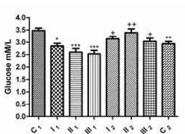
Comparing the mean values of blood glucose concentrations in the groups before dehelminthization with blood glucose concentration in the control group (C1), it was determined that the group with mild intensity of infection had signiicantly decreased (2.85±0.10 mM/L; p<0.05), while groups with moderate (2.60±0.14 mm/l) and high (2.53±0.14 mm/l) intensity of infection had a very signiicantly decreased glucose concentration (p<0.001). After treatment with abZ, concentrations of glucose in all of the studied groups were not statistically signiicantly different when compared to the C1 group (p>0.05). However,
586
figure 1. glucose concentration in sheep blood before and after dehelminthization: group with mild intensity of infection (I1 – before and I2 – after dehelminthization), group with moderate intensity of infection (II1 – before and II2 – after dehelminthization), group with high intensity of infection with S. papillosus (III1 – before and III2 – after dehelminthization); C1 – control group of sheep negative to the presence of parasite; C2 – control group of sheep negative to the presence of parasite and treated with abZ; *p < 0.05; ***p < 0.001 vs. control group (C1), +p < 0.05; ++p < 0.01 comparison of groups before and after dehelminthization (I1 vs. I2, II1 vs. II2, III1 vs. III2).
figure 2. Total protein concentration in sheep blood sera before and after dehelminthization: group with mild intensity of infection (I1 – before and I2 – after dehelminthization), group with moderate intensity of infection (II1 – before and II2 – after dehelminthization), group with high intensity of infection with S. papillosus (III1 – before and III2 – after dehelminthization); C1 – control group of sheep negative to the presence of parasite; C2 – control group of sheep negative to the presence of parasite and treated with abZ; *p < 0.05 vs. control group (C1); ++p < 0.01 comparison of groups before and after dehelminthization (I1 vs. I2, II1 vs. II2, III1 vs. III2).
acta veterinaria (beograd), vol. 63, no. 5-6, 581-600, 2013.
Comparison of the mean values of total protein concentrations in all analyzed groups (before and after dehelminthization) with the protein concentration in the control group (C1), conirmed that the group with the high intensity of infection and all groups treated with ABZ had a signiicantly decreased total protein concentration (p<0.05). analysis of the results obtained within the groups before dehelminthization conirmed that there was a signiicant statistical difference in total protein concentration between sheep with a mild and moderate infection intensity when compared to sheep with a high intensity parasitic infection (p<0.01). after dehelminthization, the analysis based on comparison between all groups did not identify any signiicant statistical difference in the total protein contents (p>0.05). It was however determined that a difference in the total protein concentration existed in the groups with mild and moderate infection intensity (80.18±1.50 and 82.07±2.07 g/l, respectively) even after dehelminthization with abZ (72.77±1.40 and 74.95±1.31 g/L) with a statistical signiicance level of p<0.01. The reduction of total protein concentration was conirmed in the C2 group, when compared
with the control group (p<0.05). It can bee seen from figure 3that antihelminthic treatment decreases the concentrations of the dominant protein fraction, albumin, in the groups with mild, moderate and high infection intensity by 25.40%, 25.00% and 17.04%, respectively. A statistically signiicant (p<0.01) reduction in the concentration of albumin by 30.91%, provoked by ABZ, was conirmed in the C2
group when compared with the C1 group. It was noticed (fig 4) that both before and after dehelminthization with abZ, the a/g ratio had a decreasing tendency and that this reached minimal values in sheep with moderate infection intensity (0.82±0.06), in comparison with the control group (C1) (1.49±0.22). The reduction of the A/G quotient was conirmed in the C2 group.
by comparing the aST activity values (fig 5) of all groups with those of the control group (C1), a statistically signiicant increase in AST activity was established before dehelminthization (p<0.05). after dehelminthization of the group with mild intensity infection, aST reached its highest value (149.9±4.88 U/l) in comparison with the enzyme activity in the control group C1 (66.50±6.50 U/l; p<0.001). Increased activity of this enzyme was also conirmed in the C2 group
(111.00±7.53 U/l; p<0.01), compared with C1 group. an increase was observed in urea concentration (fig 6) depending on the infection intensity. The group of sheep with a mild intensity infection had an increase of 40.26% (7.56±0.21 mM/L; p<0.05), the group of sheep with a moderate intensity infection had a 60.85% (8.67±0.35 mm/l; p<0.001), while the group with a high intensity infection had a 73.84% increase (9.37±0.27 mM/L; p<0.001) in comparison with the control C1
group (5.57±0.37 mm/l). after dehelminthization, urea concentration mantained the increased values, ranging from 66.79% to 62.34%. The increase of 74.82% in urea concentration was noted in the C2 group (10.06±0.18 mm/l) with a level of signiicance for p<0.001, when compared with the C1 group.
588
figure 3. albumin concentration in sheep blood sera before and after dehelminthization: group with mild intensity of infection (I1 – before and I2 – after dehelminthization), group with moderate intensity of infection (II1 – before and II2 – after dehelminthization), group with high intensity of infection with S. papillosus (III1 – before and III2 – after dehelminthization); C1 – control group of sheep negative to the presence of parasite; C2 – control group of sheep negative to the presence of parasite and treated with abZ; *p < 0.05; **p < 0.01 vs. control group (C1), +p < 0.05; +++p < 0.001 comparison of groups before and after dehelminthization (I1 vs. I2, II1 vs. II2, III1 vs. III2).
figure 4. a/g ratio in sheep blood before and after dehelminthization: group with mild intensity of infection (I1 – before and I2 – after dehelminthization), group with moderate intensity of infection (II1 – before and II2 – after dehelminthization), group with high intensity of infection with S. papillosus (III1 – before and III2 – after dehelminthization); C1 – control group of sheep negative to the presence of parasite; C2 – control group of sheep negative to the presence of parasite and treated with abZ; ***p < 0.001 vs. control group (C1), +p < 0.05; +++p < 0.001 comparison of groups before and after dehelminthization (I1 vs. I2, II1 vs. II2, III1 vs. III2).
acta veterinaria (beograd), vol. 63, no. 5-6, 581-600, 2013.
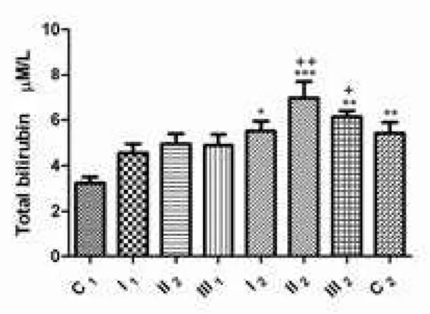
signiicance when compared to the control C1 group (3.23±0.27 µm/l; p>0.05).
after dehelminthization, an increase in the concentration of total bilirubin was determined in the group which had a mild infection intensity with the level of statistical signiicance p<0.05 (5,51±0.43 µM/L), in the group with moderate infection intensity the signiicance level was p<0.001 (6.95±0.71 µM/L), and in the group which had a high intensity of parasitic infection, the level of signiicance was p<0.01 (6.13±0.25 µm/l), when compared to the control C1 group. It is of interest to note that in the C2 control group, a rise in the concentration of total bilirubin was noted (5.42±0.48 µM/L), this value being statistically signiicant at p<0.01 when compared with the C1 group.
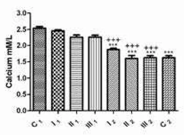
analayzing concentrations of calcium within the groups before dehelminthization, in the presence of S. papillosus, it has been determined that a fall in serum calcium concentration occurs in sheep, this being in negative correlation with the intensity of parasitic infection. This however not being of statistical signiicance compared to the control C1 group (p>0.05). after treatment
with abZ, it has been determined that there is a fall in the blood serum calcium concentration from the studied sheep, comparing within both groups before and after dehelminthization (1.87±0.04 vs 2.46±0.03; 1.59±0.09 vs 2.26±0.06; 1.62±0.06 vs 2.26±0.06 mm/l, respectively), as well as when comparing with the control C1 group (2.54±0.05 mM/L), here the level of signiicance being p<0.001. also, in the control group C2, a fall of calcium concentration was determined when compared to the C1 group (p<0.001) (fig 8).
590
figure 6. Urea concentration in sheep blood sera before and after dehelminthization: group with mild intensity of infection (I1 – before and I2 – after dehelminthization), group with moderate intensity of infection (II1 – before and II2 – after dehelminthization), group with high intensity of infection with S. papillosus (III1 – before and III2 – after dehelminthization); C1 – control group of sheep negative to the presence of parasite; C2 – control group of sheep negative to the presence of parasite and treated with abZ; *p < 0.05; ***p < 0.001 vs. control group (C1); ++p < 0.01 comparison of groups before and after dehelminthization (I1 vs. I2, II1 vs. II2, III1 vs. III2).
figure 7. Total bilirubine concentration in sheep blood sera before and after dehelminthization: group with mild intensity of infection (I1 – before and I2 – after dehelminthization), group with moderate intensity of infection (II1 – before and II2 – after dehelminthization), group with high intensity of infection with S. papillosus
(III1 – before and III2 – after dehelminthization); C1 – control group of sheep negative to the presence of parasite; C2 – control group of sheep negative to the presence of parasite and treated with abZ; *p < 0.05; **p < 0.01; ***p < 0.001 vs. control group (C1), +p < 0.05; ++p < 0.01 comparison of groups before and after dehelminthization (I1 vs. I2, II1 vs. II2, III1 vs. III2).
acta veterinaria (beograd), vol. 63, no. 5-6, 581-600, 2013.
It has been determined that the concentration of phosphorus in the group with mild parasitic infection with S. papillosus (2.87±0.08 mM/L) did not differ signiicantly
from the C1 group (2.66±0.07 mm/l; p>0.05). It may be, however, seen from figure 9, that with the rise of parasitic infection intensity, in the cases of moderate and high intensity, there is a fall of phosphorus concentrations (2.17±0.14 mm/l; p<0.05 and 1,99±0.08 mm/l; p<0.01, respectively), when compared to the control C1 group.
after therapy with abZ, there follows a fall of phosphorus concentrations in all groups (1.87±0.04; 1.59±0.09; 1.62±0.05 mm/l, respectively), the observed level of signiicance being p<0.001, when compared to the control C1 group. on
cross comparison of groups before and after dehelminthization, may be observed (fig 9) that the fall of phosphorus concentrations in the groups with mild and moderate intensity of parasitic infection, was statistically signiicant (p<0.001). It has also been determined that serum phosphorus concentration in C2 group was signiicantly lower when compared to C1 group (p<0.001).
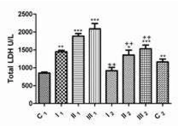
Increase lDH activity has been observed with increasing parasitic infection intensity; in the group with a mild infection, the statistical level was signiicant, p<0.01 (1447±41.04 U/l), and in the groups with moderate and high parasitic infection intensity the level of signiicance was p<0.001 (1882±72.90 and 2086±150.50 U/L, respectively) when compared to the control group C1 (849.00±28.87 U/l). after dehelminthization, lower activities of this enzyme were observed, in the group with moderate (1354±139.40 U/l; p<0.05) and high intensities (1531±102.80 U/l;
592
figure 9. phosphorus concentration in sheep blood sera before and after dehelminthization: group with mild intensity of infection (I1 – before and I2 – after dehelminthization), group with moderate intensity of infection (II1 – before and II2 – after dehelminthization), group with high intensity of infection with S. papillosus (III1 – before and III2 – after dehelminthization); C1 – control group of sheep negative to the presence of parasite; C2 – control group of sheep negative to the presence of parasite and treated with abZ; *p < 0.05; **p < 0.01; ***p < 0.001 vs. control group (C1); +++p < 0.01 comparison of groups before and after dehelminthization (I1 vs. I2, II1 vs. II2, III1 vs. III2).
figure 10. Total lDH activity in sheep blood plasma before and after dehelminthization: group with mild intensity of infection (I1 – before and I2 – after dehelminthization), group with moderate intensity of infection (II1 – before and II2 – after dehelminthization), group with high intensity of infection with S. papillosus (III1 – before and III2 – after dehelminthization); C1 – control group of sheep negative to the presence of parasite; C2 – control group of sheep negative to the presence of parasite and treated with abZ; **p < 0.01; ***p < 0.001 vs. control group (C1); ++p < 0.01 comparison of groups before and after dehelminthization (I1 vs. I2, II1 vs. II2, III1 vs. III2).
acta veterinaria (beograd), vol. 63, no. 5-6, 581-600, 2013.
p<0.001) of parasitic infection when compared to the control C1 group. on cross comparison of groups, before and after dehelminthization, statistically signiicant lower values (p<0.01) of lDH were determined after treatment with abZ. Comparing the total activities of this enzyme in the control groups (between C1 and C2 ), showed that treatment with ABZ results in a signiicant (p<0.01) increase of enzyme activity in the control C2 group (1164±78.36 U/l) as shown in figure 10.
594
Table 1. lDH isoenzyme distribution in sheep blood plasma before and after dehelminthization
lDH isoforms
(%)
C1 I1 II1 III1 I2 II2 III2 C2
± Se
lDH1 60.9±1.47 43.91±0.72*** 42.77±1.43*** 45.67±1.83*** 53.33±0.82* 58.85±1.36 58.86±1.16 56.12±0.83
lDH2 5.81±0.39 15.52±0.34*** 11.82±0.29*** 12.73±0.66*** 5.42±0.39+++ 5.56±0.49+++ 5.44±0.46+++ 3.81±0.21+
lDH3 20.71±0.76 34.94±0.65*** 27.32±1.52*** 28.58±1.20*** 32.01±0.66*** 18.21±0.54++ 18.56±0.75++ 23.58±0.11+
lDH4 4.40±0.47 2.04±0.39 2.25±0.83 2.07±0.48 3.61±0.32 4.74±0.26* 5.98±0.58*** 3.95±0.09 X
groups: mild intensity of infection (I1 – before and I2 – after dehelminthization), moderate intensity of infection (II1 – before and II2 – after dehelminthization), high intensity of infection with S. papillosus (III1 – before and III2 – after dehelminthization); C1 – control group of sheep negative to the presence of parasite; C2 – control group of sheep negative to the presence of parasite and treated with abZ
*p<0.05; ***p<0.001 vs. control group (C1); +p<0.05; +p<0.01; +++p<0.001 comparison of groups before and after dehelminthization (I1 vs. I2; II1 vs. II2; III1 vs. III2)
acta v
eterinaria (beograd), v
ol. 63, no. 5-6, 581-600, 2013.
Dimitrijević B
et al.
:
The ef
fect of the intensity of parasitic infection with
Strongyloides
papillosus
DISCUSSIoN
In clinical pathology, parasitic infections occupy a signiicant place and besides health, pose a great economic, as well as welfare, problem in sheep breeding. Sheep are highly susceptible to parasitic infections, even under conditions of very low pasture infections (Chauvin et al., 2001). Taking into account this, the aim of this study was to investigate more closely the changes in speciic biochemical parameters in the presence of parasitic infection with S. papillosus, as well as after therapy with abZ and in this way identify probable oversights in the clinical approach to the therapy of parasitic infections in sheep.
lactate dehydrogenase is a tetrameric, enzyme protein that interconverts pyruvate and lactate with concomitant interconversion of NaDH and NaD. There are normally ive isoenzymes of LDH (LDH1-5) expressed in living cells, made of
the combination between m-subunit and H-subunit. analysis of total lDH is of restricted diagnostic importance and indicates damage to the cell membrane, with consequent “outlow” and increased activity of this enzyme in blood serum/plasma (brancaccaio et al., 2010; Dimitrijević et al., 2012). In our study, we determined that with the increase of parasitic infection intensity, there is a rise in the activity of plasma LDH (Fig 10), this being a conirmation of cell membrane damage. After therapy with ABZ, the activity of this enzyme is signiicantly lower when compared to the presence of parasitic infection, although abZ itself (C2 group) leads to cell membrane damage. Namely, these results are in agreement with our previous investigation and the basic mechanism which results in cell membrane damage is the oxidative/nitrosative stress developing in the presence of xenobiotics (Dimitrijević et al., 2012). Furthermore, as it is mentioned above, this enzyme exists in ive
isoforms (depending on the species and relationship of the subunits) with a varied distribution in tissues and organs, so that the determination of levels of isoenzyme distribution may be used for evaluating the degree of damage as well as the type of tissue subjected to damage (Jaffe et al., 1996; Yoshida and Takakuwa, 1997). The irst group of LDH isoenzymes includes the so called “fast isoenzymes” LDH1 (H4
subunits) and lDH2 (H3m subunits) which are mostly present in the heart, kidneys and erythrocytes. The second group is composed of “slow isoenzymes”, lDH4 (Hm3 subunits) and lDH5 (m4 subunits) characteristic for the liver and skeletal muscles, while the third group is composed of “moderate enzymes” lDH3 (H2m2 subunits) characteristic for lungs, thyroid and adrenal glands (gamieldien and maritz, 2008). our results indicate that the presence of the S. papillosus, signiicantly increases
596
shown by changes in protein fractions of blood plasma of infected and abZ treated animals. Quantitatively, the most represented protein in the blood plasma, albumin, besides numerous physiological roles such as: transport of exogenic and endogenic substances, modulation of capillary permeability, adhesion and neutrophil activation, is signiicant as an extra-cellular antioxidant, thanks to the presence of virtue thiol groups (oettl et al., 2008). Since the liver is the exclusive place for the synthesis of albumin, the level of serum albumin represents an important biomarker for the functional state of the liver. It is possible that the fall in albumin concentration and changed a/g quotient, recorded in infected sheep, are a consequence of mechanical damage to the organs and also result from inlammation caused by the parasite migration (figs 2, 3 and 4). The trend in the fall of albumin concentration which is observable also after treatment with abZ, indicates to the hepatotoxic potential of this drug. The detection of increased aST activity (fig 5) is also a contribution to the statement that the liver is damaged, the greatest increase in activity being detected in the group with the greatest intensity of parasitic infection (Kozat and Denizhan, 2010).
The detection of increased urea concentration during our research, which increases linearly in correlation with the intensity of the parasitic infection (fig 6), may be explained by the increased production of nitrogen substances originating from secretory/excretory parasite products on one side, but also from the possible deterioration of the functional integrity of the kidneys. If one takes into account the underlying mechanism, both in parasitic infections and after drug therapy is oxidative/nitrosative stress (Saleh, 2008), by which biomarkers for oxidative stress are detected in the blood in the case of sheep chronic fascioliasis, thus indicating that the localization of damage caused by RoS/RNS is not only restricted to the location of parazitation, but has a systemic character, as the formed free radicals can by circulation reach other organ systems, thus our indings in this respect are certainly justiied. Marques et al. (2004) state that in water buffaloes infected with F. hepatica, beside the liver, kidneys can also be damaged. ganga et al. (2007) demonstrated the inluence of fascioliasis in river buffalos on their adrenal and thyroid glands, pointing out at the same time to the systemic effect of this parasite. after abZ treatment, the urea concentration remains increased when compared to the control group (C2), this leading to the conclusion that the applied antihelminthic also, through the induction of increased RoS/RNS synthesis, leads to oxidative/nitrosative stress.
The liver synhesizes, concentrates, and secretes bile acids and excretes other toxicants, such as bilirubin. It is of interest that the concentrations of total bilirubin were higher after therapy with abZ (fig 7), indicating to hepatotoxicity of this drug. Drug-induced injury to hepatocytes and bile duct cell can lead to cholestasis, which in turn, causes intrahepatic accumulation of toxic bile acids and excretion products, with further hepatic injury. fortunately, the liver has an enormous regenerative capacity, but regeneration of hepatocytes lost by necrotic and apoptotic cell death may mask detection of drug-induced injury (Jaeschke et al., 2002).
acta veterinaria (beograd), vol. 63, no. 5-6, 581-600, 2013.
In this experimental model we have also determined that with the rise in parasitic infection intensity with S. papillosus, there is a decrease in glycemia. our results are contrary to those of other authors who state that this type of parasitic infection does not have such effect on the host (Šibalić and Cvetković, 1996). Our previous results, however, conirm that the liver suffered a degree of damage caused by the parasitic infection with S. papillosus, not only due to mechanical damage during migration of the parasite, but also due to consequential development of oxidative/nitrosative stress, which leads to impaired liver gluconeogenesis. after therapy with abZ (control C2 group) lower glycemic values were detected, indicating that hepatocytes, during abZ biotransformation, also suffer damage to a certain degree (fig 1).
Considering the place where S. papillosus parasitizes (small intestines) (Kassai, 1999), it is logical to expect a fall in calcium concentration, as we have demonstrated. although the level of calcemia fell linearly with the rise in parasitic infection intensity (fig 8), it must be pointed out that calcium was within physiological values and lacking statistical signiicance when compared to the control group (C1). after therapy with abZ however, levels of calcium in the blood serum were signiicantly lower (p<0.001) than before therapy, as well as in the control C2 group. It is very probable that the functional integrity of the liver was disturbed together with consequential disturbances in hydroxylation and formation of the active form of vitamin D (braun et al., 1986) responsible for the resorption of calcium in the intestines.
phosphorus concentration also fell with the rise of parasitic infection intensity, the determined phosphorus values being statistically signiicantly lower in relation to the C1 control group. after dehelminthization, the trend of phosphorus decline continued. In the control C2 group, there have also been observed signiicantly lower values when compared to the C1 control group (p<0.001) (fig 9). from the view that under the inluence of ABZ there was a fall in the concentrations of calcium and phosphorus, with clear evidence of disturbed liver integrity, the probable reason for such results is a deicit in protein carriers (synthesized in the liver) responsible for the active transport of these macroelements (braun et al., 1986).
In conclusion, our results show that parasitic infection with S. papillosus
leads to liver damage, not only due to larval migration, but also as a consequence of the development of oxidative/nitrosative stress, which is especially pronounced after therapy with abZ. as a consequence of parasitic infection and therapy with ABZ, metabolic disturbances appear. Considering our previous studies in the ield of oxidative stress, we strictly recommend that in antiparasitic protocols, beside antihelminthics, substances with antioxidative properties should be used.
aCKNoWleDgemeNTS:
598
address for correspondence: Dr sci. med. vet. Blagoje Dimitrijević
faculty of veterinary medicine, University of belgrade Clinic for Ruminants and pigs
bul. oslobodjenja 18 11000 belgrade, Serbia e-mail: [email protected]
RefeReNCeS
1. Abbott KA, Lewis CJ, 2005, Current approaches to the management of ovine footrot, Vet. J, 169, 28-41.
2. Brancaccio P, Lippi G, Maffulli N, 2011, biochemical markers of muscular damage, Clin Chem Lab Med, 48, 757-67.
3. Braun JP, Bezille P, Rico AG, 1986, biochemical semiology of the liver in ruminants, Reprod Nutr Dev, 26, 227-43.
4. Chauvin A, Moreau E, Boulard C, 2001, Responses of Fasciola hepatica infected sheep to various infection levels, Vet Res, 32, 87-92.
5. Dimitrijević B, Borozan S, Katić-Radivojević S, Stojanović S, 2012, effects of infection intensity with Strongyloides papillosus and albendazole treatment on development of oxidative/ nitrosative stress in sheep, Vet Parasitol, 186, 364-75.
6. Eberhardt GA, Mayer, WE, Bonfoh B, Streit A, 2008, The Strongyloides (Nematoda) of sheep and the predominant Strongyloides of cattle at least two different, genetically isolated populations, Vet Parasitol, 157, 89-99.
7. Euzeby J, 1982, Diagnostic experimental des helminthoses animales (animaux domestiques - animaux de laboratoire - primates). Travaux pratiques d’Helminthologie veterinaire. Informations Techniques des Services Veterinaires, Ministere de IˇıAgriculture, paris, 360.
8. Gamieldien K, Maritz GS, 2008, Inluence of maternal nicotine exposure during gestation and
lactation on lactate dehydrogenase isoenzyme proile and transcript levels in developing neonatal rat lung, Pathophysiol,15, 1-8.
9. Ganga G, Varshney JP, Sharma RL, Varshney VP, Kalicharan A, 2007, effect of Fasciola
gigantica infection on adrenal and thyroid glands of riverine buffaloes, Res VetSci, 82, 61-7.
10. Jaeschke H, Gores GJ, Cederbaum AI, Hinson JA, Pessayre D, Lemasters JJ, 2002, mechanism of hepatotoxicity, Toxicol Sci, 65, 166-76.
11. Jaffe AS, Landt Y, Parvin CA, Abendsehein DR, Geltman EM, Ladenson JH, 1996, Comparative sensitivity of cardiac troponin I and lactate dehydrogenase isoenzymes for diagnosing acute myocardial infarction, Clin Chem, 42, 1770-6.
12. Kassai T, 1999, veterinary parasitology, butterworth-Heinemann, linacre House, Jordan Hill, oxford oX28Dp, ISbN 0 7506 3563 0.
13. Kobayashi I, Kajisa M, Samir Farid A, Yamanaka A, Horii Y, 2009, paralytic ileus and subsequent death caused by enteric parasite Strongyloides papillosus, in mongolian gerbils, Vet Parasitol, 162, 100-5.
14. Kozat S, Denizhan V, 2010, glucose, lipid, and lipoprotein levels in sheep naturally infected with Fasciola hepatica, J Parasitol, 96, 657-9.
15. Lykkesfeldt J, Svendsen O, 2007, oxidants and antioxidants in disease: oxidative stress in farm animals, Vet J, 173, 502-11.
16. Marques SMT, Scroferneker ML, Edelweiss MIA, 2004, glomerulonephritis in water buffaloes (Bubalus bubalus) naturally infected by Fasciola hepatica, Vet Parasitol, 123, 83-91.
17. Nakamura Y, Motokawa M, 2000, Hypolipemia associated with the wasting condition of rabbits infected with Strongyloides papillosus, Vet Parasitol, 88, 147-51.
acta veterinaria (beograd), vol. 63, no. 5-6, 581-600, 2013.
18. Nakamura Y, Tsuji N, Taira N, 1994, Wasting condition under normal cardiac rhythms in rabbits following Strongyloides papillosus infection, J Vet Med Sci, 56, 1005-7.
19. Nakanishi N, Nakamura Y, Ura S, Tsuji N, Taira N, Tanimura N, Kubo M, 1993, Sudden death of calves by experimental infection with Strongyloides papillosus. III. Hematological, biochemical and histological examinations, Vet Parasitol, 47, 67-76.
20. Oettl K, Stadlbauer V, Petter F, Greilberger J, Putz-Bankuti C, Hallstrom S et al., 2008, oxidative damage of albumin in advanced liver diseases, Biochim Biophys Acta, 1782, 469-73.
21. Olsen A, van Leshout L, Marti H, Polderman T, Polman K, Steinmann P et al., 2009, Strongyloidiasis - the most neglected of the neglected tropical diseases, Trans R Soc Trop Med Hyg, 103, 967-72.
22. Reinecke RK, Fonseca AH, 1992, first stage larvae per 25 ml (lI 15-1) calf faeces for the diagnosis of nematode parasites ante mortem, Pesquis Aeropecu Bras, Ser vet, 12, 43-4.
23. Rufener L, Kaminsk R, Maser P, 2009, In vitro selection of Haemonchus contortus for resistance reveals a mutation at amino acid 198 of β-tubulin, Mol Biochem Parasitol, 168, 120-2.
24. Saleh MA, 2008, Circulating oxidative stress status in desert sheep naturally infected with
Fasciola hepatica, Vet Parasitol, 154, 262-9.
25. Scion Corp, 2007, available from: http://www.scioncorp.com.
26. Šibalić S, Cvetković Lj, 1996, U: Parazitske bolesti domaćih životinja, Univerzitet u
beogradu, 31.
27. Stromberg BE, 1997, Environmental factors inluencing transmission, Vet Parasitol, 72, 247-64.
28. Taira N, Ura S, 1991, Sudden death in calves associated with Strongyloides papillosus
infection, Vet Parasitol, 39, 313-9.
29. Ura S, Taira N, Nakamura Y, Tsuji N, Hirose H, 1993, Sudden death of calves by experimental infection with Strongyloides papillosus. Iv. electrocardiographic and pneumographic observations at critical moments of the disease, Vet Parasitol, 47, 343-7.
30. Yoshida M, Takakuwa Y, 1997, method for the simultaneous assay of initial velocities of lactate dehydrogenase isoenzymes following gel electrophoresis, J Biochem Biophys Met, 34, 167-75.
uticaJ intenZiteta ParaZitsKe infeKciJe sa Strongyloides papillosus i teraPiJe albendaZolom na bioHemiJsKe Parametre u Krvi ovaca
DIMITRIJEVIĆ B, BOROZAN SUNČICA, JOVIĆ S, BACIĆ D, KATIĆ-RADIVOJEVIĆ SOFIJA, STOJANOVIĆ S i SAVIĆ MILA
SADRŽAJ
600
urea, ukupnog bilirubina, kalcijuma, fosfora, ukupne aktivnosti lDH i izoenzimske distribucije lDH1-5.
Na osnovu dobijenih rezultata utvrdili smo, preko izoenzimske distribucije lDH, da u toku parazitske infekcije sa S. papillosus dolazi do oštećenja jetre, srčanog mišića i pluća, dok nakon terapje sa ABZ jetra je organ koji je najviše oštećen. Koncentracija glukoze, ukupnih proteina i albumina linearno je opadala sa rastom intenziteta parazitske infekcije (p<0.05), a nakon terapije sa ABZ pad koncentracije ovih parametara bio je na statistički značajnom većem nivou (p<0.01). Aktivnost AST, koncentracija uree i ukupnog bilirubina takođe su linearno rasle sa intenzitetom parazitske infekcije (p<0.05). Nakon terapije sa ABZ aktivnost AST i koncentracija ukupnog bilirubina bile su na statistički značajno većem nivou (p<0.001), dok je koncentracija uree zadržala iste nivoe kao u slučaju parazitske infekcije. Vrednosti koncentracija kalcijuma (p > 0.05) i fosfora (p<0.05) takođe linearno opadaju sa rastom intenziteta parazitske infekcije. Trend pada koncentracije ovih makroelemenata, nastavlja se i nakon terapije sa albendazolom (p<0.001). Imajući u vidu naša prethodna istraživanja na polju oksidativnog stresa, fenomena koji se nalazi u osnovi ovih promena, strogo preporučujemo da se u antiparazitske protokole, pored antihelmintika koriste i preparati sa antioksidativnim osobinama.
acta veterinaria (beograd), vol. 63, no. 5-6, 581-600, 2013.