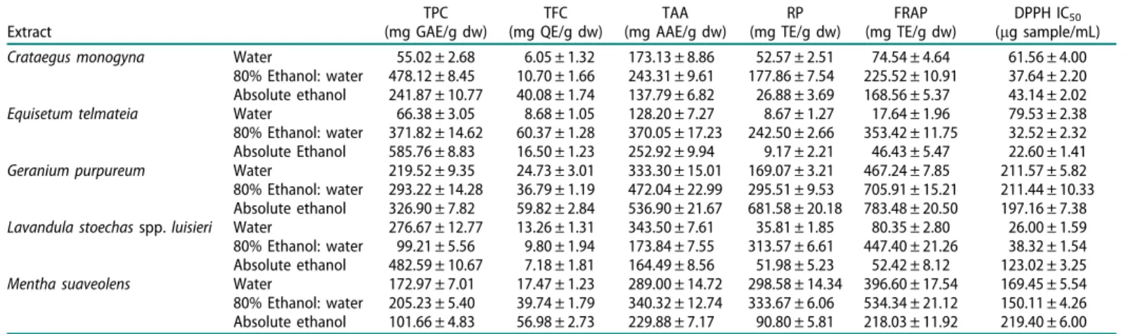
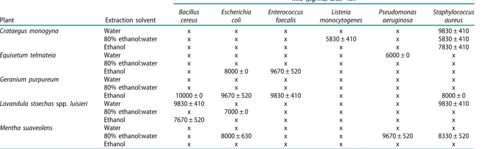
Antibacterial, antioxidant and anti-proliferative properties and zinc content of five south Portugal herbs
Texto
Imagem

Documentos relacionados
In vitro and in vivo studies evaluating the rice grains with different pericarp colour (light brown, red and black) showed potential beneficial effects on health related to
O objetivo geral deste trabalho é estudo da auto-organização do sistema de copolímero dibloco híbrido, maltoheptaose-b- polimetilmetacrilato (MH-b-PMMA) no estado
A partir da observação da autora, da variabilidade de informações que são oferecidas na maternidade em que essa profissional atua, embasada no conhecimento
ferox a partir de amostras brasileiras coletadas em diferentes locais do arquipélago de Fernando de Noronha, além de investigar a atividade das frações do extrato mais
redução importante dos coeficientes de mortalidade perinatal e infantil na década estudada, com relação à mortalidade infantil esta queda foi de apenas 28% para as
normas ambientais mais eficazes e políticas públicas que estimulem a consciência ambiental e limitem o pragmatismo econômico puro são necessárias para
Os resultados em peso (Kg) obtidos das 10 amostras submetidas à caracterização gravimétrica são apresentados na Tabela 1. Entretanto, para elaboração da média
Desde o início de minha carreira docente, lecionando matemática e desenvolvendo trabalhos com alunos de todas as séries do ensino fundamental e ensino médio,
