Received for publication 17/04/2017 - Accepted for publication 21/05/2017. The authors declare no conflicts of interests.
Corneal Astigmatism in cataract
surgery candidates
Emilio Raúl Ladeveze
1, Adriana Milena Ortiz
1, Luis Alberto Visciarelli
1, Abelardo Emilio Cavatorta
1R
ESUMOObjetivo: Relatar a prevalência e analisar os padrões de astigmatismo corneano em candidatos à cirurgia de catarata. Métodos: Os pesquisadores examinaram 2136 olhos de 1204 pacientes a cirurgia de catarata de 1 de janeiro a 31 de dezembro de 2016. Todos os candidatos foram avaliados com interferometria de coerência parcial (IOLMaster). Os resultados foram analisados estatisticamente em relação às variáveis qualitativa e quantitativa. Resultados: A média de idade dos pacientes foi de 71,9 anos, com predomínio do sexo feminino. A média de astigmatismo corneano foi de 1,0 dioptria (D). Contra a regra (ATR), o astigmatismo aumentou com a idade. As mulheres apresentaram córneas mais íngremes que os homens. Nos pacientes que tiveram os dois olhos medidos, encontramos fortes correlações entre quantidade de astigmatismo nos olhos direito e esquerdo, eixo e ceratometria. No geral, 39% dos olhos tinham astigmatismo corneano >1,00 D, e 19% > 1,50 D. Conclusões: Uma percentagem significativa da nossa população apresenta astigmatismo corneano >1,00, D com uma mudança de WTR (a favor da regra) para ATR (contra a regra) com o aumento da idade. Nossas descobertas em uma população que nunca foi descrita antes serão úteis para os cirurgiões melhorarem suas técnicas cirúrgicas, os pacientes melhorarem seus resultados visuais, os fabricantes de LIOs (lentes intraoculares) criarem projetos melhores, e os agentes de saúde otimizarem os recursos.
Descritores: Extração de catarata; Astigmatismo; Lentes intraoculares; Facoemulsificação
ABSTRACT
Purpose: To report the prevalence and analyse corneal astigmatism patterns in cataract surgery candidates. Methods: Researchers examined 2136 eyes from 1204 patients for cataract surgery from 1 January to 31 December 2016. All the candidates were evaluated with partial coherence interferometry (IOLMaster). The results were analysed statistically in relation to qualitative and quantitative variables. Results: The mean age of the patients was 71.9 years with a female predominance. Mean corneal astigmatism was 1.0 dioptre (D). Against the rule (ATR) astigmatism increased with age. Women presented with steeper corneas than men. In those patients who had both eyes measured, we found strong correlations between amount of right and left eye astigmatism, axis and keratometry. Overall, 39% of the eyes had corneal astigmatism > 1.00 D and 19% >1.50 D. Conclusions: A significant percentage of our population presents with corneal astigmatism > 1.00 D with a shift from WTR (with the rule) to ATR with increasing age. Our findings, in a population that has never been described before, will be helpful to surgeons to enhance surgical techniques, patients to improve visual outcomes, IOL (intraocular lens) manufacturers to make better designs and health agents to optimise resources.
Keywords: Cataract extraction; Astigmatism; Lenses, intraocular; Phacoemulsification
1 Centro Oftalmológico - San Nicolás, Buenos Aires, Argentina .
I
NTRODUCTIONT
homas Young, it was in 1825 that Sir George Biddell Airy for the first time successfully corrected his own astigmatism by the use of cylindrical lenses.(1)Cataract surgery is one of the most common intraocular procedures performed worldwide.(2) Advances in surgical
techniques and improvements in intraocular lens (IOL) design and calculations increase our chances to achieve the best results. At the same time, patient demands and expectations are continuously increasing. To achieve the best postoperative visual performance both spherical and cylindrical components should be corrected at the time of the surgery. (3)
The prevalence of corneal astigmatism has been widely reported in several parts of the world. To the best of our knowledge, this is the first report of corneal astigmatism in cataract surgery candidates in South America. Different techniques are available to correct the corneal astigmatism during phacoemulsification, such as incision on the steep meridian, opposite clear corneal incisions, peripheral corneal relaxing incisions and the use of a toric IOL. Each method has benefits and limitations according to the amount of correction and the possibility of regression with time. (4,5)
This study aims to identify the prevalence and patterns of pre-existing corneal astigmatism among this population. This information will be useful to cataract surgeons, patients, IOL manufacturers and health agents.
M
ETHODSThe current study was performed from 1 January to 31 December 2016. Candidates for cataract surgery at the Centro Oftalmológico (San Nicolás, Buenos Aires, Argentina) received a complete ophthalmological examination before the procedure. Trained staff took biometry measurements, and 2136 eyes from 1204 patients were included.
Optical biometry was performed using the principle of partial coherence interferometry (PCI) (IOLMaster 500, Carl Zeiss, Jena, Germany, software 5.3). The PCI has become the standard technique in IOL calculation due to its accuracy in the measurement of biometric parameters.(6-7)
Flat (K1) and steep (K2) keratometry, corneal astigmatism, axial length (AL) and anterior chamber depth (ACD) were measure five times in each eye, and the average values were included in the analysis.
Those eyes with a signal to noise ratio (SNR) < 2 were excluded from the study. The IOLMaster biometer considers reliable scans as those with a SNR > 2, so the manufacturer does not recommend the measurements with a SNR < 2.
Other exclusion criteria included: past or present corneal diseases, previous corneal refractive or intraocular surgery, dense cataract, history of ocular inflammation or patients who wore contact lenses.
For the analysis, patients were divided into groups by gender and age. Astigmatism was analysed as with the rule (WTR) when the steepest meridian was between 90° ± 30°, against the rule (ATR) when the steepest meridian was within 180° ± 30° and oblique (OB) when the steepest meridian was between 120° and 150° or 30° and 60°. The amount of astigmatism was divided into groups to allow our group to make a comparison with international publications. In those patients who had both eyes measured (n = 939), we analysed the interesting relationship.
Data analysis was performed using SAS 9.3 (Statistical Analysis System – SAS Institute, North Carolina). The quantitative variables were summarised across its mean (standard deviation) and its minimal and maximum values and the qualitative variables across percentage absolute and relative frequencies. The association between quantitative variables was analysed with the coefficient of Spearman and the association between categorical variables with the chi-square test of Pearson or the exact test of Fisher. The chance of changing to ATR type of astigmatism for every unitary increase in the age was estimated across a model of logistic regression. A p value less than 0.05 was regarded as statistically significant.
R
ESULTSA total of 2136 eyes of 1204 patients were included for analysis. The mean age was 71.9 years (Standard Deviation (SD) = 9.1 years; range: 34–94 years). Figure 1 shows the number of patients in each age group; 58.4% of the patients were women. Table 1 shows the demographic characteristics and clinical findings of the study population.
Figure 1: Distribution of entire sample for each age group.
Table 1.
Demographic characteristics and clinical findings of the study population
Characteristic Female Male Total
Patients (n) 703 (58.4%) 501 (41.6%) 1204
Eyes (n) 1259 (58.9%) 877 (41.1%) 2136
Age (years)
Mean SD) 71.9 (10.0) 71.9 (9.1) 71.9 (9.6) Range 34-94 37-93 34-94
Astgmatism (D)
Mean SD) 1.0 (0.7) 1.0 (0.7) 1.0 (0.7) Range 0.1 - 5.8 0.1 - 4.1 0.1 - 5.8
K1 (D)
Mean SD) 43.3 (1.5) 42.6 (1.5) 44.1 (1.6) Range 39.1 - 48.4 38.4 - 46.6 38.4 - 48.4
K2 (D)
Mean SD) 44.4 (1.6) 43.6 (1.5) 44.1 (1.6) Range 39.9 - 51.1 38.3 - 47.7 39.3 - 51.1
AL
Mean SD) 23.5 (1.3) 24.0 (1.2) 23.7 (1.3) Range 21.0 - 32.2 21.3 - 35.5 21.0 - 35.5
ACD
Mean SD) 3.0 (0.4) 3.1 (0.4) 3.0 (0.4) Range 2.0 - 4.2 2.1 - 4.2 2.0 - 4.2
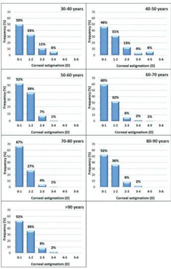
The mean astigmatism was 1.00 diopters (D) (SD = 0.7 D). We found no differences between genders. Figure 2 demonstrates the frequency distribution of corneal astigmatism for each age group. Of the entire sample, 1182 (55%) eyes presented with an astigmatism higher than 0.75 D, 841 (39%) eyes higher than 1.00 D and 398 (19%) higher than 1.50 D.
The relationship between corneal astigmatism and age was analysed. Figure 3 exhibits the dispersion diagram between this pair of variables. The correlation coeficient was not statistically significant (r = 0.0028; p = 0.202). The association between categorised variables was not statistically significant (p = 0.995).
The mean of flat K and steep K was K1: 43.0 (SD = 1.8) and K2: 44.1 (SD = 1.6). Comparing between both genders, the mean K1 and K2 in females were 43.4 (SD = 1.6) and 44.4 (SD = 1.6) against 42.6 (SD = 1.9) and 43.6 (SD = 1.5) in the male group. Steeper corneas with shorter AL were found in the female group (p = 0.001).
Table 2 shows the distribution by gender and type of astigmatism.
There was a statistically significant relationship between the type of astigmatism (ATR/OB/WTR) and age (p = 0.001). Further analysis showed an increasing prevalence of ATR astigmatism with increasing age. Even more, the chance to shift the axis of astigmatism from ATR to WTR in the 3rd decade was very low (0.55%; p = 0.001), increasing multiplicatively 6.76% (p = 0.001) per year of age. In other words, the mentioned chance increases 92.43% per decade of age (Figure 4).
In the group of patients that both eyes were evaluated (n = 939), important correlations were found.
In the first place, there was a statistically significant association in the corneal axis between the right (OD) and left eyes (OS) (p = 0.001). The majority (74%) of the patients with WTR in the OD had the same type of astigmatism in the OS. Also, 69% of the patients with ATR in the OD have the same type in the OS (Figure 5).
Figure 2: Frequency distribution of corneal astigmatism in 1.0 D steps for the 7 age groups.
Figure 3: Correlation between magnitude of corneal astigmatism and age.
Figure 4: Frequency distribution of the astigmatism type by age. WTR: With the rule, OB: Oblique, ATR: Against the rule
Table 2
Frequency distribution of type of astigmatism
Type of Astimatism Female Male Total
With the rule 637 (50.6%) 303 (34.5%) 940 (44.0%)
Oblique 215 (17.1%) 148 (16.9%) 363 (17.0%)
Against the rule 407 (32.3%) 426 (48.6%) 833 (39.0%)
Secondly, the magnitude of astigmatism also demonstrated a correlation between OD and OS. The majority (88%) of the patients that presented with a corneal astigmatism < 1 D in OD also presented with < 1 D in OS.
Considering the keratometric readings of both eyes, the correlation coefficients were K1 (r = 0.934) and K2 (r = 0.9), respectively, being in all cases significantly different from zero (p = 0.001) as is shown in figure 6.
The AL is a very important factor in calculating IOL power. The comparison between the AL of both eyes shows a positive correlation coefficient (r = 0.95; p = 0.001).
D
ISCUSSIONWith a rapidly aging population and patients expecting the best refractive results, the correction of the cylindrical component is something that surgeons cannot obviate.
The analysis of the prevalence of corneal astigmatism and characteristics of this population provides useful information for cataract surgeons.
For residents, fellows or young ophthalmologists these results demonstrate the importance of mastering more than one principal incision access during their cataract surgery program. Considering that 57% of patients older than 80 years and 42% of patients older than 70 years present with ATR astigmatism, if in the residency program residents only learn to place the main corneal wound superior, this will improve the amount of astigmatism in these eyes. The same thing will happen with younger patients if residency programs only teach temporal principal incisions.
Our study showed that a higher percentage of the patients are women, this is consistent with other published studies (with the exception of Isyaku in Nigeria).(8) The mean age of our population
(71.9 years) is similar to the international literature.
Astigmatisms lower than 1.00 D give the patients good visual acuity (VA), and the patients do not demand correction in most of the cases. On the other hand, it is well known that astigmatic refractive errors between 1.0 D and 2.0 D reduce VA to between 20/30 and 30/50. In the same way, errors between 2.0 D and 3.0 D reduce VA to between 20/70 and 20/100. (9)
The mean corneal astigmatism of our population (1.0 D) was consistent with previous studies. In this study, 39% of the eyes had corneal astigmatism > 1.0 D. This result is closer to those described by Khan (40.4%)(2) and Chen (41.3%)(10) and slightly higher than
Ferrer-Blasco (34.8%)(11) and Hoffman (36%)(12) (Table 3).
Figure 6: Correlation between right and left eyes of different parameters. (A) Corneal astigmatism; (B) K1; (C) K2; and (D) axial length.
Table 3
Summary of clinical studies of corneal astigmatism prevalence in cataract patients
Khan Ferrer-Blasco De Bernardo Hoffman Ysyaku Guan Chen Collier Wakefield Mohammadi This sudy (2) (11) (26) (12) (8) (3) (10) (15) (22) Country Argentina UK Spain Italy Germany Nigeria China China UK Iran Year 2017 2011 2009 2014 2010 2014 2012 2013 2016 2014 Patients(n) 1204 726 2415 380 15448 3169 827 2849 2247 1317 Eyes (n) 2136 230 4540 757 23239 3286 1430 4831 2247 2156 Age
Mean+SD 71.9+9.6 75.5+0.7 60.5+9.8 71.8+10.1 74 60.8+12.7 72.2+11.5 70.5+9.5 72.8+13.84 64.9+11.4 Range 34-94 30-104 32-87 33-96 - 39-97 16-98 40-95 10-109 30-88 Male/
Famale 501/703 343/403 768/1647 176/204 - 1826/1343 359/468 1090/1759 - 609/708 Corneal
Astigmatism
Mean+SD 1+0.7 1.03+0.72 0.9+0.93 1.02+0.69 0.98+0.78 1.16 1.07+0.73 1.01+0.69 1.11+0.88 1.12+1.1 Range 0.1-5.8 0.0-6.20 0.25-6.75 0.06-4.57 - 0.25-6.00 0.06-5.52 0.05-6.59 0.02-13.71 0.0 - 7.0 K1
Mean+SD 43+1.5 43.43+1.48 43.48+1.61 43.54+1.43 - 43.99 43.57+1.56 43.76+1.53 43.18+1.64 43.70+1.70 K2
Mean+SD 44.1+1.6 44.46+1.56 44.08+1.61 44.56+1.52 - 43.8 44.64+1.65 44.76+1.56 44.29+1.70 44.83+1.79 Corneal
Amesbury(5) suggested to place the main corneal wound on
the steep corneal meridian for patients with < 1.0 D of corneal astigmatism, performing corneal relaxing incisions between 1.0 D and 1.5 D and the implantation of toric IOLs in patients with more than 1.5 D of pre-existing astigmatism. Relaxing incisions are safe and inexpensive but can be unpredictable by increasing the risk of induced higher order aberrations that may affect the final vision.
(13) Considering this, 19% of our population would benefit from the
implantation of toric IOLs. In addition, other studies have shown toric IOLs to be effective at correcting 1.0 D or 0.75 D levels of corneal astigmatism.(14) According to these criteria, between 39%
and 55% of our population could be effectively corrected if toric lens were available.
Collier Wakefield et al.(15) found a relationship between
the amount of astigmatism and age. This group demonstrated a reduction in the prevalence of astigmatism < 1.0 D with age. Additionally, they describe an increased prevalence of eyes with > 3.0 D with age. At the same time, it is the first study group to report the relationship between the magnitude of preoperative corneal astigmatism and the type of astigmatism (WTR/ATR/OB). They demonstrated that ATR astigmatism is increasingly prevalent with increasing magnitude of astigmatism. In our study group, we did not find statistically significant values analysing such variables.
Data from our analysis demonstrated that ATR corneal astigmatism is increasingly prevalent with increasing age. This shift is well documented in other studies. WTR astigmatism is more prevalent in young patients. As corneal astigmatism continues to change towards ATR with age, young patients should be aware about this progression in the next years. Although it is not our case, some study groups suggest to treat ATR astigmatism more aggressively, as its magnitude is likely to increase with age. (16)
In contrast, Kim et al.(17) in their analysis of patients that had
phacoemulsification with a 3 mm clear corneal incision with a 10-year follow-up did not find changes in the axis with time.
Several factors had been proposed to explain the corneal changes with age, some of them with contrasting results. Grosvenor(18) in 1978 proposed in his theory that the pressure from
the upper eyelid alters the corneal shape steepening in the cornea’s vertical meridian. This results in WTR astigmatism, typically in young patients. A reduction in lid tension explains the shift towards ATR astigmatism. Marin-Amat(19) also suggests that a reduced
action of the medial rectus muscle in elderly patients may lead to ATR astigmatism. However, Vihlen(20) some years later found
no association between eyelid tension and corneal astigmatism. There are also structural corneal factors that have been proposed to explain the changes in corneal toricity, which include a decrease in the interfibrillar spacing, thickening of the stromal collagen bundles and structural changes that alter the rigidity and elasticity of the cornea. (21)
When comparing the keratometries between males and females, we found that women had steeper corneas than men. Mohammadi(22) in Iran in 2016 had found the same results. Goto(23)
suggests that these differences of corneal curvature between genders may be due to the influence of sex hormones.
In our study, we found a statistically significant correlation between right and left keratometries and AL in patients where both eyes could be evaluated. There are not many reports that compare both eyes in the same patient. This interesting correlation supports the idea of also utilising bilateral biometry in candidates for unilateral surgery. In case we find a large discrepancy between OD and OS measurements, the biometry should be repeated in order to correct any mistakes in time.
After this report (in case we assume an astigmatic threshold for toric IOL insertion of 1.5 D), we realise that 19% of the patients of this population would have benefited from the implantation of toric IOLs. This data is important for health boards of South America.
Eye dimensions described is an important cohort, as the analysed might be helpful for IOL manufacturers to suit their designs to a particular population.
Finally, as it was described by Pineda(24), the correction of
astigmatism with toric lenses reduces the long-term potential costs (practically for life), diminishing the dependence of spectacles or contact lenses in those patients with corneal astigmatism > 1.5 D.
The limitation of this study is that, as we used the PCI to take the measurements, we could not measure the posterior corneal astigmatism. Now, it is known that it does not correlate with the anterior surface, and those studies that investigated the posterior surface found amounts between 0.18 D and 0.31 D. (21,25)
In conclusion, our study provides information about the characteristics of corneal astigmatism in a population that, to the best of our knowledge, has not been described before. These findings will be helpful in improving surgical techniques and visual outcomes of patients as well as designing new toric IOLs.
WHAT WAS KNOWN
• It is important to correct astigmatism in cataract surgery candidates to achieve the best postoperative visual outcomes.
• Against the rule astigmatism is increasingly prevalent with increasing age.
WHAT THIS PAPER ADDS
• To our knowledge, this is the first report of corneal astigmatism prevalence and characteristics in a South American country.
• The significant correlation between right and left eye biometric values in the same person.
R
EFERENCES1. Levene JR. Sir George Biddell Airy, F.R.S. (1801–1892) and the discovery and correction of astigmatism. Notes Rec R Soc Lond. 1966;21(2):180–99.
2. Khan MI, Muhtaseb M. Prevalence of corneal astigmatism in patients having routine cataract surgery at a teaching hospital in the United Kingdom. J Cataract Refract Surg. 2011;37(10):1751–5.
3. Guan Z, Yuan F, Yuan YZ, Niu WR. Analysis of corneal astigmatism in cataract surgery candidates at a teaching hospital in Shanghai, China. J Cataract Refract Surg. 2012;38(11):1970–7.
4. Wang L, Misra M, Koch DD. Peripheral corneal relaxing incisions combined with cataract surgery. J Cataract Refract Surg. 2003;29(4):712–22.
5. Amesbury EC, Miller KM. Correction of astigmatism at the time of cataract surgery. Curr Opin Ophthalmol. 2009;20(1):19–24. 6. Eleftheriadis H. IOLMaster biometry: refractive results of 100
consecutive cases. Br J Ophthalmol. 2003;87(8):960–3.
7. Salouti R, Nowroozzadeh MH, Zamani M, Ghoreyshi M, Salouti R. Comparison of the ultrasonographic method with 2 partial coherence interferometry methods for intraocular lens power calculation. Optometry. 2011;82(3):140–7.
8. Isyaku M, Ali SA, Hassan S. Preoperative corneal astigmatism among adult patients with cataract in Northern Nigeria. Indian J Ophthalmol. 2014;62(11):1094–5.
10. Chen W, Zuo C, Chen C, Su J, Luo L, Congdon N, et al. Prevalence of corneal astigmatism before cataract surgery in Chinese patients. J Cataract Refract Surg. 2013;39(2):188–92.
11. Ferrer-Blasco T, Montés-Micó R, Peixoto-de-Matos SC, González-Méijome JM, Cerviño A. Prevalence of corneal astigmatism before cataract surgery. J Cataract Refract Surg. 2009;35(1):70–5.
12. Hoffmann PC, Hütz WW. Analysis of biometry and prevalence data for corneal astigmatism in 23,239 eyes. J Cataract Refract Surg. 2010;36(9):1479–85.
13. Montés-Micó R, Muñoz G, Albarrán-Diego C, Rodríguez-Galietero A, Alió JL. Corneal aberrations after astigmatic keratotomy combined with laser in situ keratomileusis. J Cataract Refract Surg. 2004;30(7):1418–24.
14. Sheppard AL, Wolffsohn JS, Bhatt U, Hoffmann PC, Scheider A, Hütz WW, et al. Clinical outcomes after implantation of a new hydrophobic acrylic toric IOL during routine cataract surgery. J Cataract Refract Surg. 2013;39(1):41–7.
15. Collier Wakefield O, Annoh R, Nanavaty MA. Relationship between age, corneal astigmatism, and ocular dimensions with reference to astigmatism in eyes undergoing routine cataract surgery. Eye (Lond). 2016;30(4):562–9.
16. Lyall DA, Srinivasan S, Ng J, Kerr E. Changes in corneal astigmatism among patients with visually significant cataract. Can J Ophthalmol. 2014;49(3):297–303.
17. Kim H, Whang WJ, Joo CK. Corneal astigmatism in patients after cataract surgery: a 10-year follow-up study. J Refract Surg. 2016;32(6):404–9.
18. Grosvenor T. Etiology of astigmatism. Am J Optom Physiol Opt. 1978;55(3):214–8.
Corresponding author:
Emilio Raúl Ladeveze, MD
Francia 130 – San Nicolas – Argentina Postal Code: 2900
Phone: 54 - 0341 – 156539047 E-mail: [email protected]
19. Marin-Amat M. Les variations physiologiques de la courbure de la cornée pendant la vie; leur importance et transcendance dans la réfraction oculaire. Bull Soc Belge Ophtalmol. 1956;113:251–93. 20. Vihlen FS, Wilson G. The relation between eyelid tension, corneal
toricity, and age. Invest Ophthalmol Vis Sci. 1983;24(10):1367–73. 21. Hayashi K, Hayashi H, Hayashi F. Topographic analysis of the changes
in corneal shape due to aging. Cornea. 1995;14(5):527–32.
22. Mohammadi M, Naderan M, Pahlevani R, Jahanrad A. Prevalence of corneal astigmatism before cataract surgery. Int Ophthalmol. 2016;36(6):807–17.
23. Goto T, Klyce SD, Zheng X, Maeda N, Kuroda T, Ide C. Gender- and age-related differences in corneal topography. Cornea. 2001;20(3):270–6.
24. Pineda R, Denevich S, Lee WC, Waycaster C, Pashos CL. Economic evaluation of toric intraocular lens: a short- and long-term decision analytic model. Arch Ophthalmol. 2010;128(7):834–40.
25. Ho JD, Liou SW, Tsai RJ, Tsai CY. Effects of aging on anterior and posterior corneal astigmatism. Cornea. 2010;29(6):632–7.