Flexible intramedullary nails with traction versus plaster
cast for treating femoral shaft fractures in children:
comparative retrospective study
Hastes intramedulares lexíveis com tração
versus
aparelho gessado para o tratamento
de fraturas diaisárias do fêmur em crianças: estudo comparativo retrospectivo
Fabiano Prata do Nascimento
I, Cláudio Santili
II, Miguel Akkari
III, Gilberto Waisberg
IV, Susana dos Reis Braga
V, Patrícia Maria
Moraes de Barros Fucs
VIPediatric Orthopedic Surgery Division, Department of Orthopedics and Traumatology, Faculdade de Ciências Médicas, Santa Casa de
Misericórdia de São Paulo (SCMSP), Brazil
ABSTRACT
CONTEXT AND OBJECTIVE: Femoral fractures are common in children between 2 and 12 years of age, and 75% of the lesions afect the femoral shaft. Traction followed by a plaster cast is universally accepted as conservative treatment. However, in some situations, a surgical approach is recommended. The objec-tive here was to compare treatments for femoral shaft fractures using intramedullary nails (titanium elastic nails, TEN) versus traction and plaster casts in children. The hypothesis was that TEN might provide better treatment, with good clinical results in comparison with plaster casts.
DESIGN AND SETTING: This retrospective comparative study was conducted in a public university hospital.
METHODS: Sixty children with femoral fractures were evaluated; 30 of them underwent surgical treat-ment with TEN and 30 were treated conservatively using plaster casts. The patients’ ages ranged from 5 to 13 years (mean of 9 years).
RESULTS: The mean duration of hospitalization was nine days for the surgical group and 20 days for the conservative group. The incidence of overgrowth in the patients treated with TEN was 60.0% and, for those treated conservatively, 13.3%. Partial weight-bearing was allowed after 3.5 weeks in the surgical group and after 9.6 weeks in the conservative group. New hospitalization was required for 90.0% in the surgical group and 16.7% in the conservative group. Patients treated with plaster casts presented higher incidence of complications, such as loss of reduction.
CONCLUSIONS: The surgical method presented better results for children.
RESUMO
CONTEXTO E OBJETIVO: Fraturas femorais são comuns em crianças entre 2 e 12 anos de idade, e 75% das lesões acometem a diáise. Tração seguida de aparelho gessado (“gesso”) é universalmente aceita como tratamento conservador. Entretanto, em algumas situações o tratamento cirúrgico é recomendado. O ob-jetivo foi comparar o tratamento de fraturas diaisárias do fêmur com hastes intramedulares (titanium elastic nails, TEN) com tração e gesso em crianças. A hipótese era de que TEN pode ser melhor tratamento, com bons resultados clínicos em comparação com o gesso.
TIPO DE ESTUDO E LOCAL: Este estudo retrospectivo e comparativo foi conduzido num hospital público universitário.
MÉTODOS: Sessenta crianças com fraturas de fêmur foram avaliadas, 30 delas foram submetidas a trata-mento cirúrgico com TEN e 30 foram tratadas de forma conservadora usando gesso. A idade dos pacientes variou de 5 a 13 anos (média de 9 anos).
RESULTADOS: O tempo médio de internação foi de 9 dias para o grupo cirúrgico e 20 dias para o grupo conservador. A incidência de crescimento excessivo nos pacientes tratados com TEN foi de 60,0% e, para aqueles tratados de forma conservadora, 13,3%. Sustentação parcial de peso foi permitida após 3,5 se-manas no grupo cirúrgico e após 9,6 sese-manas no grupo conservador. Houve 90,0% de novas internações no grupo cirúrgico e 16,7% no grupo conservador. Pacientes tratados com gesso apresentaram maior incidência de complicações, tais como perda de redução.
CONCLUSÃO: O método cirúrgico apresentou melhores resultados para as crianças.
IMD, PhD. Head of the Pediatric Orthopedic Staf in the Itaim and Anália Franco Units of Hospital São Luiz, São Paulo, and Coordinator of the Orthopedic Staf in Hospital Estadual de Santo André, Faculdade de Medicina do ABC, Santo André, São Paulo, Brazil.
IIMD, PhD. Adjunct Professor, Faculdade de Ciências Médicas, Irmandade da Santa Casa de Misericórdia de São Paulo, and Attending Physician, Pediatric Orthopedic Group, Santa Casa de Misericórdia de São Paulo, São Paulo, Brazil. IIIMD, PhD. Head of the Pediatric Orthopedic Group in Santa Casa de Misericórdia de São Paulo, São Paulo, Brazil.
IVMD. Attending Physician in the Pediatric Orthopedic Group, Santa Casa de Misericórdia de São Paulo, São Paulo, Brazil.
VMD, MSc. Attending Physician in the Pediatric Orthopedic Group, Santa Casa de Misericórdia de São Paulo, São Paulo, Brazil.
VIMD, PhD. Adjunct Professor, Faculdade de Ciências Médicas, Irmandade da Santa Casa de Misericórdia de São Paulo, and Head of the Neuromuscular Disorders Group, Santa Casa de Misericórdia de São Paulo, São Paulo, Brazil.
KEY WORDS: Femur. Femoral fractures. Bone nails. Child. Orthotic devices.
PALAVRAS-CHAVE: Fêmur.
Fraturas do fêmur. Pinos ortopédicos. Criança.
INTRODUCTION
Femoral fractures are most common in children around the age of two years and around 12 years old, therefore with two peaks of incidence, and 75% of the lesions afect the femoral shat.1 Skin or
skeletal traction followed by a plaster cast is universally accepted as a conservative treatment.
However, in open and multiple fractures in children over 10 years of age, a surgical approach has been recom-mended. Intramedullary rods (rigid or semi-rigid),2,3 elastic
sta-ble intramedullary nails (ESIN) or simply titanium elastic nails (TEN) have been used very successfully in children under the age of 12 years. Intramedullary nails are better suited for transverse and/or short oblique fractures than for long oblique or commi-nuted fractures, which respond better to external ixation or to traction followed by a cast.1
We previously reported our first results using TEN in cases of femoral fractures in children from 5 to 14 years of age.4 This study compares those results with conservative
treatment, in children of the same ages, especially focusing on the duration of hospitalization, discrepancies and deformities and the time taken to achieve weight bearing and the return to daily activities. The setting was a public university hospi-tal in Brazil, where cost of treatment can be an issue and sur-gical treatment was only incorporated in the routine service ten years ago. The conservative method was represented by traction followed by a cast, and the surgical method by medullary fixation with TEN. The hypothesis was that intra-medullary nails would offer an earlier return to daily activities with fewer complications compared with plaster casts.
OBJECTIVE
To compare treatments for femoral shat fractures using TEN,
versus traction and plaster casts in children. he hypothesis was
that TEN might provide better treatment, with good clinical results in comparison with plaster casts.
METHODS
Setting and patients
his retrospective comparative study was conducted in a pub-lic university hospital, covering the period between January 1995 and February 2004. All children between the ages of 5 and 14 years who were treated during this period for femoral shat frac-tures using TEN or traction and cast, and with at least 24 months of follow-up, were included.
Before 2000, TEN was not used in Brazil and it was thus not available in public services: femoral shat fractures were there-fore treated conservatively. With the advent of TEN, it became possible to undertake a retrospective analysis comparing those
previously treated with casts with those treated with nails. A total of 60 children (a convenience sample) with unilateral fracture of the femoral shat during this period were evaluated. During the irst part of the study period, all children underwent skin or skel-etal traction before treatment. When nails became available in our public service, most children underwent surgery.
In this manner, 30 patients treated using TEN and 30 patients treated using traction followed by a cast during the study period were selected. Patients who presented diseases that could afect the normal anatomical and physiological characteristics of the skeleton, such as osteometabolic diseases, bone dysplasia or pathological fractures associated with neuromuscular syndromes or alterations were excluded. he institution’s Ethics Committee approved the study.
In 60.0% of the cases, the fracture was caused by pedestrian-automobile collisions; 21.6% were due to falls from heights; 10% were the result of motor-vehicle accidents; in 5%, objects fell onto the thigh; 1.7% of the patients sufered bicycle accidents; and 1.7% were child abuse victims. Associated lesions were found in 22% of the patients with femoral fractures. Of these, 30.7% had tibial fractures.
Regarding the patients treated with TEN, the fracture was transverse in 60.0% of the cases and oblique in 26.7%. In the conservative group, 36.7% of the fractures were transverse and 40.0% were oblique (P = 0.264). he transverse and oblique frac-tures represented 81.7% of all the fracfrac-tures. here were only two open fractures (grade II of Gustilo and Anderson), in the surgical group. Treatments casts were placed at least ive days ater frac-ture occurrence (mean: 18.7 days).
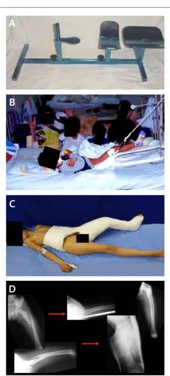
The cast for the conservative treatment was constructed on a children’s orthopedic table, with the hips flexed at between 30 and 45 degrees, with semi-flexion of the knee and inclusion of the foot. Following this, radiographs were taken for reduction control. The cast was then made after evaluating the adjacent soft tissues and the radiographic results (i.e. to investigate shortening or formation of bone callus) (Figure 1).
In the subsequent evaluations, in addition to physical examination, radiographs of the affected limb in the ante-rior-posterior and lateral views were produced, and scanom-etry of the lower limbs was performed. Angular deformities (in degrees), discrepancies (in centimeters), initial and final shortening and excessive growth were measured. Other vari-ables were also analyzed, such as the duration of traction and hospitalization, time taken for the patient to be able to bear weight on the limb and return to his daily activities, time taken for consolidation to be achieved, presence of acute and late complications, number of subsequent hospitalizations, com-plaints and length of follow-up. Between six and eight months after fracture healing and remodeling, the nails were removed surgically, in a single-day hospitalization period, and the chil-dren could walk freely thereafter. Chilchil-dren treated using casts had the cast removed after consolidation, which took approxi-mately two months to achieve. They were then allowed to bear weight partially two weeks later. Use of crutches was ended after fracture healing.
Figure 1. Children’s orthopedic table (A) for traction (B) and cast (C), for children with femoral shaft fractures; and radiographs showing the reduction control before and after the cast was applied (D).
A
B
C
D
fashion, two to three centimeters proximally from the femo-ral distal growth cartilage (Figure 2). No casts were used for complementary immobilization. Depending on the fracture characteristics and its reduction, early weight-bearing and joint movement were allowed, especially of the knee, and the patients were encouraged to do so from the first postopera-tive day onwards.
Figure 2. Fixation of lexible intramedullary titanium elastic nails (TEN) in a child with femoral shaft fracture: procedure on a radiotransparent table, with the patient in supine position (A) and radiographs taken initially (B), in the immediate postoperative period (C) and inally (D).
A
Data analysis
he data were analyzed as absolute and relative frequencies for quantitative measurements, and were presented as means, fre-quencies (%) and standard deviations. Comparisons were made using the Fisher, Mann-Whitney tests, Barlett and Pearson chi-square test. Analysis of variance (ANOVA) was also used.
RESULTS
Patients’ ages
he patients’ ages ranged from 5.0 to 13.5 years (mean: 9 years). he patients in the surgical group had a mean age of 9.6 years and the patients in the conservative group had a mean age of 8 years (analysis of variance, ANOVA; P = 0.004). here were 41 male patients (68.4%). In the surgical group, the gender distribution was even, but in the conservative group, there was a preponderance of male patients (83.3%).
Lengths of hospitalization and follow-up and time taken to return to activities
he minimum length of follow-up was 24 months for the surgical patients (mean: 35.4 months) and 59.0 months for those treated using casts. he mean duration of hospitalization was signiicantly diferent between the groups: 9.4 days for the surgically treated children and 20.5 days for the conservatively treated children (ANOVA; P < 0.001). he times taken for healing were also signiicantly diferent: 7.7 weeks for consolidation in the surgical group and 9.3 weeks in the conserva-tive group (ANOVA; P = 0.005). he mean duration of traction was 5.3 days for the surgical group, with a maximum of 14 days and a mean of 18.4 days for the conservative group, with a maximum of 40 days. he mean time taken for the patients to return to their activities was 3.7 weeks (ranging from one week to ten weeks) for the surgical group and 9.5 weeks (ranging from six weeks to 16 weeks) for the conserva-tive group (P < 0.001). Partial loading was allowed ater 3.5 weeks, on average, for the surgical group and ater 9.6 weeks for the conservative group (Mann-Whitney; P < 0.001). here was a relationship between increasing age and longer time taken for weight-bearing on the frac-tured limb to be allowed among the patients who were treated con-servatively (i.e. with casts). On the other hand, for those treated with TEN, the time taken for this remained relatively constant: for younger patients (5 to 9 years of age), the average was 3.4 weeks, and for those aged 10 to 14 years, 3.8 weeks (ANOVA; P = 0.000). he average time taken for total weight-bearing to be allowed was 8.8 weeks for the patients treated with nails and 11.3 weeks for the patients treated with casts (ANOVA; P = 0.007). For the conservative method, the total time taken for weight-bearing to be allowed increased with increasing age; for the surgical method, the opposite was seen (Graph 1). he percentage of patients hospitalized was 90.0% in the surgical group and 16.7% in the conservative group. None of the patients sufered a
repeated fracture in either group. Most hospitalizations ater surgical treatment were preplanned, in order to remove the nail.
In the surgical group, one patient presented migration of the nail (the tip of the nail was cut of in the new surgery); another patient fell, thereby losing his reduction (new surgery was indicated); a third patient sufered sot-tissue irritation (the nail had to be removed early on). Among these three, only the case of migration required new hos-pitalization that had not been preplanned. he patient who fell was a girl who tried to walk during the irst hospitalization and fell from her bed. In the conservative group, three patients lost their reduction: one at six weeks; one who started walking before consolidation had been achieved; and one case in which the patient removed the pelvic portion of the cast. hese events required a second hospital sion and, in the case of the surgically treated patients, a third admis-sion. A signiicant diference was found between the groups regarding the total number of hospitalizations, including those for nail removal (Pearson’s chi-square test; P < 0.001). All the surgical patients under-went closed reduction. his was possible because of the traction that was irstly performed on all the patients (28 patients underwent skel-etal traction and two patients underwent cutaneous traction).
Shortening, overgrowth and deformities
he mean initial shortening, prior to treatment, was 2 cm, ranging from 0.5 to 4.5 cm for the entire group, from 0.5 to 3 cm in the conser-vative group and from 0.5 to 4.5 cm in the surgical group. Shortening, ater a period of at least 24 months, occurred in 6.7% of the patients in the surgical group and in 63.3% of the patients in the conserva-tive group (Pearson’s chi-square test; P < 0.001). In the surgical group, the mean shortening was 0.25 cm and in the conservative group the mean was 1.14 cm (ANOVA; P = 0.133), with no signiicant difer-ence between the means of the two groups. here was overgrowth in 60.0% of the patients in the surgical group and 13.3% in the conserva-tive group (Pearson’s chi-square test; P < 0.001). he mean overgrowth
Graph 1. Mean and standard error of load (in weeks), according to the treatment (surgical or with cast), for femoral shaft fractures in children in two age groups.
4 6 8 10 12 14 16
5 to 10 years 10 to 14 years
M
ea
n
o
f
to
ta
l w
ei
g
h
t
b
ea
ri
n
g
(
w
ee
ks
)
was not statistically diferent (ANOVA; P = 0.072) and was 0.66 cm (range: 0.25 to 1.50 cm) in the surgical group and 1.06 (range: 0.05 to 1.50 cm) in the conservative group.
Every patient presented some type of deformity. he average posterior angulation and varus and valgus deformities did not exceed 10 degrees. he mean and standard deviation of the ante-rior angulation deformity was 6.5 degrees for patients treated sur-gically and 12.1 degrees for the patients treated using traction and cast. Complaints about the treatment were registered in the medi-cal records, from 10% of the patients treated with nails and from 16.6% of the patients treated with casts.
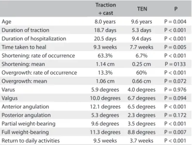
Table 1 summarizes the results.
DISCUSSION
Age and indication for treatments
Conservative treatment using an early cast is indicated for children with femoral fractures under the age of 11 years, 5-10 but some authors advocate this treatment method only
for children under six years of age.11,12 Traction may be used
before applying the cast, in view of the possibility of signifi-cant limb shortening and bad alignment in children over the age of five or six years.13 Among the disadvantages from
con-servative methods are limb discrepancies or angle deviations, compartment syndrome due to skin traction and possible psy-chological harm. Moreover, longer hospitalization periods and consequent higher costs are attributed to conservative treatment.2,14,15 The advantages of such treatment are that it
is available in all healthcare services and that it eliminates the risks that are inherent to surgery.
Surgical treatment in our service was only recently intro-duced into routine practice, such that observation of the dates of treatment (conservative cases from 1995 to 2004 and sur-gical cases from 2000 to 2004) in our sample allow us to say that there has been a gradual and natural transition towards choosing surgery. Today, surgery is the first choice in our pediatric service and this study provides an evaluation of the benefits that this change of approach towards femoral shaft fractures has brought.
Treatments using nails for fixation have been indicated for patients between the ages of 4 and 17 years, although Bopst et al.16 more recently reported an indication for children as
young as 1.5 years of age and Simanovsky et al.17 for those
aged three years and over. This age group includes the phase at which these patients go to school, and thus, independence during treatment is important for these patients. By reduc-ing hospitalization time, children may return to school earlier, thereby avoiding social isolation and the need for extra care, such as the care needed to maintain hygiene when individuals
are treated using casts.4,18-21 TEN or semi-rigid Ender rods for
use in cases of transverse and short oblique fractures has been advocated as the best approach for children over the age of five years. These nails make it possible to control limb length and avoid growth cartilage lesions, and they also shorten the dura-tion of hospitalizadura-tion and allow faster recovery.10,18-22
In our study, TEN was mainly indicated for transverse and short oblique fractures, and in patients over the age of ive years. Some patients had comminuted and spiral fractures and, in order to control the alignment of one of the comminuted fractures, three nails had to be applied.
he length of time under traction is directly associated with the duration of hospitalization, in cases in which a femoral fracture is the most signiicant lesion. Newton and Mubarak23
reported that their minimum hospitalization time prior to cast placement was 20.6 days for skin traction, 20.8 days for skeletal traction, 8.5 days for intramedullary nails and 2.5 days for early cast placement. Ligier et al.20 and Heinrich et al.18 reached
dif-ferent results: they analyzed fractures treated using lexible rods and found that the duration of hospitalization ranged from 4.5 to 8 days. Our patients treated with traction and cast presented signiicantly longer mean hospitalization times (20.5 days ver-sus 9.4 days). One of the reasons for this may have been that, in
our hospital, at least ive days pass between fracture occurrence and cast placement.
It also should be taken into consideration that all cases treated surgically required another period of hospitalization in order to remove the nails. We chose to remove the nails six to eight months ater their implantation, which is in accor-dance with reports from other authors, such as Flynn et al.19
and Buford et al.24 At this time, the fracture presents very solid Table 1. Demographic and clinical data on the 60 children treated for femoral shaft fractures with cast or TEN (titanium elastic nails)
Traction
+ cast TEN P
Age 8.0 years 9.6 years P = 0.004
Duration of traction 18.7 days 5.3 days P < 0.001 Duration of hospitalization 20.5 days 9.4 days P < 0.001 Time taken to heal 9.3 weeks 7.7 weeks P = 0.005 Shortening: rate of occurrence 63.3% 6.7% P < 0.001 Shortening: mean 1.14 cm 0.25 cm P = 0133 Overgrowth: rate of occurrence 13.3% 60% P < 0.001 Overgrowth: mean 1.06 cm 0.66 cm P = 0.072
Varus 5.9 degrees 4.0 degrees P = 0.976
union. Ligier et al.20 recommends nail removal three months
ater surgery.
In addition to the hospitalization for nail removal, we had one case that needed yet another hospitalization in order to cut the tip of one nail that had migrated. In the conservative group, additional hospitalizations were needed because of loss of posi-tioning and reduction, as well as one for cast removal. A signif-icant diference between the groups was found regarding the number of hospitalizations: some of the hospital admissions were not expected, but the majority were due to nail removal. An analysis by Santili et al.25 that included pediatric patients within
our setting showed that conservative treatment of femoral shat fractures was 22.5% more expensive than surgical treatment with lexible nails. We did not perform a cost analysis in this study, but the incidence of infections and other complications, which could have an impact on cost, was zero in our sample of surgi-cal patients.
With regard to consolidation and return to activities, Ligier et al.20 and Saseendar et al.26 reported that elastic movement
promoted faster and more abundant bone callus formation. Stans et al.27 reported that consolidation was faster using
lex-ible rods than using external ixation. In our patients, the mean time taken for fracture consolidation to be achieved was 1.6 weeks shorter in the surgical cases than in the conservatively treated cases. Some authors (Stans et al.,27 Reeves et al.28 and
Staheli and Sheridan8) considered that the fracture had healed
and then removed the cast ater eight weeks, on average, while others11 reported removing it ater six weeks. he time taken to
return to school among the patients treated with TEN, walking with the aid of crutches (i.e. partial loading), has ranged from two days to four weeks, and for full loading, from three to 11 weeks.4,14,15,20,21,26,29 Concerning weight-bearing and the return
to normal activities in the present study, the surgical method allowed partial loading before consolidation and was earlier than with conservative treatment. his diference was highly signiicant (ANOVA; P < 0.001). Partial and full weight-bearing were allowed, on average, ater 3.5 and 8.8 weeks for the sur-gical group, and ater 9.6 and 11.3 weeks for the conservative group, respectively.
Therefore, there was a difference in partial load-bear-ing between the two methods, of approximately six weeks (ANOVA; P < 0.001). Concerning age, the time taken for par-tial and total loading to be allowed increased with increas-ing age among the patients treated with casts, while there was a reduction in the time taken for total loading to be allowed among those treated with TEN. The explanation for this is that the older patients probably assimilated the orientations for crutch use better. The definition for the return to daily activities was taken to be when the patient was able to walk on
his own, with crutches, which exactly corresponded to when partial weight-bearing was allowed.
Shortening, overgrowth and deformities
A consensus exists in the literature that the inal shortening of the limb is produced by the initial shortening combined with the patient’s potential for growth, which is greater in younger chil-dren. Cadman and Neer29 considered that a maximum of 3 cm of
shortening was acceptable, Czertak and Hennrikus11 considered
that up to 2.5 cm was acceptable and Staheli30 and Buehler et al.5
considered that up to 1.5 cm was acceptable. In our group, the method using traction and cast caused greater shortening (mean of 1.14 cm), occurring in 63.3% of the patients in this group, in comparison with the surgical group, in which this occurred in only 6.7% of the patients (mean of 0.25; Pearson’s chi-square test, P < 0.001). Despite the diferences between the groups, these val-ues were clinically very well tolerated (ANOVA = 0.133). Only one patient had a inal shortening of 4.0 cm. As observed in the literature,18,20 signiicantly greater overgrowth also occurred
in our patients treated with TEN. his was present in 60.0% of the cases, whereas it was only present in 13.3% of the cases in the conservative group.
However, even though the frequency of overgrowth was dif-ferent between the groups, there was no statistically signiicant diference in the amount of overgrowth between the two methods used (P = 0.072), with a mean of 0.66 cm for the surgical group and 1.06 cm for the conservative group. hese results are com-patible with what has been reported in the literature.13,18-20,29,31,32
All the patients in the present study presented some type of deformity. he means for the varus, valgus, posterior and rota-tional deviations were less than ten degrees with both meth-ods. According to Flynn et al.,19 angles of less than ten degrees
are considered satisfactory; therefore, we can consider that the results from our patients are in conformity with the literature standards. We had high incidence of anterior angulation, with means of 6.5 degrees for the surgical group and 12.1 degrees for the conservative group. It must be emphasized that in the sagittal plane, the measurement included the physiological angulation of the femur. Some authors have reported losses of reduction and angular deviation when treating children with more than 45 kg of body weight. Care is required when indi-cating lexible intramedullary nails for patients who are obese and closer to skeletal maturity, and an indication for an inter-locking pediatric nail with a lateral entry point should be con-sidered.5,33-35 In order to provide better control over the
his study had a retrospective design, and used a conve-nience sample, which were limitations of the study. he diferent characteristics of the two groups of patients were also a limita-tion. Nonetheless, this study points towards the important and already-suspected hypothesis that femoral shat fractures in chil-dren can be better treated with surgery. his is a proper scenario within which a randomized controlled trial could be developed in order to obtain reliable answers, without bias.
CONCLUSION
Patients older than ive and younger than 14 years of age, with femoral shat fractures treated using a lexible intramedul-lary method, returned to daily activities and to school earlier, with shorter periods of traction and hospitalization and less limb shortening and a lower rate of loss of reduction, compared with those treated with casts. Both methods showed few compli-cations or problems relating to alignment.
REFERENCES
1. Flynn JM, Schwend RM. Management of pediatric femoral shaft fractures. J Am Acad Orthop Surg. 2004;12(5):347-59.
2. Kirby RM, Winquist RA, Hansen ST Jr. Femoral shaft fractures in
adolescents: a comparison between traction plus cast treatment and closed intramedullary nailing. J Pediatr Orthop. 1981;1(2):193-7.
3. Mann DC, Weddington J, Davenport K. Closed Ender nailing
of femoral shaft fractures in adolescents. J Pediatr Orthop.
1986;6(6):651-5.
4. Nascimento FP, Santili C, Akkari M, et al. Short hospitalization period with elastic stable intramedullary nails in the treatment of femoral shaft fractures in school children. J Child Orthop. 2010;4(1):53-60.
5. Buehler KC, Thompson JD, Sponseller PD, et al. A prospective study of early spica casting outcomes in the treatment of femoral shaft fractures in children. J Pediatr Orthop. 1995;15(1):30-5.
6. Irani RN, Nicholson JT, Chung SM. Long-term results in the treatment
of femoral-shaft fractures in young children by immediate spica immobilization. J Bone Joint Surg Am. 1976;58(7):945-51.
7. Martinez AG, Carroll NC, Sarwark JF, et al. Femoral shaft fractures in children treated with early spica cast. J Pediatr Orthop. 1991;11(6):712-6.
8. Staheli LT, Sheridan GW. Early spica cast management of femoral shaft fractures in young children. A technique utilizing bilateral ixed skin traction. Clin Orthop Relat Res. 1977;(126):162-6.
9. Sugi M, Cole WG. Early plaster treatment for fractures of the femoral
shaft in childhood. J Bone Joint Surg Br. 1987;69(5):743-5.
10. Templeton PA, Wright JG. Femoral shaft fractures: North American and European perspectives. Current Orthopaedics. 1998;12(3):153-8. Available from: http://www.journals.elsevierhealth.com/periodicals/ycuor/article/
PIIS0268089098900196/abstract. Accessed in 2012 (Jun 1).
11. Czertak DJ, Hennrikus WL. The treatment of pediatric femur fractures with early 90-90 spica casting. J Pediatr Orthop. 1999;19(2):229-32.
12. Illgen R 2nd, Rodgers WB, Hresko MT, et al. Femur fractures in children: treatment with early sitting spica casting. J Pediatr Orthop.
1998;18(4):481-7.
13. Viljanto J, Linna MI, Kiviluoto H, Paananen M. Indications and results of operative treatment of femoral shaft fractures in children. Acta Chir Scand. 1975;141(5):366-9.
14. Kissel EU, Miller ME. Closed Ender nailing of femur fractures in older children. J Trauma. 1989;29(11):1585-8.
15. Timmerman LA, Rab GT. Intramedullary nailing of femoral shaft fractures in adolescents. J Orthop Trauma. 1993;7(4):331-7.
16. Bopst L, Reinberg O, Lutz N. Femur fracture in preschool children: experience with lexible intramedullary nailing in 72 children. J Pediatr Orthop. 2007;27(3):299-303.
17. Simanovsky N, Porat S, Simanovsky N, Eylon S. Close reduction and
intramedullary lexible titanium nails ixation of femoral shaft fractures in children under 5 years of age. J Pediatr Orthop B. 2006;15(4):293-7. 18. Heinrich SD, Drvaric DM, Darr K, MacEwen GD. The operative stabilization
of pediatric diaphyseal femur fractures with lexible intramedullary
nails: a prospective analysis. J Pediatr Orthop. 1994;14(4):501-7. 19. Flynn JM, Hresko T, Reynolds RA, et al. Titanium elastic nails for
pediatric femur fractures: a multicenter study of early results with analysis of complications. J Pediatr Orthop. 2001;21(1):4-8.
20. Ligier JN, Metaizeau JP, Prévot J, Lascombes P. Elastic stable intramedullary nailing of femoral shaft fractures in children. J Bone Joint Surg Br. 1988;70(1):74-7.
21. Vrsansky P, Bourdelat D, Al Faour A. Flexible stable intramedullary
pinning technique in the treatment of pediatric fractures. J Pediatr Orthop. 2000;20(1):23-7.
22. Huber RI, Keller HW, Huber PM, Rehm KE. Flexible intramedullary nailing as fracture treatment in children. J Pediatr Orthop.
1996;16(5):602-5.
23. Newton PO, Mubarak SJ. Financial aspects of femoral shaft fracture treatment in children and adolescents. J Pediatr Orthop. 1994;14(4):508-12.
24. Buford D Jr, Christensen K, Weatherall P. Intramedullary nailing of femoral fractures in adolescents. Clin Orthop Relat Res. 1998;(350):85-9.
25. Santili C, Akkari M, Waisberg G, et al. Tratamento incruento das
fraturas diaisárias do fêmur nas crianças [Bloodless treatment of femoral diaphyseal fractures in children]. Acta Ortop Bras. 2005;13(5):249-52.
26. Saseendar S, Menon J, Patro DK. Treatment of femoral fractures in
children: is titanium elastic nailing an improvement over hip spica casting? J Child Orthop. 2010;4(3):245-51.
27. Stans AA, Morrissy RT, Renwick SE. Femoral shaft fracture treatment in patients age 6 to 16 years. J Pediatr Orthop. 1999;19(2):222-8.
29. Cadman EF, Neer CS 2nd. Treatment of fractures of the femoral shaft in children. J Am Med Assoc. 1957;163(8):634-7.
30. Staheli LT. Femoral and tibial growth following femoral shaft fracture in childhood. Clin Orthop Relat Res. 1967;55:159-63.
31. Aronson J, Tursky EA. External ixation of femur fractures in children. J Pediatr Orthop. 1992;12(2):157-63.
32. Kregor PJ, Song KM, Routt ML Jr, et al. Plate ixation of femoral shaft fractures in multiply injured children. J Bone Joint Surg Am. 1993;75(12):1774-80.
33. Hunter JB. Femoral shaft fractures in children. Injury. 2005;36
Suppl 1:A86-93.
34. Keeler KA, Dart B, Luhmann SJ, et al. Antegrade intramedullary nailing of pediatric femoral fractures using an interlocking pediatric femoral nail and a lateral trochanteric entry point. J Pediatr Orthop.
2009;29(4):345-51.
35. Li Y, Stabile KJ, Shilt JS. Biomechanical analysis of titanium elastic nail ixation in a pediatric femur fracture model. J Pediatr Orthop. 2008;28(8):874-8.
36. Nectoux E, Giacomelli MC, Karger C, Gicquel P, Clavert JM. Use of end caps in elastic stable intramedullary nailing of femoral and tibial unstable fractures in children: preliminary results in 11 fractures. J Child Orthop. 2008;2(4):309-14.
Sources of funding: None
Conlict of interest: None
Date of irst submission: October 17, 2011
Last received: April 12, 2012
Accepted: June 12, 2012
Address for correspondence:
Fabiano Prata do Nascimento Rua Barão do Rio Branco, 450 — casa 15 Vila Eldízia — Santo André (SP) — Brasil