The Contribution of Color during Object
Recognition: Behavioral, Electrophysiological and
Neuroimaging Evidence
O Papel da Cor no Reconhecimento Visual de Objectos: Um
Contributo de Estudos Comportamentais, Electrofisiológicos e de
Neuroimagem
Inês Bramão
Doutoramento em Psicologia
Tese Orientada por:
Professora Doutora Alexandra Reis
Professor Doutor Karl Magnus Petersson
Universidade do Algarve, Faculdade de Ciências Humanas e Sociais
2011
The research reported in this thesis was carried out at the Cognitive Neuroscience Research Group of the University of Algarve, with the financial support from the Fundação para a Ciência e Tecnologia (FCT/SFRH/BD/27381/2006).
AGRADECIMENTOS
A realização deste trabalho não teria sido possível sem o apoio de várias pessoas com quem tive a sorte e o privilégio de poder contar…
Em primeiro lugar agradeço à minha orientadora, a Professora Doutora Alexandra Reis. Muito obrigada pela excelente orientação deste projecto, e pelo acompanhamento de todas as suas fases (as melhores e as piores). Muito obrigada por todos os comentários e sugestões que permitiram a evolução deste trabalho e mais ainda pelo seu optimismo e encorajamento que foram sem dúvida uma ajuda preciosa nos momentos menos bons. É um privilégio poder trabalhar consigo, espero que continuemos a trabalhar juntas durante muito tempo!
A conclusão deste trabalho não teria sido possível sem a excelente co-orientação do Professor Doutor Karl Magnus Petersson. Tenho a agradecer-lhe a oportunidade de trabalho no Donders Institute em Nijmegen na Holanda. Foi uma experiência bastante enriquecedora onde aprendi a resolver uma série de problemas metodológicos… Alguns deles que muito possivelmente ainda estaria a resolver se não estivesse lá estado. Mais ainda, o meu muito obrigada por todo o tempo dispendido na ajuda da escrita dos artigos e pela enorme paciência com o meu (nem sempre bom) inglês…
Ao Professor Doutor Luís Faísca, agradeço-lhe todo o interesse mostrado por este trabalho. Muito obrigada por todos os seus comentários que muito ajudaram a amadurecer este projecto. Muito obrigada ainda por todas as dicas e ensinamentos de Matlab e pela excelente orientação e tempo dispendido na meta-análise apresentada no capítulo 7. Todas estas tarefas tornaram-se ao seu lado bem mais simples e (claro) bem mais divertidas! É sem dúvida um privilégio poder trabalhar diariamente ao seu lado!
A todos os colegas do Grupo de Neurociências Cognitivas da Universidade do Algarve! Muito obrigada a todos não só pela partilha de conhecimentos, mas também pela partilha de emoções, de ansiedades, de alegrias e tristezas que também fizeram parte da concretização deste trabalho! Em particular agradeço à Filomena Inácio e à Susana Araújo, não só pelos anos de convívio, mas também por toda a ajuda prestada em momentos chave deste trabalho. À Ana Francisco, agradeço, em especial, a ajuda prestada no estudo apresentado no capítulo 5. À Ana Carolina Sousa e ao Christian Forkstam, muito obrigada pela partilha do dia-a-dia no período que vivemos na Holanda (e também pelos cozinhados…). A vossa companhia tornou tudo bem melhor! Christian: muito obrigada pela ajuda preciosa com a análise de dados de ressonância magnética funcional. Ana Carolina: muito obrigada por partilhares comigo os momentos de desespero na conquista do FiedlTrip. Muito obrigada a todos, por estas e por tantas outras coisas que me ajudaram ao longo deste percurso!!!
Queria ainda agradecer a todo o pessoal da Clínica Fernando Sancho em Faro onde foram realizados os exames de ressonância magnética que fazem parte do estudo apresentado no capítulo 6 deste trabalho. Muito obrigada pela amabilidade e simpatia com que sempre nos receberam. Em particular agradeço ao Dr. Vasco da Câmara Pires por, a troco de nada, se ter disponibilizado para nos receber. À Lambertine Tackenberg e ao Paulo Tinoco agradeço todo o apoio e ajuda técnica prestada na realização dos exames de ressonância magnética. Muito obrigada pela vossa compreensão e disponibilidade na realização dos exames!
Deixo também um agradecimento especial ao Professor Doutor Peter Hagoort e a todo o pessoal técnico e administrativo do Donders Institute pela forma acolhedora e simpática com que me receberam. Agradeço ainda a todos os estudantes e investigadores que com tive a sorte de me cruzar durante o período que passei neste instituto. Muito obrigada pela forma sempre disponível e amável com que
responderam às minhas dúvidas (acredito que algumas bem bem básicas…). O período que passei na Holanda foi sem dúvida muito enriquecedor e determinante na forma como este trabalho foi conduzido. Muito Obrigada!
Àqueles que não tão directamente contribuíram para a realização deste trabalho, mas que são sem dúvida parte do motivo pelo qual o concluí. São eles aqueles sempre amigos e a entusiasta família! Por serem um pilar e uma fonte de inspiração, por de perto ou de longe acompanharem mais uma fase da minha vida. Muito obrigada pelos anos e anos de vida: pelo amor, pela amizade, por todas as vossas palavras e pela compreensão relativa às minhas ausências! Sem vocês seria com certeza uma pessoa pior! Muito Obrigada! Tack så mycket!
O meu último agradecimento para vai para a Fundação para a Ciência e Tecnologia que financiou este projecto e a todos os participantes dos estudos aqui apresentados sem os quais não teria sido possível concretizar este trabalho.
Table of Contents
Abstract 9
Chapter 1 11
General Introduction
Chapter 2 35
The influence of surface color information and color knowledge information in object recognition
Chapter 3 53
The interaction between surface color and color knowledge: Behavioral and electrophysiological evidence
Chapter 4 77
The influence of color information on the recognition of color diagnostic and non-color diagnostic objects
Chapter 5 97
Electrophysiological evidence for color effects on the recognition of color diagnostic and non-color diagnostic objects
Chapter 6 115
Cortical brain regions associated with color processing: An FMRI study
Chapter 7 135
The influence of color information in object recognition: A review and meta-analysis
Chapter 8 163
Summary and Discussion
Resumo em Português 185
References 199
Abstract
In this thesis, we present six studies that investigated the role of color information during visual object recognition. The interactions between surface color and color knowledge information were investigated in two studies (chapters 2 and 3). In chapters 4 and 5, we present data that identify the visual processing stage at which color information improves color and non-color diagnostic object recognition. In chapter 6, the neural pathways supporting color object recognition were investigated. Additionally, in an attempt to bring some consistency to the literature, we performed a systematic meta-analysis on the effects of color on object recognition in chapter 7.
Chapter 2 and 3 provided data suggesting that surface color information is more influential than color knowledge information during object recognition. Chapter 4 and 5 showed that color information improves the recognition of color and non-color diagnostic objects at different stages of visual processing. Although color information is an important cue for both of these types of objects in the early visual processes, it is also important in later stages of visual processing for color diagnostic object recognition. In chapter 6, we observed that colored objects, when compared with black and white objects, activated a more extensive brain network related to visuo-semantic activation and retrieval. Finally, the meta-analysis in chapter 7 conclusively showed a significant effect of color information during object recognition.
In summary, the general picture that emerges from this body of work is that color information takes part in object recognition processes at multiple levels of representation.
Keywords: surface color information, color knowledge information, color diagnostic objects, non-color diagnostic objects, object recognition and identification.
Chapter 1
General Introduction
1.1 Overview
The cognitive processes involved in object recognition remain a mystery to the cognitive sciences. The visual system recognizes objects via multiple features. The effortless way in which features are constructed to recognize objects seems to be almost magic. Color is one of the features of the environment that our visual system can extract and use. Humans possess trichromatic color vision that most likely developed for specialized uses. For example, color vision could be used to detect ripe fruit amongst foliage (Gegenfurtner, 2003; Surridge, Osorio, & Mundy, 2003). This thesis attempts to clarify the functional role of color information during object recognition processing.
The first models of object recognition emerged in the field of cognitive psychology 35 years ago. Although there is evidence to support the hypothesis that color information participates in object recognition, there is still no consensus regarding the type of objects and the viewing conditions that are affected by this visual attribute. This thesis outlines six studies that were designed to further elucidate the way in which color and shape information are combined to recognize familiar objects. In chapters 2 and 3, we clarify the interactions between surface color and color knowledge information during object recognition. In chapter 4 and 5, we investigate the visual processing level at which color participates in the recognition of color and non-color diagnostic objects. Chapter 6 presents the neural correlates associated with the recognition of colored objects. Finally, in chapter 7, we perform a meta-analysis on the effects of color on object recognition. Before turning to the results of these studies, the themes that are relevant to the topics that are discussed in this thesis will be shortly introduced. First, we will briefly introduce the major models of object recognition and its neural basis. Next, we will present the current state of the art concerning the role of color information in object recognition.
Chapter 1
14
1.2 Visual Object Recognition
Object recognition is an amazing human ability. We can effortlessly recognize and identify the objects around us within a fraction of a second. If we assume that the only information available to recognize the objects is a static two-dimensional image on the retina, a problem immediately arises in the explanation of visual recognition. Depending on the angle, lighting conditions and distance, there are an infinite number of possible retinal images that can correspond to a particular object, yet object recognition is enormously flexible and largely unaffected by these dramatic changes in object appearance.
In a pioneering study, Thorpe and collaborators (Thorpe, Fize, & Marlot, 1996) allowed observers only 20 milliseconds to determine whether an animal was present in a natural scene. Event-related potentials (ERPs) measured during the performance of this task revealed that, approximately 150 milliseconds after stimulus onset, there was a significant difference between the neural responses for trials in which there was an animal and trials in which there was not. Such data indicate that the visual system processes complex natural scenes quite rapidly and with only the briefest of inputs. Not surprisingly, how the human brain enables this to happen is currently an open problem for cognitive neuroscience.
Object Recognition Models
Most of the significant work in theorizing about object recognition came from Marr and Nishihara (1978), which was further developed a few years later by Biederman (1987). Marr and Nishihara (1978) developed a computational theory to explain how the human visual system recognizes an object. The authors introduced the idea of structural representations based on three-dimensional volumes and their spatial relations. In particular, they proposed that objects can be described as a set of generalized cones. A generalized cone is the surface created by moving a cross-section of constant shape but with variable size along an axis. Shapes that are elongated or that have a natural axis are more easily described in terms of
General Introduction
generalized cones, and Marr and Nishihara (1978) limited their investigation to these types of objects. Generalized cones include forms such as spheres or cubes but can also include arms and legs. These powerful representational units have the potential to discriminate between objects that have only subtle shape differences. Objects with more complex shapes are often described by more than one generalized cone. Objects can be described as hierarchical organized structural models, meaning that their parts are related to each other by spatial relations at multiple scales. That is, a given representation can be refined to the shape and the details of configuration necessary to distinguish it from other objects of similar shape. For example, two different faces might have subtly different relations between the angles of their noses and eyes and subtly different generalized cones representing the shapes of the noses.
One of the most challenging issues in object recognition is the fact that, when rotated in depth, three-dimensional objects change their two-dimensional retinal projection. This problem, called viewpoint invariance, must be addressed by theories of object recognition. Marr and Nishihara (1978) proposed that object parts, encoded as generalized cones, are represented in an object-centered manner, i.e., in a coordinate system that decouples the orientation of the object from the position of the viewer. The significance of this assumption is that the same generalized cone can be recovered from the image regardless the orientation of the object generating that image. Consequently, object recognition performance should be independent of both observer position and object orientation. However, this proposal is based on the era of the computer vision models, and Marr and Nishihara (1978) offered no empirical support for their theory.
By far, the most well-known model of object recognition is the recognition-by-components (RBC) proposed by Biederman (1987). In this model, objects are described as spatial arrangements of a restricted set of roughly 30 basic component shapes, such has wedges and cylinders, called geons. This idea suggests an analogy with words, which are constructed from a restricted set of phonemes.
Chapter 1
16
Biederman (1987) suggested that the first stage of object recognition involves the segmentation of the contour in regions of sharp concavity. This segmentation divides the contour into a number of parts that then are matched against the set of geons. Like Marr and Nishihara (1978), Biederman (1987) used view-invariant representations. According with the RBC model, geons are defined by properties that are invariant over different views. Object representations are simply assemblies of geons constructed by inferring the qualitative spatial relations between them. Because geons and the relationships between them are viewpoint-invariant, the recognition process is likewise viewpoint-invariant. Experimental support, both for the importance of the geons in object recognition (Biederman, 1987; Biederman & Cooper, 1991; Biederman & Gerhardstein, 1993; Hummel & Biederman, 1992; Vogels, Biederman, Bar, & Lorincz, 2001) and the idea that object recognition is viewpoint-invariant (Biederman & Cooper, 1992), has been published.
A final issue raised by Biederman (1987) in the RBC model is that object recognition typically occurs at a basic level (Rosch, Mervis, Gray, Johnson, & Boyes-Braem, 1976). More specifically, the first and fastest label that is applied to most objects is their category label (e.g., dog). The exception to this rule is visually idiosyncratic category exemplars (e.g., penguin). RBC only explains how observers recognize objects at the category level, making no attempt to account for how we arrive at either superordinate (e.g., animal) or subordinate (e.g., poodle) labels. Thus, there is no particular theory that can explain how such a wide variety of visual recognition tasks are accomplished.
The Contribution of Cognitive Neuropsychology
Individuals with cerebral damage have been the basis of some of the strongest and earliest research of the processing stages that are involved in object recognition. Much of this work comes from case studies of patients who, after suffering cerebral lesions, showed impairments in their ability to recognize stimuli presented in the
General Introduction
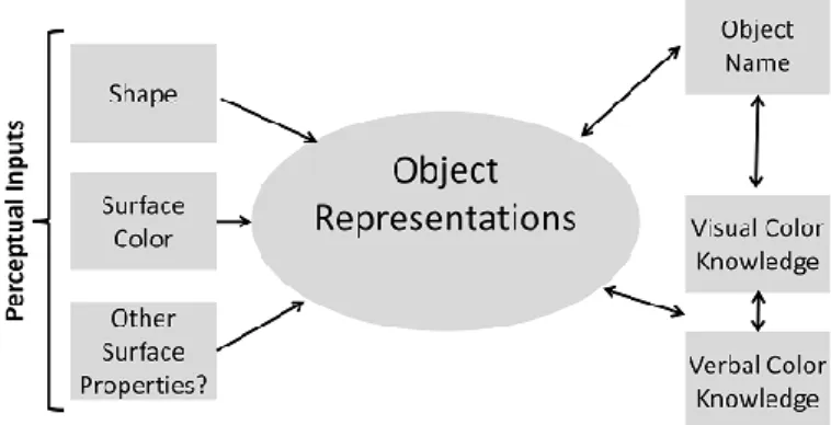
visual modality (i.e., visual agnosia). The study of such patients led Humphreys and colleagues (Humphreys, Price, & Riddoch, 1999; Humphreys, Riddoch, & Quinlan, 1988; Riddoch & Humphreys, 1987b) to propose a hierarchical model of object recognition. According to this model, object recognition involves a set of separate processes arranged in a hierarchical fashion. This quasi-modular decomposition of object recognition is presented in Figure 1.1.
Figure 1.1. A schematic framework illustrating the stages of processing involved in
object naming. Adapted from Humphreys, Price and Riddoch (Humphreys, Price, & Riddoch, 1999).
When we see an object, early visual processes encode the shape information and, possibly, other surface details present in the object image. To recognize an object, the encoded perceptual information must be matched against different forms of stored information: knowledge about the form of the object (i.e., its structural description), knowledge about functional and associative properties of the object (i.e., its semantic description), and finally knowledge about the object name (i.e., its phonological description). Access to these various types of knowledge constitutes distinct stages in the recognition process. The first stage is the access to object’s structural description. The encoded perceptual information must be matched against a known form stored in the long-term memory. Evidence
Chapter 1
18
for the existence of this separate stage comes from patients who show good early visual processing but have difficulty performing object decision tasks. The performance in these tasks can be assessed with familiarity discrimination tasks between pictures of real objects and non-objects generated by combining parts of different real objects. The same patients are good at retrieving functional and associative properties of the objects via other modalities, indicating that their problem is restricted to visual knowledge about the form of the object (Gainotti & Silveri, 1996; Sartori & Job, 1988). The second stage in object recognition requires access to functional and associative knowledge of the objects. Patients with deficits in retrieving stored semantic representations from the visual modality demonstrate access to stored visual knowledge, as indicated by their ability to perform object decision tasks. However, the same patients may show impairments in matching tasks that require access to semantic knowledge from vision (e.g., match a hammer to a nail or a screw) and object naming (Hillis & Caramazza, 1995; Riddoch & Humphreys, 1987a; Sheridan & Humphreys, 1993). Despite this deficit, these patients demonstrate good performance on tests that require access to semantic knowledge from other modalities (Riddoch & Humphreys, 1987a). Thus, poor object naming cannot be attributed to general deficits in semantic knowledge but, rather, to impaired visual access to semantic knowledge following intact access to stored visual knowledge. These evidences clearly indicate the existence of a separate system that supports long-term visual knowledge about objects, isolated from the functional and associative semantic knowledge system. Finally, the last stage in object recognition is the access to the object’s name representation. Evidence for this separate stage comes from patients who are able to make accurate judgments about the visual and semantic properties of objects but cannot readily retrieve phonological information (Kay & Ellis, 1987).
The contribution of cognitive neuropsychology to the study of object recognition was important for the identification of several independent cognitive processes involved in object recognition tasks.
General Introduction
The Neural Basis of Object Recognition
Neuroimaging techniques, specifically functional magnetic resonance imaging (FMRI), offer an opportunity to investigate the neural and cognitive mechanisms underlying object recognition. Objects are represented in a large portion of the visual ventral stream, the processing pathway that extends from the occipital to the inferior temporal lobe. Indeed, FMRI studies have revealed a constellation of object selective brain regions in the lateral and ventral occipito-temporal cortices, referred together as the lateral occipital complex (LOC; Grill-Spector, 2003; Malach et al., 1995; Peissig & Tarr, 2007). The LOC responds more strongly to pictures of objects than to their scrambled counterparts (Grill-Spector et al., 1999; Kourtzi & Kanwisher, 2001; Malach et al., 1995) and shows a number of response properties that characterize an effective object recognition system that subserves perceptual object constancy. First, the LOC responds similarly to objects defined by luminance, texture, motion and other cues, thus representing objects independently of the precise physical cues that define an object (Grill-Spector, Kushnir, Edelman, Itzchak, & Malach, 1998; Kourtzi & Kanwisher, 2000). Second, the LOC represents objects invariant of changing external viewing conditions, such as viewpoint or transformations of object size (Grill-Spector et al., 1999; James, Humphrey, Gati, Menon, & Goodale, 2002; Sawamura, Georgieva, Vogels, Vanduffel, & Orban, 2005; Vuilleumier, Henson, Driver, & Dolan, 2002). These neuroimaging studies are in agreement with early monkey neurophysiological studies in which visual object recognition was mapped to the responses of single neurons in the inferior temporal cortex. For example, Gross and colleagues (Gross, Bender, & Rocha-Miranda, 1969; Gross & Rocha-Miranda, 1972) reported that neurons in the inferior temporal cortex of macaques responded strongly to complex visual stimuli, such as hands and faces. Interestingly, these higher-level areas of the inferior temporal cortex showed very little response to simple stimuli, suggesting that this and related areas are critical to complex visual processing, such as object recognition.
Chapter 1
20
Ungerleider and Mishkin (1982), based on the pattern of behavior following lesions to dorsal and ventral regions of the monkey cortex, suggested that the visual cortex can be broken down into two pathways. The ventral pathway, including areas of the inferior temporal cortex, is involved in the identification of visual objects. The dorsal pathway, including areas of the posterior parietal cortex, is related to spatial properties of vision. An alternative description of the two pathways exists in terms of vision for perception (ventral stream) and vision for action (dorsal stream; Goodale & Milner, 1992). Although neural representations of object information have been extensively studied in the ventral pathway, little is known about the role of the dorsal pathway in object processing. The functional role of the dorsal pathway in object recognition has been frequently attributed to modulation of attention and action guidance (Grill-Spector et al., 1999; Kourtzi & Kanwisher, 2000). However, in a recent neuroimaging study, Konen and Kastner (2008) challenged this idea. The authors found representations for a variety of different object stimuli in the human parietal posterior cortex when action planning was not involved and when attention was drawn away from the stimuli. These results indicate that basic object information related to shape, size and viewpoint may be represented similarly in two parallel and hierarchically organized neural systems in the ventral and in the dorsal pathways.
1.3 Color Processing in the Human Brain
Given that the brain has developed specialized mechanisms to handle color perception information in the visual environment, it is a fair question to ask what functional role color might play in everyday vision, namely during object recognition. Although other mammals possess dichromatic or monochromatic color vision, only primates have trichromatic color vision. What is the ecological advantage of having trichromatic color vision? Primates evolved trichromacy from their dichromatic ancestors approximately 40 million years ago following the duplication of a gene coding for the L-cone (Jacobs, 1993; Jacobs & Rowe, 2004;
General Introduction
Yokoyama, 2000). The dominant view is that trichromatic color vision emerged as a specific adaptation for finding fruits and young leaves against a background of mature leaves. Because fruits and leaves play an important role in the primate diet, trichromacy could have evolved as a specific adaptation for finding food (e.g., Osorio & Vorobyev, 1996; Regan et al., 2001). Alternatively, color vision in primates could have evolved for discriminating the spectral modulations on the skin of conspecifics, probably for the purpose of discriminating emotional states, socio-sexual signals and threat displays (Changizi, Zhang, & Shimojo, 2006). Therefore, social and sexual selection could also have played a role in evolution of primate trichromacy. Given that color plays a prominent role in our subjective experience of the visual world, it makes sense to investigate how color information contributes to object recognition.
Cortical Stages of Color Processing in the Human Visual Brain
Several physiological and anatomical studies have established the human color center in the V4 area located in the posterior part of the fusiform gyrus. However, the color center is just part of a more broadly distributed cortical network responsible for color processing that includes V1, V2, V4, and regions beyond the inferior temporal cortex (e.g., Bartels & Zeki, 2000; Lueck et al., 1989; McKeefry & Zeki, 1997; Zeki & Bartels, 1999; Zeki et al., 1991). Nevertheless, it is unclear what role these areas play within the color processing system. Evidence suggests that the first stage of color processing, located in the V1 and V2, primarily registers the presence and intensity of different wavelengths. A second stage, located in the V4, is involved in automatic color constancy operations (Zeki & Marini, 1998). Color constancy is a property of the human visual system that ensures that the perceived color on a surface remains relatively constant under varying illumination conditions. A very interesting case study reported by Zeki and colleagues (Zeki, Aglioti, McKeefry, & Berlucchi, 1999) shows the specific roles of V1, V2 and V4 within the color processing system. After an electric shock that led to vascular
Chapter 1
22
insufficiency, the patient PB became virtually blind, although he retained the capacity to see colors consciously. The psychophysical results suggested that color constancy mechanisms were severely deficient in the patient and that his color vision was merely wavelength-based. The imaging studies showed that, when he viewed and recognized colors, significant increases in activity were restricted to V1 and V2, while no activation of V4 was observed.
Corroborating these initial processing states of color perception is the finding that achromatopsia, a condition in which patients report no experience of color, results from lesions in V4. Achromatopsic patients can discriminate between different wavelengths, but they cannot attribute colors to them (e.g., Beauchamp, Haxby, Rosen, & DeYoe, 2000; Kennard, Lawden, Morland, & Ruddock, 1995; Tranel, 2001; Zeki, 1990). Bouvier and Engel (2006) performed a meta-analysis of 92 case reports of achromatopsia in the literature. Lesion overlap analyses revealed a relatively small region of high overlap in the ventral occipital cortex, close to areas that are important for color perception. However, the behavioral deficits in the achromatopsic patients were often incomplete and were not restricted to color vision. Notably, most of the cases reported have concomitant deficits in spatial vision. This observation led the authors to suggest that some visual areas, outside those commonly damaged in achromatopsia, also participate in the color processing stream. This meta-analysis indicates that color perception arises from a stream of processing that flows through multiple visual areas and that achromatopsia likely results from damage to one critical step in the many stages supporting color perception.
Finally, a third and final stage in color processing involves object colors and is supported by the inferior temporal and probably also by the frontal cortex (Zeki & Marini, 1998). Little is known about the neural mechanisms underlying higher-level aspects of color processing. According to the review of the literature, the cortical brain regions believed to be important for color perception are shown in Figure
General Introduction
Figure 1.2. Schematic view of the human brain. The regions that are important for
various aspects of color perception are shown. These regions include the lingual gyrus and the posterior portion of fusiform gyrus, located below the calcarine fissure.
1.4 Does Color Information Improve Object Recognition?
The role that color plays in object recognition has been a point of contention in the literature. Initially, object recognition theories state that objects are recognized based only on shape information, largely ignoring the influence of color information (Biederman, 1987; Marr & Nishihara, 1978). More recently, a large body of behavioral, neuroimaging and neurophysiological studies indicate that color might contribute to object recognition. Tanaka and colleagues (Tanaka, Weiskopf, & Williams, 2001) proposed the Shape + Surface model of object recognition that takes into consideration the recent evidence for the role of color information in object recognition (Figure 1.3). The model recognizes that object recognition is primarily a shape-driven system (e.g., blue strawberries are still recognized as strawberries); however, color and possibly other surface properties, such as texture, are perceptual inputs for the object representation system. The Shape + Surface model draws a distinction between surface color at the input level and stored color knowledge and considers object recognition to be jointly determined by the bottom-up influence of surface color and the top-down
Chapter 1
24
influence of color knowledge. According to this model, visual color knowledge can be triggered either by the perceptual object during object recognition or by its lexical label during mental imagery. Finally, the model maintains a separation between linguistic and visual representations of object color. For example, it is possible to know that strawberries are red without having to consult a visual representation.
Figure 1.3. The Shape + Surface model of object recognition. Adapted from Tanaka,
Weiskopf and Williams (2001).
By examining whether there is an advantage to recognizing the typical colored version of an object (e.g., a red strawberry) over its black and white or atypical color version (e.g., a purple strawberry), it is possible to verify whether color information contributes to object recognition. However, this relatively straightforward test has yielded mixed results. Some studies have shown that recognition times are essentially unaffected by color information (Biederman & Ju, 1988; Davidoff & Ostergaard, 1988; Ostergaard & Davidoff, 1985). However, other studies have found that objects presented in their typical color version are recognized faster than when individuals are presented with their black and white or atypical color versions (e.g., Humphreys, Goodale, Jakobson, & Servos, 1994; Price & Humphreys, 1989; Therriault, Yaxley, & Zwaan, 2009; Wurm, Legge, Isenberg, & Luebker, 1993). Different explanations have been proposed for these apparently contradictory results. For instance, color information may facilitate the recognition
General Introduction
of objects within structurally similar categories (e.g., animals, fruits) but not structurally dissimilar categories (e.g., body parts, musical instruments, tools). Objects belonging to structurally similar categories activate a larger set of structural representations, leading to a higher competition within the visual system, and thus color can help resolve this competition (Price & Humphreys, 1989). Other studies have proposed that color can provide useful information when objects are strongly associated with a color (i.e., color diagnostic objects; Nagai & Yokosawa, 2003; Tanaka & Presnell, 1999). Although the color red might be useful to recognize strawberries or fire engines, the red color might not be useful to recognize combs or shoes. Additionally, it has been suggested that color might provide important information for people with low visual acuity (Boucart, Despretz, Hladiuk, & Desmettre, 2008; Wurm, Legge, Isenberg, & Luebker, 1993) and patients suffering from visual object agnosia (Humphreys, Goodale, Jakobson, & Servos, 1994; Mapelli & Behrmann, 1997).
Surface Color and Color Knowledge Information
Perceiving that a strawberry is red as opposed to knowing and recalling that a strawberry is red are distinct cognitive operations. The surface color of an object can be defined as the percept generated by the color present in the object image (e.g., the color red in a picture of a red strawberry), while the color knowledge is represented in the semantic information about the prototypical color of an object (e.g., the knowledge that strawberries are typically red).
To study how surface color and color knowledge might interact during object recognition, Joseph and collaborators (Joseph, 1997; Joseph & Proffitt, 1996) manipulated perceptual color input independently of color knowledge in a series of verification tasks. The authors found that color knowledge significantly influenced object recognition. For example, a purple apple was more likely to be mistaken for a cherry than for a blueberry. This interference effect occurs because both apples and cherries are typically red, not because the apple was colored in purple, the
Chapter 1
26
typical color of a blueberry. The same pattern of results was obtained when uncolored pictures were used. These findings suggest that the conceptual processing of color does not depend on the presence of a surface color and that automatic color knowledge is more powerful than the perceptual surface color processing during object recognition. However, they allowed participants to verify a target object against three types of different distractors: a distractor similar in shape but not color, a distractor similar in shape and color, and a distractor dissimilar in shape and color. Given that object recognition is a shape-driven system (Tanaka, Weiskopf, & Williams, 2001), a fourth distractor, similar in color and dissimilar in shape, should have been included to exclude the possible interference of shape. Moreover, the effects of color and shape might not be additive; shape and color similarity might yield super additive effects.
At the neuroanatomical level, several studies have tried to clarify whether there are distinct neural regions that process surface color perception and color knowledge retrieval. For example, Martin and colleagues (Martin, Haxby, Lalonde, Wiggs, & Ungerleider, 1995) used a property production task to activate color and action knowledge associated with objects. Subjects were presented with black and white pictures or the written names of objects and were required to generate words describing an action or a color associated with the presented objects. The type of information that was retrieved modulated activity in the posterior temporal cortex. Relative to action words, color words generation activated the fusiform gyrus anterior to regions associated with color perception and object perception. Activation of the ventral temporal cortex when retrieving color information has been replicated several times using property production (Chao & Martin, 1999; Wiggs, Weisberg, & Martin, 1999) and verification tasks (Goldberg, Perfetti, & Schneider, 2006; Oliver & Thompson-Schill, 2003; Simmons et al., 2007). These results indicate that the ventral temporal cortex is important for color knowledge retrieval. However, it is unclear whether it is also the system that supports color perception. Chao & Martin (1999) addressed this question by evaluating both
General Introduction
processes in the same experiment. Color word generation activated the posterior ventral temporal cortex, as previously reported, while passive viewing of colored stimuli activated the lingual gyrus in the occipital cortex. This finding is consistent with studies of color imagery in normal subjects (Howard et al., 1998) and in color-word synesthetes who experience vivid color imagery when hearing color-words (Paulesu et al., 1995). In both studies, color imagery was associated with activity in the same ventral temporal sites identified in the studies discussed above but not in occipital sites that are active during color perception (e.g., Zeki & Bartels, 1999; Zeki et al., 1991). In addition, neuropsychological studies have reported dissociations between surface color and color knowledge in the ventral occipitotemporal cortex. Although lesions in the posterior fusiform gyrus result in achromatopsia without sacrifice of color knowledge (Bouvier & Engel, 2006), lesions in the ventral temporal cortex result in color agnosia without sacrifice of color perception (Miceli et al., 2001). Coupled with neuropsychological reports of a double dissociation between color perception and color imagery (De Vreese, 1991; Shuren, Brott, Scheft, & Houston, 1996), these data suggest that distinct neural regions appear to be differentially engaged during the processes of color perception and the retrieval of object color knowledge. Information about object color is stored in the ventral temporal cortex. This region is close to, but does not include, the sites in the occipital cortex that selectively respond to the presence of color.
However, the dissociation between perception and knowledge retrieval mechanisms does not necessarily implicate that these two abilities are completely independent. Some neuroimaging studies have claimed that color knowledge modulates regions that are involved in color perception (Goldberg, Perfetti, & Schneider, 2006; Howard et al., 1998; Kellenbach, Brett, & Patterson, 2001; Simmons et al., 2007; Ueno et al., 2007). Some neuroimaging studies have provided additional direct evidence for this claim. Beauchamp and colleagues (Beauchamp, Haxby, Jennings, & DeYoe, 1999) showed that neural activity is limited to the occipital lobes when color perception was tested by passive viewing; however,
Chapter 1
28
when the task was made more demanding by requiring subjects to judge subtle differences in hue, activity associated with color perception extended from the occipital cortex into the fusiform gyrus in the ventral temporal cortex. Additionally, Simmons and colleagues (2007), using a task demanding high levels of attention to evaluate color perception and a verbal property verification task to assess color knowledge, found that retrieving information about object color activated the same region of the fusiform gyrus that is activated during color perception (Simmons et al., 2007). Thus, these data support the idea that information about a particular object property, such as its typical color, is stored in the same neural system that is activated when that property is perceived. Therefore, passive color perception may be mediated by occipital cortical regions located early in the visual processing stream, whereas active color perception seems to require more extensive neural activity extending anteriorly into the fusiform gyrus. In a recent review, Martin (2007) suggested that the fusiform gyrus is to provides a neural substrate for acquiring new object-color associations and representing those associations during conceptual processing.
The Color Diagnosticity Hypothesis
The level of color diagnosticity refers to the degree to which a particular object is associated with a specific color. For example, a color diagnostic object, such as a strawberry, is strongly associated with the color red. A comb, however, which is a non-color diagnostic object, is not strongly associated with any particular color. According to the color diagnosticity hypothesis, color diagnostic objects are the most likely candidates to show an advantage due to color information in object recognition tasks (Nagai & Yokosawa, 2003; Tanaka & Presnell, 1999). According to this hypothesis, Tanaka and Presnell (1999) showed that the presence of color information has a significant impact on the recognition of high color diagnostic objects and no effect on the recognition of objects with low color diagnosticity. In a control condition, when high and low color diagnostic objects were matched for
General Introduction
structural complexity, reliable color effects were still found, indicating that color made a unique contribution to recognition in a manner that is independent of shape. Similar results were found in the recognition of everyday scenes (Oliva & Schyns, 2000). Scenes that are rich in color diagnostic content (e.g., coast, forest) are best recognized in their typical color versions when compared to black and white or atypical color versions. On the other hand, non-color diagnostic scenes (e.g., city, shopping area) showed no difference in recognition performance across the typical, black-and-white and atypical color versions (Oliva & Schyns, 2000). Thus, the concept of color diagnosticity generalizes to the recognition of both objects and scenes.
However, recent studies have failed to replicate this finding and have documented that color information, independent of the color diagnosticity status of the object, improves its recognition (Rossion & Pourtois, 2004; Uttl, Graf, & Santacruz, 2006). For example, Rossion and Pourtois (2004) colored the 260 line-drawings from the Snodgrass and Vanderwart (1980) set with texture and shadow details. Norms for the color diagnosticity level of the objects were collected and correlated with the advantage provided by color alone in the naming responses. The authors did not report a significant correlation between these two measures (r = 0.05), showing that color information improves object recognition independently of its color diagnosticity level.
The effects of color diagnosticity and its interactions with the observed advantage due to color information in object recognition are not well understood, and the reasons for the apparently contradictory results reported in the literature are not obvious. One possibility is that color information helps the recognition of color and non-color diagnostic objects at different levels of visual processing. To recognize an object, different processing stages must be resolved (Humphreys, Price, & Riddoch, 1999). First, the perceptual input must be encoded and matched against a template form stored in the long-term memory. Next, the semantic object representations are accessed, and, finally, the object name is activated. Color
Chapter 1
30
information might be useful for recognition of both color and non-color diagnostic objects in the early stages of the visual processing. Specifically, this information could be used to match the perceptual input with a known shape representation or, at an even earlier visual processing stage, segregate and organize of the visual input. However, in the later stages of the recognition process, color information might play different roles depending upon the color diagnosticity status of the specific objects. Although color information might be important for semantic representation of a color diagnostic object, color information is probably not as important for semantic representation of a non-color diagnostic object. When we think about the properties of a strawberry, the property red is one of the first that comes to mind; however, if we think about the features of a comb, its color is certainly not one of the first properties one might think of.
1.5 Specific Aims of the Thesis
The general picture that emerges from the literature is that the role of color information is not well understood. Theories of object recognition have traditionally ignored the role of color information in higher-level vision (Biederman, 1987; Biederman & Ju, 1988). More recently, data from behavioral studies, neuroimaging, and neuropsychological studies have suggested that surface color features and color knowledge information might also contribute to object recognition. However, the conditions under which color information improves object recognition are not well understood. This thesis contributes to this discussion by clarifying some open questions found in the literature. One of the main questions addressed here is the interaction between surface color and color knowledge information during object recognition. It was previously suggested that that object recognition is jointly determined by the bottom-up influence of surface color and the top-down influence of color knowledge information (Tanaka, Weiskopf, & Williams, 2001). However, the way that these two sources of color
General Introduction
information interact and which plays the most important role during object recognition is unclear.
Moreover, the color effects on object recognition might depend on the color diagnosticity status of the specific objects. The color diagnosticity status of the objects is probably the most investigated object property in studies that examine the role of color information in object recognition. Color diagnostic objects have a strong association with a particular color; on the other hand, non-color diagnostic objects do not have any specific color association (Tanaka & Presnell, 1999). Thus, we proposed that color information might participate in the recognition of color and non-color diagnostic objects at different levels of visual processing. More specifically, we hypothesize that color information participates in the recognition of both types of objects in the early visual perceptual stages, helping both segmentation and organization of the perceptual input. Studies have indicated that color information is an important cue in the early visual processing stages (Gegenfurtner & Rieger, 2000; Wurm, Legge, Isenberg, & Luebker, 1993); however, these studies did not control for or manipulate the color diagnosticity level of the presented objects. Color information is expected to play an additional role during the recognition of color diagnostic objects at the semantic levels of visual processing. Color is an intrinsic property of these objects. For example, Naor-Raz and Tarr (2003), using a variation of the stroop paradigm, asked participants to name the displayed color of objects and words. They found that color is an intrinsic property of color diagnostic objects at multiple levels. Thus, the presence of color information in an image of a color diagnostic object might be important for the activation of semantic object representation and recognition of the object.
The discussion about which type of objects might benefit from color information does not end with the object’s color diagnosticity status. Another object property which effects have been investigated is the object’s semantic category. If color vision developed in humans species to find ripe fruit amongst foliage (Gegenfurtner, 2003; Surridge, Osorio, & Mundy, 2003), it would make
Chapter 1
32
sense that recognition of biological objects would benefit more from color information than artifacts. In fact, some studies support this hypothesis (Humphreys, Goodale, Jakobson, & Servos, 1994; Price & Humphreys, 1989). However, more recent data suggest that color information affects object recognition independently of the semantic category (Rossion & Pourtois, 2004; Uttl, Graf, & Santacruz, 2006).
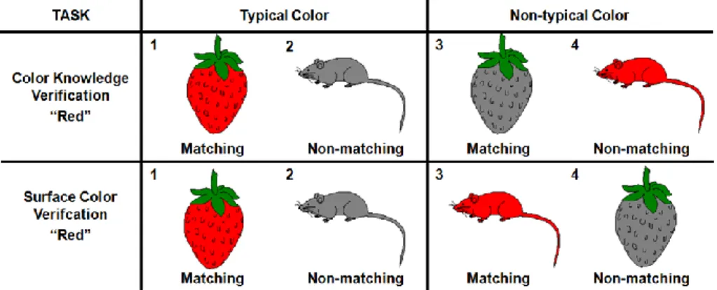
In this thesis, we present six studies that attempt to clarify these open questions in the literature. In chapters 2 and 3, we try to clarify which type of color information is the most important during object recognition. This question was previously investigated by Joseph and collaborators (Joseph, 1997; Joseph & Proffitt, 1996). In a series of verification tasks, the authors found that color knowledge is more influential than surface color during object recognition (Joseph, 1997; Joseph & Proffitt, 1996). However, during verification tasks, the role of color information was not controlled independently of the role of shape information. Thus, in chapter 2, we try to replicate these findings while independently controlling color and shape information. Participants performed a computerized name-object verification task where the relationship between the color and shape information provided by the object name and by the object picture was manipulated in four conditions: different shape/different color, different shape/same color, same shape/different color, and same shape/same color. If the contribution of color knowledge during recognition is independent of the presence of the appropriate surface color, interference during the non-matching trails should be higher whenever the color knowledge activated by the name and by object picture is the same, not only when pictures are presented in the typical color version, but also in black-and-white and atypical color versions. In chapter 3, we use event-related potentials (ERPs) to further explore this question. Participants performed two color-object verification tasks: a surface color verification task, where they were asked to verify the color of the objects depicted in the image; and a color knowledge verification task, where they were asked to verify the color of
General Introduction
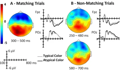
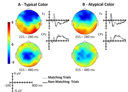
the objects in the real world. The surface color of the objects was manipulated to cause interference of the color knowledge information during the surface color task and to cause surface color interference during the color knowledge task. By comparing the ERPs elicited by typical and atypical color presentations in both tasks, we were able to identify at which point during the recognition process subjects recruit surface color and color knowledge information to recognize the presented objects.
In chapter 4 and 5, we examine the interaction between the color diagnosticity status of objects and the manner in which color affects recognition. In chapter 4, participants performed three object recognition tasks with different cognitive demands at the perceptual, semantic and phonological levels. Color and black-and-white versions of color and non-color diagnostic objects were used. By comparing the performance in the three recognition tasks, we identified the visual processing stage at which color information is recruited to recognize color and non-color diagnostic objects. In chapter 5, we used ERPs to further explore this question. In contrast to behavioral measures, the ERPs permits the analysis of cognitive processes with a temporal resolution in a range of milliseconds and represents an optimal approach to study the level at which visual processing of color information improves object recognition. In a recognition task, subjects were presented with color and black-and-white versions of color and non-color diagnostic objects. Color effects were investigated in an early visual ERP component, N1, and in two visual ERP components modulated by higher visual processes, N350 and N400. The study of color information in object and scene recognition has been previously examined using the ERP technique; however, these studies used only high color diagnostic objects or scenes (Goffaux et al., 2005; Lu et al., 2010). For example, Goffaux and colleagues (2005) reported that a color effect can be visualized after 150 milliseconds of the stimuli onset, showing an early role of color information in visual scene recognition (Goffaux et al., 2005).
Chapter 1
34
In chapter 6, we explore whether color information plays different roles when we recognize biological and artifacts objects. More specifically, we investigated whether the neural correlates of color information are the same for biological, artifacts and nonsense objects. Functional magnetic resonance imaging (FMRI) responses were collected during a covert naming task where natural, artifacts and nonsense objects were presented in color and in black and white. The literature suggests that color information is more important for the recognition of objects belonging to natural categories than for the recognition of artifacts (e.g., Price & Humphreys, 1989). Accordingly, different brain regions are expected to be activated during the recognition of colored natural objects and artifacts.
Finally, in chapter 7, we present a review and a meta-analysis that aim to comprehensively integrate and discuss the behavioral literature on the effect of color information during object recognition. We drew some conclusions regarding the moderator role of several variables (e.g., color diagnosticity status and the semantic category of the objects) that are typically manipulated in studies that examine the influence of color information on object recognition.
Chapter 2
The influence of surface color information and
color knowledge information in object recognition
Based on:
Bramão, I., Faísca, L., Petersson, K. M., & Reis, A. (2010). The influence of surface color information and color knowledge information in object recognition. American Journal of Psychology, 123, 459-468.
36
Abstract
In order to clarify whether the influence of color knowledge information in object recognition depends on the presence of the appropriate surface color, we designed a name-object verification task. The relationship between color and shape information provided by the name and by the object photo was manipulated in order to assess color interference independently of shape interference. We tested three different versions for each object: typically colored, black and white, and atypically colored. The response times on the non-matching trials were used to measure the interference between the name and the photo. We predicted that the more similar the name and the photo are, the longer it would take to respond. Overall, the color similarity effect disappeared in the black-and-white and atypical color conditions, suggesting that the influence of color knowledge on object recognition depends on the presence of the appropriate surface color information.
Surface color information versus color knowledge information
2.1 Introduction
The role of surface color in object recognition (i.e., the color present in the image of an object) is an unresolved issue in cognitive science. For example, theories differ on the role shape plays in object recognition (Biederman, 1987; Marr & Nishihara, 1978) and whether other object features, such as surface details, texture, and color, contribute to object recognition (Tanaka, Weiskopf, & Williams, 2001; Tarr, Williams, Hayward, & Gauthier, 1998). Different studies have suggested different roles for color in object recognition. For example, color serves as a perceptual input to early stages of visual processing (Davidoff, Walsh, & Wagemans, 1997; Wurm, Legge, Isenberg, & Luebker, 1993) and is part of the structural representation system of the objects (Price & Humphreys, 1989) or of the semantic system (Davidoff, Walsh, & Wagemans, 1997; Tanaka, Weiskopf, & Williams, 2001). Moreover, color serves as an important cue in object retrieval processes (Jones, 2005; Jones & Nakabayashi, 2009; Vernon & Lloyd-Jones, 2003).
Although it is not yet clear at which level surface color facilitates object recognition, there is a consensus that colored objects and visual scenes are recognized faster than corresponding black and white versions (e.g., Oliva & Schyns, 2000; Rossion & Pourtois, 2004). In order for surface color to be a useful cue for recognition, the participants must decide whether a color is appropriate for a particular object, and it seems plausible that semantic object information (including stored color knowledge) has to be accessed for this to occur. This suggests that prior color knowledge plays a role in object recognition in addition to surface color input, because the color input must in some sense be checked against the activated prototypical color of the object.
In order to study how surface color input and prior color knowledge interact, Joseph and Proffitt (Joseph, 1997; Joseph & Proffitt, 1996) manipulated color knowledge and surface color input independently in a series of verification tasks. The authors found that prior color knowledge was more influential than perceptual
Chapter 2
38
input color; for example, a purple apple was more likely to be mistaken for a cherry (typically red) than for a blueberry (typically purple). It was argued that the interference effect is explained by the fact that apples and cherries are prototypically red and not because the apple was colored in purple, the typical color of blueberries. The same pattern of results was obtained when uncolored pictures were used, suggesting that the semantic processing of color is independent of the presence of a perceptual input color.
However, the authors did not fully control whether the interference was caused by prior shape knowledge. In their verification tasks the participants were asked to verify a target object against three different types of distractors: a distractor similar in shape but not similar in color, a distractor similar in shape and color, and a distractor that was dissimilar in both shape and color. To rule out a possible shape interference effect, it is important to include a fourth distractor type that is similar in color and dissimilar in shape. Because shape information is needed for object identity, strong similarity in shape will influence the verification decision. Thus it is important to investigate the previous findings (Joseph, 1997; Joseph & Proffitt, 1996) by controlling color knowledge interference fully independent of shape knowledge interference.
In this study we investigated whether prior color knowledge information takes place in object recognition independently of the presence of the appropriate surface color, controlling the shape information. We designed a verification task in which an object name was presented before an object picture. Two types of trials were included: matching (the name matches the picture) and non-matching (the name does not match the picture). On non-matching trials, the name might activate shape and color knowledge that interferes with shape and color information provided by the picture. To test whether the role of color knowledge information in object recognition is dependent on the presence of the appropriate surface color, three different versions of each object were tested: typically colored, black and white, and atypically colored. If color knowledge information contributes
Surface color information versus color knowledge information
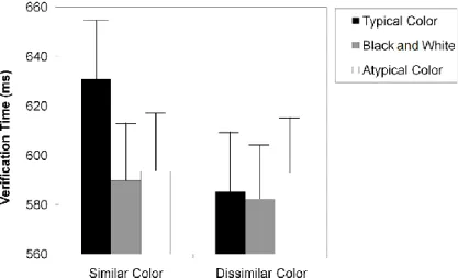
to the recognition process, independently of the presence of the appropriate surface color, it should be more difficult to say “no” whenever the color knowledge activated by the name and by object picture is the same, not only when pictures are presented in their typical color version but also when black-and-white and atypical color versions are presented. In order to assess color interference independently of shape interference, the relationship between color and shape information provided by the name and by the picture was manipulated to assess four possible mismatches: dissimilar shape and dissimilar color, dissimilar shape and similar color, similar shape and dissimilar color, and similar shape and similar color. The interference in the response was measured by the longer response times (Joseph, 1997; Joseph & Proffitt, 1996).
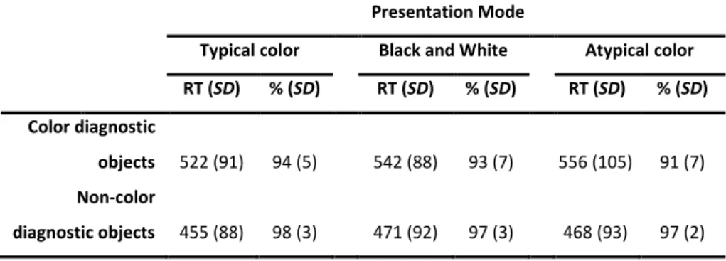
A second aim of this study was to explore the role of color diagnosticity in object recognition. Color diagnosticity is the degree to which a particular object is associated with a specific color. For example, a strawberry – a color diagnostic object – is clearly associated with the red color, whereas a comb – a non-color diagnostic object – is not strongly associated with any specific color. According to the color diagnosticity hypothesis (Tanaka & Presnell, 1999) surface color information improves the recognition of color but not non-color diagnostic objects (see also Nagai & Yokosawa, 2003). However, Rossion and Pourtois (2004) documented that colored objects, independent of the diagnosticity status, were named faster than their noncolored versions (see also Biederman & Ju, 1988; Uttl, Graf, & Santacruz, 2006; Wurm, Legge, Isenberg, & Luebker, 1993). Although color diagnosticity is an important aspect to control when the influence of color information is being studied in object recognition, its role is not well understood. In an attempt to clarify this question, we used in our verification task both color and non-color diagnostic objects. If surface color information is engaged during recognition of both color and non-color diagnostic objects, then the name-picture matching should be faster with typical colored than with black-and-white and atypical color pictures, for both color and non-color diagnostic objects.
Chapter 2
40
2.2 Methods
ParticipantsTwenty-eight Portuguese graduate students with normal or corrected-to-normal vision volunteered to participate in the experiment (mean age [± SD] = 22 ± 4 years, range 18-34 years; mean school years [± SD] = 14.5 ± 1 years, range 13-16 years).
Stimuli
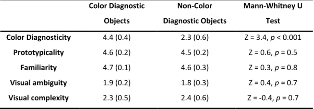
The initial pool of pictures consisted of 62 photos of common objects selected from the Reis, Faísca, Ingvar, and Petersson (2006) set. An independent group of 30 participants named and rated the initial set according to prototypicality, familiarity, visual ambiguity, visual complexity, and color diagnosticity. Each photo was presented for 1 min, and participants were asked to write down the name of the object. If they did not know the name, they were asked to mark one of the following categories: do not know name, do not know object, or tip-of-the-tongue. Participants were also asked to evaluate the prototypicality of each photo “according to the degree that the presented picture represents a typical exemplar of the concept” and rated the degree of agreement between the presented photo and their mental image of the concept using a 5-point scale, where 1 indicated low agreement and 5 indicated high agreement. The familiarity of each photo was judged “according to how usual or unusual the object is in your realm of experience”, and the participants were asked to rate the concept itself, rather than the photo, using a 5-point rating scale (1 = very unfamiliar, 5 = very familiar). The visual ambiguity of each photo was evaluated “according to how large is the group of different objects that are visually similar with the presented object” (5-point rating scale: 1 = completely nonambiguous object, 5 = completely ambiguous object). Visual complexity was defined as “the amount of detail or intricacy of line in the photo”, and the participants were told to rate the photo itself rather than the real-life object (5-point scale: 1 = very low visual complexity, 5 = very complex picture). Color diagnosticity was defined as “the degree to which the object is
Surface color information versus color knowledge information
associated with a specific color” and was also rated on a 5-point scale (1 = low diagnostic color, 5 = high diagnostic color). These instructions are similar to the ones typically used in rating studies (Rossion & Pourtois, 2004; Snodgrass & Vanderwart, 1980; Ventura, 2003).
Following the analysis of the rating scores, we selected only the photos that showed at least 80% name agreement between participants. From these, we selected 16 photos to be used in the experiment: 8 representatives of color diagnostic objects (apple, tomato, carrot, orange, pineapple, pear, onion, and lemon) and 8 representative of non-color diagnostic objects (book, glasses, bowl, pencil, water, can, ruler, and comb). The only significant mean difference between the two groups of objects was color diagnosticity. The mean comparisons between diagnostic and nondiagnostic items on the other rating variables were nonsignificant (p > 0.5; Table 2.1).
Each colored photograph was used to create a black and white version (using Adobe Photoshop 7.0 “grayscale mode” command, which preserves luminance while discarding color) and an atypically colored version1 (using Adobe Photoshop 7.0 “variations” command, until a complete transformation of object color was obtained, which preserves luminance). Stimuli luminance was measured using Adobe Photoshop 7.0. We did not find any statistical difference between the diagnostic and nondiagnostic items for the three color versions concerning the luminance values (overall, Mann-Whitney U test: |Z| = 0.7, p > 0.30).
1
For the non-color diagnostic objects we did not construct an atypical color version but just another color version of the same object, because these objects do not have an atypical color associated with them. When we refer to an atypical color version of the non-color diagnostic objects we just mean a second color version of the same object.