1357
COGNITION AND MEMORY FUNCTION OF TARAXACUM COREANUM IN AN IN VIVO
AMYLOID-Β-INDUCED MOUSE MODEL OF ALZHEIMER’S DISEASE
AH YOUNG LEE1, NORIKO YAMABE1, KI SUNG KANG2, HYUN YOUNG KIM3, SANGHYUN LEE4*
and EUN JU CHO1*
1 Department of Food Science and Nutrition & Kimchi Research Institute, Pusan National University, Busan 609-735,
Republic of Korea
2 Natural Medicine Center, Korea Institute of Science and Technology, Gangneung 210-340, Republic of Korea 3 Department of Food Science, Gyeongnam National University of Science and Technology, Jinju 660-758, Republic of Korea
4 Department of Integrative Plant Science, Chung-Ang University, Anseong 456-756, Republic of Korea
*Corresponding author: slee@cau.ac.kr
Abstract - We investigated whether the ethyl acetate fraction of Taraxacum coreanum (ETC) had a protective efect against memory impairment in an amyloid beta (Aβ)-induced mouse model of Alzheimer’s disease (AD). he formation of Aβ in the brain is a hallmark of AD. We examined whether oxidative stress contributes to learning and memory deicits us-ing the T-maze test, the object recognition test, and the Morris water maze test in mice injected with Aβ. Cognition and memory function were signiicantly impaired in mice injected with Aβ, as compared to the normal group. However, mice that received ETC orally at doses of 50 or 100 mg/kg/day for 2 weeks showed high recognition behavior of tasks. ETC may
have prevented oxidative stress to the brain tissue by reducing lipid peroxidation levels and a NO scavenger. ETCcould be
useful for the prevention and treatment of AD.
Key words: Taraxacum coreanum; Alzheimer’s disease; cognition, memory
INTRODUCTION
Alzheimer’s disease (AD) is the most common neu-rodegenerative disorder in the aged population and the number of people with AD is expected to reach 81 million by 2040 (Ferri et al., 2006). AD is a pro-gressive neurodegenerative disorder, which results in memory loss, confusion, and a variety of cognitive disabilities (Khachaturian, 1985). Sites in the AD brain where neurodegeneration occurs and where oxidative stress exists are associated with increased amyloid beta (Aβ) deposits (Hensley et al., 1995). he extent of Aβ protein deposition correlates with the
and generates free radicals that cause lipid peroxida-tion and protein oxidaperoxida-tion (Varadarajan et al., 2000). herefore, antioxidants may help treat Aβ-induced neurotoxicity and improve memory in patients with AD.
Plants in the genus Taraxacum have been used since ancient time as medicinal herbs to treat dyspep-sia, heartburn, the spleen and hepatitis anorexia. Of the nearly 400 plants in this genus, the Korean dande-lion (Taraxacum coreanum) is one of most common species in Korea and Japan. T. coreanum has been used for its diuretic and anti-inlammatory activities (Koo et al., 2004). In addition, the extracts from T. coreanum have been shown to protect low-density li-poprotein from oxidation (Yang and Jeon, 1996). he efects of T. coreanum are related to its phytochemi-cal constituents, including phenols and lavonoids, which are important sources of natural antioxidants. hese characteristics have led to increased demand for the use of T. coreanum as a dietary supplement, as well as in pharmaceutical products. However, no previous study has examined whether T. coreanum protects against AD or improves cognition impair-ment in AD. herefore, in this study, we examined if T. coreanum protected against AD in an in vivo Aβ 25-35-injected animal model.
MATERIALS AND METHODS
Plant materials
Aerial parts of T. coreanum Nakai were collected in 2007 near the Westcoast Express Highway in Korea. A voucher specimen (No. LEE 2007-01) was depos-ited at the Herbarium of Department of Integrative Plant Science, Chung-Ang University, Korea.
Instruments and reagents
Aβ25-35 and malondialdehyde (MDA) were obtained
from Sigma Aldrich (Saint Louis, Missouri, USA). Dimethyl sulfoxide and sodium chloride (NaCl) were purchased from Bio Basics Inc. (Ontario, Canada). hiobarbituric acid (TBA) was obtained from Lan-caster Synthesis (Ward Hill, USA). Phosphoric acid
and 1-butanol were acquired from Samchun Pure Chemical Company (Pyeongtaek, Korea).
Preparation of the ethyl acetate (EtOAc) fraction from T. coreanum (ETC)
Freeze-dried T. coreanum was extracted with metha-nol (MeOH) for 3 h and the MeOH extraction proc-ess was repeated eight times. he extract was con-centrated using a rotary evaporator and suspended in water. he concentrate was suspended in distilled water and partitioned with n-hexane, chloroform (CHCl3), EtOAc, and n-butanol (n-BuOH),
succes-sively. he various fractions, namely n-hexane (99 g), CHCl3 (12 g), EtOAc (23 g), and n-BuOH (25 g)
frac-tions, were collected.
Animals and experimental protocols
Male ICR mice (5-weeks-old; Orient Inc. Seongnam, Republic of Korea) weighing 25-27 g were housed in plastic cages with free access to food and water, and were maintained in a controlled environment (20±2°C, 50±10%, 12-h light/dark cycle). he ani-mal protocol used in this study was reviewed and approved by Pusan National University-Institutional Animal Care and Use Committee (PNU-IACUC). Mice were divided into four groups comprising 10 individuals in each of the four cages. he groups were deined as follows: Normal = 0.9% NaCl injec-tion + oral administrainjec-tion of water; Control = Aβ 25-35 injection + oral administration of water; ETC 50
= Aβ25-35 injection + oral administration of ETC (50
mg/kg/day); ETC 100 = Aβ25-35 injection + oral
ad-ministration of ETC (100 mg/kg/day) for 14 days us-ing a sonde. here were no signiicant diferences in initial body weight among the groups.
Aβ25-35-infused mouse model
Aβ25-35 was aggregated according to the procedure
outlined by Maurice et al., (1996). In brief, the pep-tide was dissolved and diluted in sterile distilled wa-ter to achieve a concentration of 1 mg/ml, aliquoted into tubes, and then dissolved. Aβ25-35 was incubated
aggre-gation. Distilled water containing aggregated Aβ25-35
was injected into mice according to the procedure established by Laursen and Belknap (1986). Mice were lightly anesthetized with ether, and the solution was injected 0.8 mm posterior to the bregma and 1.5 mm lateral to the sagittal suture. All injections were made with a 10 μl Hamilton microsyringe itted with a 26-gauge needle that was inserted 2.2 mm beneath the surface of the brain. Animals were injected with 5 μl of sterile distilled water or 5 nmol of Aβ25-35
ag-gregate in each cerebral lateral ventricle at a rate of 1 μl/min. he needle was let in the injection site for 1 min.
T-maze test
he T-maze test was conducted according to the pro-cedure established by Montgomery (1952). he maze apparatus was T-shaped, and the walls were made of black boards (length of start and goal stems = 50 cm, width = 13 cm, height = 20 cm) that were glued to a square blackboard bottom. he maze consisted of a start box, let arm and right arm, with a door to separate the two sides. he mice were placed at the start box, and the number of touches and exploration times of the right arm of the T-maze were recorded during a 10-min period (training session). he mice were then placed back into the same apparatus 24 h ater the training session. hey were allowed to ex-plore the right and let sides of the maze freely for 10 min, and the number of touches and exploration times were recorded (test session). Space perceptive ability (%) was calculated as the ratio of the number of let or right maze entries to the number of total maze entries multiplied by 100.
Novel object recognition test
he object recognition test (Bevins and Besheer, 2006) was performed in a square black open-ield apparatus (40 × 30 × 20 cm). Two identical objects (plastic bottles) were placed at ixed distances within the square ield. he mice were then placed at the center of the square ield, and the number of touches of each object was recorded during a 10 min period (training session). he mice were placed back into
the same ield 24 h ater the training session, but this time one of the objects used during the training ses-sion was replaced with a new object (another plastic bottle). he mice were allowed to explore freely for 10 min, and the number of touches was recorded (test session). Object cognitive ability (%), a ratio of the amount of time spent exploring any one of the two original objects (training session) or the novel object (test session) over the total time spent exploring both objects, was used to measure cognitive function.
he Morris water maze test
per-centage of time spent in the target quadrant during a 60 s trial.
Measurement of lipid peroxidation
MDA levels were measured by the method described by Ohkawa et al., (1979). Ater completion of the be-havioral observations, mice were anesthetized with ether. Mice brains, livers, and kidneys were removed immediately and placed on ice. he dissected tissue was homogenized in saline solution, and mixed with 1% phosphoric acid and 0.67% TBA solution. Ater boiling for 45 min, the solution mixture was cooled in an ice bath, and 2 mL of 1-butanol was added fol-lowed by centrifugation at 3 000 rpm for 10 min. he absorbance values of the supernatant were measured at 535 and 520 nm. he level of lipid peroxidation was calculated using a MDA standard curve.
Nitric oxide (NO) scavenging activity
he NO concentration in tissues was determined by the method described by Schmidt et al., (1992). One hundred ity microliters of supernatant from the li-pid peroxidation procedure was mixed with 130 μl of distilled water, and 20 μl of dilution was added to the same amount of phosphoric acid and 0.1% n-(1-naphthyl) ethylenediamide dihydrochloride. he ab-sorbance value of the mixture was measured at 540 nm. he NO yield was calculated using a sodium ni-trite (NaNO2) standard curve.
Statistical analysis
Results are expressed as means ± SD. Statistical sig-niicance was determined using one-way ANOVA, followed by Duncan’s post-hoc tests. Signiicance was set at P<0.05.
RESULTS
T-maze test
To investigate short-term memory, a T-maze test was carried out ater oral administration of ETC for 1 week to mice injected with Aβ25-35. he efect of ETC
on preventing impaired spatial cognition is shown in Fig. 1. Mice in the normal group had a shorter la-tency to ind the platform using new routes than the control group, which means that injection of Aβ 25-35 resulted in functional impairment of short-term
memory function. However, the cognitive ability of mice treated with ETC (50 and 100 mg/kg/day) to ind a new route improved dose-dependently com-pared with the control group.
Novel object recognition test
he novel object recognition test was conducted on the day following the T-maze test. he same two objects were explored during the acquisition phase. Ater 24 h of training, mice in the normal group touched the new object frequently, while mice inject-ed with Aβ25-35 showed less ability to recognize the
new object than mice in the normal group. In con-trast, groups that received ETC (50 and 100 mg/kg/ day) touched the new object signiicantly more than control mice and spent longer exploring the novel object than the control group mice. In particular, mice that received 100 mg/kg/day ETC had an object cognition ability of 61.43% versus an object cogni-tion ability of 48.42% in the control group. hese re-sults demonstrated that oral administration of ETC protected against the Aβ25-35 injection-induced
im-pairment in spatial cognition ability (Fig. 2).
he Morris water maze test
he Morris water maze test was conducted on days 16-19 ater the injections. Results are shown in Figs. 3 and 4. During the training session, latency to reach the platform decreased in all groups over a period of 3 days. However, the control group took a relatively longer time to reach the platform than the two groups that received ETC. hese results indicated that oral administration of ETC signiicantly improved Aβ 25-35-induced cognitive deicits. he exercise and visual
mo-tor function. here were no signiicant diferences in latency to reach the exposed platform among groups. However, when the platform was hidden, the control group took longer to ind the platform than the nor-mal and ETC-treated groups. hese data indicated that damage induced by Aβ25-35 did not afect
swim-ming or visual ability, but did afect recognition abil-ity. hese results indicate that ETC not only protects against cognitive impairment, but also has a strong positive inluence on learning and memory abilities.
Measurement of lipid peroxidation
Lipid peroxide levels in the brain, liver, and kidney of mice injected with Aβ25-35 are shown in Table 1.
In-jection of Aβ25-35 into the cerebral ventricle increased
lipid peroxide levels in the brain. he MDA levels in the brain of normal and control groups were 22.69 and 27.69 nmol/mgprotein, respectively. he MDA values in the ETC (50 and 100 mg/kg/day) groups
were 26.13, and 25.19 nmol/mgprotein, respectively, demonstrating that oral administration of ETC in-hibited the formation of MDA in the brain. In addi-tion, when we examined lipid peroxidation levels in the kidney, we found that the MDA value of the con-trol group was 51.92 nmol/mg protein, while that of the 50 and 100 mg/kg/day ETC groups was 46.15 and 40.38 nmol/mg protein, respectively. Furthermore, the MDA concentrations in the liver of the groups that received ETC were signiicantly lower than that of the control group. hese results indicate that ad-ministration of ETC protected the brain, kidney, and liver from lipid peroxidation.
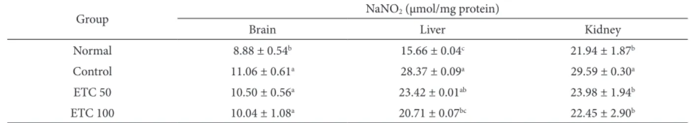
NO scavenging activity
As shown in Table 2, ETC signiicantly inhibited NO production in a dose-dependent manner. he NO level in the normal group was 8.88 μmol/mg protein, while in the control group it was signiicantly
high-Table 1. Protective activity of ETC against lipid peroxidation in mice injected with Aβ25-35.
Group MDA (nmol/mg protein)
Brain Liver Kidney
Normal 22.69 ± 0.91c 59.61 ± 0.95b 30.77 ± 2.95d
Control 27.69 ± 1.55a 59.04 ± 3.05a 51.92 ± 5.02a
ETC 50 26.73 ± 0.98ab 62.88 ± 0.53b 46.15 ± 3.27b
ETC 100 25.19 ± 1.26b 62.50 ± 3.89b 40.38 ± 5.58c
ETC 50: Oral administration of ETC (50 mg/kg/day) ETC 100: Oral administration of ETC (100 mg/kg/day) Values are means ± SD.
Diferent letters are signiicantly diferent (P<0.05) according to Duncan’s multiple range test.
Table 2. Efects of oral administration of ETC on Aβ25-35-induced nitric oxide formation.
Group NaNO2 (μmol/mg protein)
Brain Liver Kidney
Normal 8.88 ± 0.54b 15.66 ± 0.04c 21.94 ± 1.87b
Control 11.06 ± 0.61a 28.37 ± 0.09a 29.59 ± 0.30a
ETC 50 10.50 ± 0.56a 23.42 ± 0.01ab 23.98 ± 1.94b
ETC 100 10.04 ± 1.08a 20.71 ± 0.07bc 22.45 ± 2.90b
ETC 50: Oral administration of ETC (50 mg/kg/day) ETC 100: Oral administration of ETC (100 mg/kg/day) Values are means ± SD.
Fig. 4. Latency to reach hidden and exposed platform in the Morris water maze test.
he time to ind hidden and exposed platform was recorded on the inal test day of the water maze test: Normal = 0.9% NaCl in-jection + oral administration of water; Aβ25-35 = Aβ25-35 injection
+ oral administration of water; ETC 50 = Aβ25-35 injection + oral
administration of ETC (50 mg/kg/day); ETC 100 = Aβ25-35
injec-tion + oral administrainjec-tion of ETC (100 mg/kg/day). Values are reported as means ± SD. he mean latency to ind the exposed platform was not show signiicantly diferent among experimen-tal groups. a-cMeans with diferent letters indicate signiicant
dif-ferences between groups in the time taken to reach the hidden platform (P<0.05).
Fig. 1. Spatial perceptive ability scores by group as assessed by the T-maze test.
Ater training to explore the right arm of the T-maze for 10 min, the number of touches and exploration times of the right and let sides of the maze were calculated. he groups were deined as follows: Normal = 0.9% NaCl injection + oral administration of water; Aβ25-35 = Aβ25-35 injection + oral administration of
wa-ter; ETC 50 = Aβ25-35 injection + oral administration of ETC (50
mg/kg/day); ETC 100 = Aβ25-35 injection + oral administration
of ETC (100 mg/kg/day). Values are reported as means ± SD. Spatial perception of the old route was not signiicantly difer-ent among experimdifer-ental groups. a-bMeans of the space
percep-tive ability to ind new routes indicated with diferent letters are signiicantly diferent (P<0.05) between groups.
Fig. 2. Percentage change in object cognitive ability test scores. Ater training with two identical objects, the mice were allowed to explore one familiar object from training and one novel ob-ject. he time that the mice spent with the novel object was re-corded: Normal = 0.9% NaCl injection + oral administration of water; Aβ25-35 = Aβ25-35 injection + oral administration of water;
ETC 50 = Aβ25-35 injection + oral administration of ETC (50 mg/
kg/day); ETC 100 = Aβ25-35 injection + oral administration of
ETC (100 mg/kg/day). Values are reported as means ± SD. he ability to recognize the old object was not signiicantly difer-ent among experimdifer-ental groups. However, there were signiicant diferences in ability to recognize a novel object among groups;
a-c means with diferent letters are signiicantly diferent (P<0.05)
from each other.
Fig. 3. Efect of ETC on spatial learning in the Morris water maze test.
Mice were trained to swim and ind the platform for 3 days. he latency time to reach the platform during training and on the i-nal test day was calculated: Normal = 0.9% NaCl injection + oral administration of water; Aβ25-35 = Aβ25-35 injection + oral
admin-istration of water; ETC 50 = Aβ25-35 injection + oral
administra-tion of ETC (50 mg/kg/day); ETC 100 = Aβ25-35 injection + oral
er at 11.06 μmol/mg protein. Interestingly, the NO levels in the ETC groups administered ETC (50 and 100 mg/kg/day) were 10.50 and 10.04 μmol/mg pro-tein, revealing its inhibitory activity that was higher than in the control group. Moreover, NO levels in the kidney were also elevated by Aβ25-35 injection. Mice
that received ETC at oral doses of 50 and 100 mg/ kg/day had a NO level of 23.98 and 22.45 μmol/mg protein, respectively, in comparison with the NO concentration in the control group: 29.59 μmol/mg protein (P<0.05). In addition, the level of NO in the liver of the normal group was 15.66 μmol/mg pro-tein, whereas that of the control group was higher at 28.37 μmol/mg protein. he liver NO levels of the ETC groups were lower than in the control group, exhibiting a signiicant dose-dependent relationship. hese results indicate that administration of ETC had a strong inhibitory efect on NO generation in the brain, kidney, and liver.
DISCUSSION
AD is the most common cause of progressive cogni-tive decline and dementia in aged humans (Meziane et al., 1998). Although the causes of AD are not well known, one widely discussed hypothesis is that de-posits of Aβ are the causative agents of AD (Hardy and Allsop 1991). he “amyloid theory” is based on the close correlation between Aβ production and the neurodegenerative process of AD. Neuroi-brillary tangles and Aβ deposits have been found primarily in regions of the brain associated with memory and cognition in both AD patients and AD transgenic mice (Manczak et al., 2006). In the brain, neuropathological hallmarks of AD lesions include difuse and neuritic extracellular Aβ peptides gen-erated by endoproteolytic cleavage of APP, reactive microglial cells, dystrophic neuritis, and bundles of astrocytic processes and intracellular neuroibril-lary tangles (Tran et al., 2002; Kar et al., 2004). Fur-thermore, excessive reactive oxygen species (ROS) production can lead to neuronal apoptosis in neu-rodegenerative disorders, observed as Aβ-induced neuronal apoptosis (Butterield et al., 2001; Fukui et al., 2005). According to the oxidative stress hypoth-esis of AD, Aβ inserts into the neuronal membrane
bilayer and generates oxygen-dependent free radi-cals that cause lipid peroxidation, protein oxidation and ROS formation (Fu et al., 2006). Furthermore, antioxidants have been shown to have a beneicial efect in neurodegenerative disorders (Calabrese et al., 2007) and Aβ-induced neurotoxicity (Sultana et al., 2004).
Plants of the genus Taraxacum are members of the plant family Asteraceae. Several studies have demonstrated that T. coreanum had beneicial bio-logical efects, including antioxidant, free-radical scavenging and anti-inlammatory properties. Ac-cording to Choi et al., (2012), ETC showed very strong radical scavenging activity and protective ac-tivity against oxidative stress in a cellular system. In a previous chromatographic study of ETC, two lavo-noids were identiied as active compounds: luteolin and luteolin 7-O-glucose. hey both exhibit strong aldose reductase inhibitory activity (IC50 values, 0.15
and 1.05 μM, respectively) (Mok et al., 2011). he total content of luteolin and luteolin 7-O-glucose in dandelions (T. coreanum, 15.8 mg/g; T. oicinale, 12.6 mg/g; T. ohwianum, 8.5 mg/g) was determined by high-performance liquid chromatography (Lee et al., 2011). his inding provides a logical basis for the use of T. coreanum as a functional food. However, no studies have investigated whether ETC protects against oxidative stress-related deicits in cognition using an animal model. To determine whether ETC can inhibit neurodegenerative disorders such as AD and improve hippocampal-dependent learning and memory, we used a mouse model of AD.
explor-ing a novel object versus a previously seen object novel, the percentage of novel objects recognized by the groups that received ETC was higher than that of the control group, suggesting that ETC improved cognition ability. Furthermore, ETC improved short-term memory in the object recognition task when administered ater the irst trial. hese experiments demonstrate that ETC protects against Aβ-induced impairments in learning and memory function in mice.
he Morris water maze is a well-known experi-mental method to study long-term memory. In the training session, the latency times of mice adminis-tered ETC decreased remarkably ater training for 3 days, but the latency time of the control group did not decline with training. Furthermore, the time it took the mice to reach the exposed platform was not signiicantly diferent among groups, while the time was shorter in the ETC group than in the control group when the platform was hidden. hese results indicate that mice that received ETC retained memo-ries signiicantly longer and therefore had shorter la-tencies in the water maze test than the control group, suggesting that ETC improves long-term memory ability.
he lipid bilayer of the brain is rich in polyunsat-urated fatty acids (PUFA) and oxygen, and the inter-action of polyunsaturated fatty acids with free radi-cals results in lipid peroxidation. Lipid peroxidation occurs in several neurodegenerative diseases (Reed 2011). To determine the level of brain lipid peroxi-dation in AD, we measured MDA levels, as MDA is widely used as an index of oxidative stress. MDA is produced during the oxidative degradation of some macromolecules. Because peroxidation of PUFA is its major source, MDA can be considered a marker of lipid peroxidation. Increased peroxynitrite forma-tion and membrane lipid peroxidaforma-tion are directly associated with degenerating neurons in AD patients (Behl et al., 1994), suggesting that peroxynitrite-in-duced lipid peroxidation may play a key role in the cell death process induced by Aβ in AD. In our Aβ mice, MDA increased and so did NO levels. When we measured lipid peroxidation, we found that
injec-tion of Aβ induced free radical damage in the neu-rons. However, the groups that received ETC had a lower concentration of free radicals, which in turn decreased lipid peroxidation, leading to a signiicant reduction in MDA in tissues.
NO is linked to many neuropathological condi-tions as it plays many roles in the central nervous system as a messenger molecule. Either it can have a neuroprotective or a neurotoxic function, depending on the concentration; when generated in excess, NO can be neurotoxic (Mark et al., 1996). Aβ can activate NO synthase, stimulating excessive NO release (Hu et al., 1998). We found that treatment of AD mice with ETC attenuated oxidative stress through inhi-bition of lipid peroxidation and NO production. In comparison with the control group, the groups that received ETC showed a signiicant decrease in NO levels. hese results suggest that the administration of ETC protected mice against Aβ-induced memory deicits and attenuated oxidative stress. hus, neuro-degenerative diseases, including those that involve learning and cognitive impairment, may be inhibited by increased dietary intake of ETC.
We found that ETC protected against Aβ-induced memory deicits in mice and signiicantly reduced oxidative stress. Moreover, administration of 100 mg/kg/day of ETC was more efective than admin-istration of 50 mg/kg/day. hese results suggest that ETC could protect against progressive neurological damage associated with AD.
Acknowledgments - his research was supported by Basic
Sci-ence Research Program through the National Research Foun-dation of Korea (NRF) funded by the Ministry of Education, Science and Technology (2011-0026053).
REFERENCES
Behl, C., Davis, J.B., Lesley, R. and D. Schubert (1994). Hydrogen peroxide mediates amyloid beta protein toxicity. Cell77, 817-827.
Butterield, D.A., Drake, J., Pocernich, C. and A. Castegna (2001). Evidence of oxidative damage in Alzheimer’s disease brain: central role of amyloid beta-peptide. Trends. Mol. Medi.7, 548-554.
Calabrese, V., Guagliano, E., Sapienza, M., Panebianco, M., Calaf-ato, S., Puleo, E., Pennisi, G., Mancuso, C., Allan, B.D. and
A.G. Stella (2007). Redox regulation of cellular stress re-sponse in aging and neurodegenerative disorders: role of vitagenes. Neurochemi. Res. 32, 757-773.
Choi, J.M., Choi, M.J., Lee, S., Yamabe, N., Lee, S. and E.J. Cho
(2012). Protective efects of ethylacetate fraction from
Taraxacum coreanum against peroxynitrite-induced oxi-dative damage under cellular system. Can. Prev. Res.17, 251-256.
Coyle, J.T. and P. Puttfarcken (1993). Oxidative stress, glutamate, and neurodegenerative disorders. Science262, 689-695.
Ferri, C.P., Prince, M., Brayne, C., Brodaty, H., Fratiglioni, L., Ganguli, M., Hall, K., Hasegawa, K., Hendrie, H., Huang, Y., Jorm, A., Mathers, C., Menezes, P.R., Rimmer, E. and M. Scazufca (2006). Global prevalence of dementia: a Delphi consensus study. he Lancet366, 2112-2117.
Fu, A.L., Dong, Z.H. and M.J. Sun (2006). Protective efect of n-acetyl-l-cysteine on amyloid β-peptide-induced learning and memory deicits in mice. Brain Res.1109, 201-206.
Fukui, K., Takatsu, H., Shinkai, T., Suzuki, S., Abe, K. and S. Ura-no (2005). Appearance of amyloid beta-like substances and delayed-type apoptosis in rat hippocampus CA1
re-gion through aging and oxidative stress. J. Alzheimer’s Dis. 8, 299-309.
Hardy, J. and D. Allsop (1991). Amyloid deposition as the central event in the aetiology of Alzheimer’s disease. Trends Phar-macolo. Sci.12, 383-388.
Hensley, K., Hall, N., Subramaniam, R., Cole, P., Harris, M., Ak-senov, M., Aksenova, M., Gabbita, P., Wu, J.F., Crney, J.M., Lovell, M., Markesbery, W.R. and D.A. Butterield (1995). Brain regional correspondence between Alzheimer’s dis-ease histopathology and biomarkers of protein oxidation.
J.Neurochem.65, 2146-2156.
Hu, J., Akama, K.T., Krat, G.A., Chromy, B.A. and L.J. Van Eldik
(1998). Amyloid-beta peptide activates cultured astrocyte: morphological alterations, cytokine induction and nitric oxide release. Brain Res. 785, 195-206.
Inestrosa, N.C. and A.E. Reyes (1998). Acetylcholinesterase in-duces amyloid formation and increases neurotoxicity of Alzheimer’s ibrils. Neurobiol. Aging19, S44.
Kar, S., Slowikowski, S.P., Westaway, D. and H.T. Mount (2004). Interactions between beta-amyloid and central cholinergic neurons: Implications for Alzheimer’s disease. J. Psy. and Neurosci. 29, 427-441.
Khachaturian, Z.S. (1985). Diagnosis of Alzheimer’s disease. Ar-chives of Neurology42, 1097-1105.
Koo, H.N., Hong, S.H., Song, B.K., Kim, C.H., Yoo, Y.H. and H.M. Kim (2004). Taraxacum oicinale induces cytotoxicity through TNF-α and IL-1α secretion in Hep G2 cells. Life Sci.74, 1149-1157.
Laursen, S.E. and J.K. Belknap (1986). Intracerebroventricular injections in mice. Some methodological reinements. J. Pharmacol. Met.16, 355-357.
Lee, S., Han, S., Kim, H.M., Lee, J.M., Kim, J., Park, C.G. and S. Lee (2011). Simultaneous determination of luteolin and luteoloside in dandelions using HPLC. Horticult., Envi-ron., and Biotechnol.52, 536-540.
Manczak, M., Anekonda, T.S., Henson, E., Park, B.S., Quinn, J.
and P.H. Reddy (2006). Mitochondria are a direct site of Aβ accumulation in Alzheimer’s disease neurons: implica-tions for free radical generation and oxidative damage in disease progression. Hum. Mol. Gen.15, 1437-1449.
Mann, D.M., Yates, P.O. and B. Marcyniuk (1985). Correlation between senile plaque and neuroibrillary tangle counts in cerebral cortex and neuronal counts in cortex and subcor-tical structures in Alzheimer’s disease. Neuroscie. Let.56, 51-55.
Mark, R.J., Blanc, E.M. and M.P. Mattson (1996). Amyloid β-peptide and oxidative cellular injury in Alzheimer’s dis-ease. Mol. Neurob.12, 211-224.
Maurice, T., Lockhart, B.P. and A. Privat (1996). Amnesia induced in mice by centrally administered β-amyloid peptides in-volves cholinergic dysfunction. Brain Res. 706, 181-193.
Meziane, H., Dodart, J.C., Mathis, C., Little, S., Clements, J., Paul, S.M. and A. Ungerer (1998). Memory-enhancing efects of secreted forms of the of β-amyloid precursor protein in normal and amnestic mice. Pro. Nati. Acad. Sci. 95, 12683-12688.
Mok, S.Y., Lee, S., Kim, H.M., Lee, J.M., Lee, D.G., Ahn, Y.H., Park, C.G., Cho, E.J. and S. Lee (2011). Inhibition of rat lense aldose reductase by lavonoids from dandelions. Nat. Prod. Sci.17, 130-134.
Montgomery, K.C. (1952). A test of two explanations of spon-taneous alternation. J. Comparat. Physiolo. Psycholo.45, 287-293.
Morris, R. (1984). Developments of a water-maze procedure for studying a spatial learning in the rat. J. Neurosci. Meth.11, 47-60.
Ohkawa, H., Ohishi, N. and K. Yagi (1979). Assay for lipid per-oxides in animal tissues by thiobarbituric acid reaction.
Reed, T.T. (2011). Lipid peroxidation and neurodegenerative dis-ease. Free Radical Biol. Med.51,1302-1319.
Schmidt, H.H., Warner, T.D., Nakane, M., Forstermann, U. and F. Murad (1992). Regulation and sub cellular location of ni-trogen oxide synthases in RAW264.7 macrophages. Mole. Pharmacol. 41, 615-624.
Sultana, R., Newman, S., Mohmmad-Abdul, H., Keller, J.N. and
D.A. Butterield (2004). Protective efect of the xanthate, D609, on Alzheimer’s amyloid beta-peptide (1-42)-in-duced oxidative stress in primary neuronal cells. Free Rad-ical Res.38, 449-458.
Tran, M.H., Yamada, K. and T. Nabeshima (2002). Amyloid be-ta-peptide induces cholinergic dysfunction and cognitive deicits: A minireview. Peptides23, 1271-1283.
Varadarajan, S., Yatin, S., Aksenova, M. and D.A. Butterield
(2000). Review: Alzheimer’s amyloid β-peptide-associated free radical oxidative stress, and neurotoxicity. Journal of Structural Biolo.130, 184-208.
Yang, K.S. and C.M. Jeon (1996). Efect of Taraxacum coreanum