eScholarship provides open access, scholarly publishing services to the University of California and delivers a dynamic research platform to scholars worldwide.
Dermatology Online Journal
UC Davis
Peer Reviewed Title:
Plasmablastic lymphoma: an atypical cutaneous presentation of a rare entity Journal Issue:
Dermatology Online Journal, 22(5) Author:
Mota, Fernando, Dermatology Department, Centro Hospitalar do Porto, Porto, Portugal Mesquita, Bruno, Hematology Department, Centro Hospitalar do Porto, Porto, Portugal Carvalho, S, Dermatology Department, Centro Hospitalar do Porto, Porto, Portugal
Coelho, André, Anatomopathology Department, Centro Hospitalar do Porto, Porto, Portugal Velho, Glória, Dermatology Department, Centro Hospitalar do Porto, Porto, Portugal Unit for Research in Dermatology, Centro Hospitalar do Porto Instituto de Ciências Biomédicas Abel Salazar, Universidade do Porto
Lima, Margarida, Hematology Department, Centro Hospitalar do Porto, Porto, Portugal Multidisciplinary Consultation for Cutaneous Lymphomas, Centro Hospitalar do Porto Instituto de Ciências Biomédicas Abel Salazar, Universidade do Porto
Selores, Manuela, Dermatology Department, Centro Hospitalar do Porto, Porto, Portugal Unit for Research in Dermatology, Centro Hospitalar do Porto Instituto de Ciências Biomédicas Abel Salazar, Universidade do Porto
Publication Date: 2016
Permalink:
http://escholarship.org/uc/item/2x218828 Keywords:
plasmablastic lymphoma, B-cell lymphomas, Human herpesvirus 8, HIV infection Local Identifier(s):
doj_30944 Abstract:
Plasmablastic lymphoma is a very rare B-cell lymphoma typically associated with immunosuppression: It occurs primarily in the oral cavity, although some cases were reported in other organs and tissues.To date, only 10 cases of primary cutaneous plasmablastic lymphoma have been described. Clinically, primary cutaneous plasmablastic lymphoma presents as non-specific cutaneous lesions (purple nodules, erythematous infiltrated plaques). In previously described cases, as in this case, histology and immunohistochemistry are required to make the diagnosis. Owing to the rarity of this entity, there is no established therapy, which makes its management an individualized, patient-based decision.
eScholarship provides open access, scholarly publishing services to the University of California and delivers a dynamic research platform to scholars worldwide.
Copyright Information:
Copyright 2016 by the article author(s). This work is made available under the terms of the Creative Commons Attribution-NonCommercial-NoDerivs4.0 license, http:// creativecommons.org/licenses/by-nc-nd/4.0/
Volume 22 Number 5
May 2016
Case Presentation
Plasmablastic lymphoma: an atypical cutaneous presentation of a rare entity
Fernando Mota
1, Bruno Mesquita
2, S Carvalho
1, André Coelho
3, Glória Velho
1,4,6, Margarida Lima
2,5,6,
Manuela Selores
1,4,6Dermatology Online Journal 22 (5): 6
1
Dermatology Department, Centro Hospitalar do Porto, Porto, Portugal
2
Hematology Department, Centro Hospitalar do Porto, Porto, Portugal
3
Anatomopathology Department, Centro Hospitalar do Porto, Porto, Portugal
4
Unit for Research in Dermatology, Centro Hospitalar do Porto, Porto, Portugal
5
Multidisciplinary Consultation for Cutaneous Lymphomas, Centro Hospitalar do Porto, Porto, Portugal
6
Instituto de Ciências Biomédicas Abel Salazar, Universidade do Porto, Porto, Portugal
Correspondence:
Fernando Jorge Rocha Mota
Rua Central, 180 Branzelo, 4515-498 Melres, Portugal
Tel. 00351914529411
Email: [email protected]
Abstract
Plasmablastic lymphoma is a very rare B-cell lymphoma typically associated with immunosuppression: It occurs primarily in the oral cavity, although some cases were reported in other organs and tissues.To date, only 10 cases of primary cutaneous plasmablastic lymphoma have been described. Clinically, primary cutaneous plasmablastic lymphoma presents as non-specific cutaneous lesions (purple nodules, erythematous infiltrated plaques). In previously described cases, as in this case, histology and immunohistochemistry are required to make the diagnosis. Owing to the rarity of this entity, there is no established therapy, which makes its management an individualized, patient-based decision.
Keywords: plasmablastic lymphoma; B-cell lymphomas; Human herpesvirus 8; HIV infection.
We describe a plasmablastic lymphoma manifesting as a rapidly growing tumor in a chronic leg ulcer, previously diagnosed as a venous ulcer, in a patient who was subsequently shown to be infected with human immunodeficiency virus type 1.
Introduction
The first publication of a plasmablastic lymphoma (PBL) was made in 1997 by H.J. Delecluse et al 1. Six years later, Nicol et al reported the first case of a PBL with a cutaneous presentation 2. To date, 27 cases of plasmablastic lymphoma with cutaneous involvement have been described, with only 13 cases of primary cutaneous PBL (PCPBL) 3,4,5.
Case synopsis
We report a 65-year-old man with a 2-year history of relapsing venous ulcers on the right leg unresponsive to conservative therapy. Before surgical treatment (skin grafting), he was referred to the dermatology department owing to a tumor growth in a previously ulcerated plaque with 6 weeks of evolution.
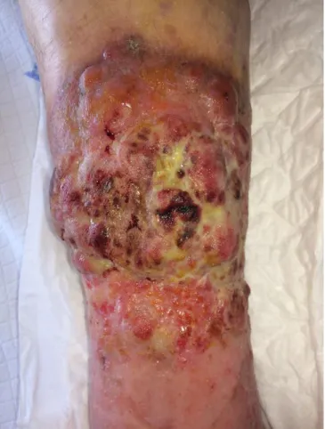
On physical examination we observed multiple coalescent nodules forming a vegetating mass of 15x10cm in the right leg and a peripheral ill-defined erythematous plaque, with some associated bleeding and seropurulent discharge (Figure. 1). There were no systemic symptoms and the physical examination was otherwise normal.
Figure 1. – Initial clinical aspect of the lesions.
The tumor biopsy showed a diffuse infiltrate composed by medium/large cells, with plasmacytoid features and high number of mitoses (Figure. 2, a). These cells were strongly positive for CD38, CD138, CD56 and immunoglobulin kappa light chains, faintly positive for CD79a and CD117, and negative for CD20 and PAX5 (Figure. 2, b – e). The in situ hybridization for Epstein-Barr virus (EBV)-encoded RNAs (EBER) was positive (Figure. 2, f).
Peripheral blood counts were normal and lymphocyte immunophenotyping showed a low CD4+
T-no other relevant changes, and subsequent investigation revealed seropositivity for human immuT-nodeficiency virus type 1 (HIV1). The bone marrow (BM) biopsy revealed a normocellular marrow with two tumor islets, strongly positive for CD138, dimly positive for CD117 and negative for CD3, CD20, and CD79a. The computed tomography scan of the thorax, abdomen, and pelvis provided no evidence for extracutaneous involvement.
Figure 2. Histology and immunohistochemistry: (a) – hematoxilin-eosin 400x; (b) – strong positivity for CD138; (c) – strong positivity for
CD38; (d) – dim positivity for CD79a; (e) – negative staining for CD20; (f) – positive in situ hybridization for EBV-encoded RNAs
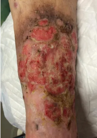
Treatment was started with highly active antiretroviral therapy (HAART). Two weeks later, while waiting for radiotherapy, it was decided to perform chemotherapy with the CVP (cyclophosphamide, vincristine and prednisone) in an attempt to control the extremely rapid tumor growth, with extensive necrosis. After the first cycle the lesion decreased in size and infiltration (Figure. 3). Unfortunately, the patient died 4 weeks later with sepsis owing to pulmonary infection.
Discussion
Plasmablastic lymphoma is a very rare B-cell lymphoma typically associated with immunosuppression: 69% of the patients have HIV infection and 33% of the HIV-negative patients have iatrogenic immunosuppression 6. Coexistent EBV infection is found in 60% of the cases 3. It occurs primarily in oral cavity, mostly in males, although some cases were reported in other organs and tissues 7,8,9.
Clinically, PCPBL presents as non-specific cutaneous lesions (purple nodules, erythematous infiltrated plaques).
It cannot be proven if this is a PBL presenting as an ulcerative skin lesion that was erroneously misdiagnosed as a venous ulcer or if this PBL arose in a chronic venous ulcer. In the second case, this might happen owing to the association of
immunodeficiency and local chronic inflammation that might further stimulate lymphocytic proliferation, thereby facilitating the accumulation of genetic defects and lymphomagenesis.
In previously described cases, as in this case, tumor cells are medium/large plasmacytoid cells (abundant cytoplasm, eccentric nucleus with granular chromatin and prominent central nucleolus). Multiple mitotic figures sometimes produce a “starry-sky” appearance 6. The immunohistochemistry shows typical plasmacytoid markers (CD38, CD138 and MUM1) and little to no expression of CD45 and B-cell markers (CD20, CD79a and PAX5). The association with EBV infection is usually
documented through positive in situ hybridization for EBER, with a higher proportion in HIV- affected individuals 6. Despite the presence of two tumor islets in the bone marrow, our patient did not have signs of bone marrow failure and the cutaneous tumor was the only clinical manifestation. Therefore, we assume this case is an atypical cutaneous presentation of a PBL.
Owing to the rarity of this entity, there is no established therapy, which makes its management an individualized, patient-based decision. In localized cutaneous lesions without systemic involvement, wide excision is recommended and radiation therapy is optional 5. In the case of systemic involvement, treatment consists of chemotherapy with or without consolidation radiation, and hematopoietic stem cell transplantation 10. Various regimens options include cyclophosphamide, doxorubicin, vincristine, and prednisone (CHOP), R-CHOP and cyclophosphamide, vincristine, doxorubicin, high-dose
methotrexate/ifosfamide, etoposide, and high-dose cytarabine (CODOX-M/IVAC) 10. In the HIV-infected population, the addition of HAART improves prognosis. In this case, although only one cycle was completed, there was a good local response to the HAART plus chemotherapy.
References
1. Delecluse H, Anagnostopoulos I, Dallenbach F, et al. Plasmablastic lymphomas of the oral cavity: a new entity associated with the human immunodeficiency virus infection. Blood. 1997;89:1413-1420 (PMID: 9028965) 2. Nicol I, Boye T, et al. Post-transplant plasmablastic lymphoma of the skin. Br J Dermatol. 2003 Oct;149(4):889-91
(PMID: 14616390)
3. Horna P, Hamill JR Jr, Sokol L, Glass LF. Primary cutaneous Plasmablastic lymphoma in an immunocompetent patient. J Am Acad Dermatol. 2013 Nov;69(5):e274-6 (PMID: 24124865)
4. Tiong IS, Strauss M, Lau MB, Chiruka S. Cutaneous plasmablastic lymphoma in an immunocompetent patient with long-term pyrimethamine use for essential thrombocythemia: a case report and literature review. Case Rep Hematol. 2013;541783:1-6 (PMID: 23476834)
5. Heiser D, Muller H, Kempf W, Eisendle K, Zelger B. Primary cutaneous plasmablastic lymphoma of the lower leg in an HIV-negative patient. J Am Acad Dermatol. 2012;67:e202-5 (PMID: 23062913)
6. Castillo JJ, Reagan JL. Plasmablastic lymphoma: a systematic review. ScientificWorldJournal. 2011 Mar 22;11:687-96 (PMID: 21442146)
7. Lin Y, Rodrigues GD, Turner JF, Vasef MA. Plasmablastic lymphoma of the lung: report of a unique case and review of the literature. Arch Pathol Lab Med. 2001 Feb;125(2):282-5 (PMID: 11175653)
8. Pruneri G, Graziadei G, Ermellino L, Baldini L, Neri A, Buffa R. Plasmablastic lymphoma of the stomach. A case report. Haematologica. 1998 Jan;83(1):87-9 (PMID: 9542326)
9. Ojanguren J, Collazos J, Martínez C, Alvarez J, Mayo J. Epstein-Barr virus-related plasmablastic lymphomas arising from long-standing sacrococcygeal cysts in immunosuppressed patients. AIDS. 2003 Jul 4;17(10):1582-4 (PMID: 12824797)
10. Elyamany G, Al Mussaed E, Alzahrani AM. Plasmablastic Lymphoma: A Review of Current Knowledge and Future Directions. Adv Hematol. 2015;2015:315289 (PMID: 26357515)