UNIVERSIDADE DE LISBOA
FACULDADE DE MEDICINA VETERINÁRIA
HIGH-THROUGHPUT PRODUCTION AND CHARACTERIZATION OF CARBOHYDRATE-ACTIVE ENZYMES FOR ANIMAL NUTRITION
VÂNIA ALEXANDRA DA SILVA CARDOSO LOPES
Orientadores: Professor Doutor Carlos Mendes Godinho de Andrade Fontes
Doutora Joana Luís Armada Brás
Tese especialmente elaborada para obtenção do grau de Doutor em Ciências Veterinárias na especialidade Produção Animal
2
2019
UNIVERSIDADE DE LISBOA
FACULDADE DE MEDICINA VETERINÁRIA
HIGH-THROUGHPUT PRODUCTION AND CHARACTERIZATION OF CARBOHYDRATE-ACTIVE ENZYMES FOR ANIMAL NUTRITION
VÂNIA ALEXANDRA DA SILVA CARDOSO LOPES
Orientadores: Professor Doutor Carlos Mendes Godinho de Andrade Fontes Doutora Joana Luís Armada Brás
Tese especialmente elaborada para obtenção do grau de Doutor em Ciências Veterinárias na especialidade Produção Animal
Júri
Presidente: Professor Doutor Luís Filipe Lopes da Costa Vogais:
- Professor Doutor Luís Manuel dos Anjos Ferreira
- Professor Doutor Carlos Mendes Godinho de Andrade Fontes - Professora Doutora Teresa Sacadura Santos Silva
- Professora Doutora Maria Isabel Ferraz de Oliveira
2
2019
DECLARAÇÃO RELATIVA ÀS CONDIÇÕES DE REPRODUÇÃO DA TESE Nome: Vânia Alexandra da Silva Cardoso Lopes
Título da Tese ou Dissertação: High-Throughput production and characterization of Carbohydrate-Active enZYmes for animal nutrition
Ano de conclusão (indicar o da data da realização das provas públicas): 2020 Designação do curso de
Mestrado ou de
Doutoramento: Doutoramento em ciências veterinárias na especialidade produção animal Área científica em que melhor se enquadra (assinale uma):
Clínica Produção Animal e Segurança Alimentar Morfologia e Função Sanidade Animal
Declaro sobre compromisso de honra que a tese ou dissertação agora entregue corresponde à que foi aprovada pelo júri constituído pela Faculdade de Medicina Veterinária da ULISBOA.
Declaro que concedo à Faculdade de Medicina Veterinária e aos seus agentes uma licença não-exclusiva para arquivar e tornar acessível, nomeadamente através do seu repositório institucional, nas condições abaixo indicadas, a minha tese ou dissertação, no todo ou em parte, em suporte digital.
Declaro que autorizo a Faculdade de Medicina Veterinária a arquivar mais de uma cópia da tese ou dissertação e a, sem alterar o seu conteúdo, converter o documento entregue, para qualquer formato de ficheiro, meio ou suporte, para efeitos de preservação e acesso.
Retenho todos os direitos de autor relativos à tese ou dissertação, e o direito de a usar em trabalhos futuros (como artigos ou livros).
Concordo que a minha tese ou dissertação seja colocada no repositório da Faculdade de Medicina Veterinária com o seguinte estatuto (assinale um):
1. Disponibilização imediata do conjunto do trabalho para acesso mundial;
2. Disponibilização do conjunto do trabalho para acesso exclusivo na Faculdade de Medicina Veterinária durante o período de 6 meses, 12 meses, sendo que após o tempo assinalado autorizo o acesso mundial*; * Indique o motivo do embargo (OBRIGATÓRIO)
Aguarda publicação de artigos científicos
Nos exemplares das dissertações de mestrado ou teses de doutoramento entregues para a prestação de provas na Universidade e dos quais é obrigatoriamente enviado um exemplar para depósito na Biblioteca da Faculdade de Medicina Veterinária da Universidade de Lisboa deve constar uma das seguintes declarações (incluir apenas uma das três):
1. É AUTORIZADA A REPRODUÇÃO INTEGRAL DESTA TESE/TRABALHO APENAS PARA EFEITOS DE INVESTIGAÇÃO, MEDIANTE DECLARAÇÃO ESCRITA DO INTERESSADO, QUE A TAL SE COMPROMETE.
Faculdade de Medicina Veterinária da Universidade de Lisboa, 16 de janeiro de 2020
AGR ADECIMENTOS
Esta tese não é apenas o resultado de muitos anos trabalho, mas também o resultado da colaboração de muitas pessoas. Nesta longa viagem, de quatro anos, as pessoas que me acompanharam foram fundamentais desde o primeiro até ao último dia desta jornada. Por isso quero agradecer:
À Fundação para a Ciência e Tecnologia pelo financiamento de parte da minha bolsa de doutoramento;
À NZYTech por me ter acolhido e permitido fazer o doutoramento em empresa. Por me ter financiado a segunda parte da minha bolsa. Pelos meios físicos e materiais que me disponibilizou para a realização de todo o trabalho;
À Faculdade de Medicina Veterinária e ao CIISA por me terem aceite como estudante de doutoramento e por terem disponibilizado meios físicos e materiais para a realização de todo o trabalho;
Ao Professor Doutor Carlos Fontes, por ter aceite ser meu orientador. Foi um privilégio ser sua aluna, não só por ser um investigador excecional e profissional dedicado, mas também por ter sido o melhor orientador que podia ter tido. Um grande e sincero obrigada;
À Doutora Joana Brás por me ter orientado sempre que precisei. Pelo apoio e ensinamentos em todas as tarefas laboratoriais e científicas, principalmente no que tocas às técnicas HTP. Obrigada;
Ao Professor Doutor Luís Ferreira, pela sua orientação e pelas palavras sábias no momento certo;
Ao Professor Doutor José Prates, pelos ensinamentos que me transmitiu pela sua disponibilidade e simpatia constantes;
À Teresa Ribeiro por ter sido o suporte mais importante no início da minha jornada pela investigação;
Ao Pedro Bule pelo apoio nas tarefas laboratoriais, pelos ensinamentos e pela troca de ideias;
À Vânia Fernandes pela amizade, companheirismo, entreajuda e suporte, em especial nas tarefas laboratoriais;
À Andreia Peixoto, João Belchior, Mara Ramires e Rosa Marques pela vossa amizade e companhia, mas a cima de tudo, pelo sentido de humor e as risadas que me provocaram;
À Virgínia Pires e Immacolata Venditto pela amizade e companheirismo;
À Patrícia Ponte, Filipa Sequeira e Nádia Rodrigues por todas as partilhas de conhecimentos e orientação no funcionamento de uma empresa;
Ao Diogo Comprido, Pedro Rio e José Albuquerque pela vossa capacidade de trabalho e pela boa disposição e simpatia;
Ao Instituto Superior de Agronomia na cedência de instalações para os ensaios de frangos e a toda a equipa envolvida nos mesmos;
À minha família, em especial aos meus pais, João e Fátima Cardoso, pelo apoio constante, motivação, amor e carinho. Sem eles não teria conseguido chegar onde cheguei. Agradeço-vos muito por tudo o que já fizeram por mim;
Ao meu marido Pedro Lopes pela motivação, apoio incondicional e toda a paciência. Por toda a assistência informática e bioinformática que me deu, desde o desenvolvimento de ferramentas bioinformáticas (p.e. Gene alignment algorithm) à resolução das pequenas dificuldades encontradas no dia-a-dia. Por nunca ter duvidado das minhas capacidades. Principalmente por me aturar;
À minha filha Leonor Lopes por me ter feito tanta companhia durante o processo de escrita e por involuntariamente me ter dado tanta motivação e inspiração para finalizar esta etapa da minha vida.
This work was co-funded by Fundação para a Ciência e a Tecnologia, grant SFRH/BDE/101026/2014 from Ministério da Ciência, Tecnologia e Ensino Superior, and by NZYTech, Lda, genes & enzymes
RESUMO
Na natureza, a biodegradação dos hidratos de carbono da parede celular vegetal é realizada por enzimas microbianas, geralmente conhecidas como CAZymes. Os animais monogástricos produzem um reportório limitado de enzimas para degradação destes hidratos de carbono, não conseguindo usar eficientemente alguns ingredientes da dieta, que muitas vezes manifestam propriedades anti nutritivas. Assim, é sabido que a suplementação com CAZymes exógenas melhora o valor nutritivo das dietas e aumenta o desempenho produtivo dos animais. Este trabalho revelou que as enzimas mais apropriadas para suplementar dietas ricas em 1,3-1,4-β-glucanos são as 1,3-1,4-β-glucanases e não as celulases. Além disso, verificou-se que a suplementação com β-xilanases melhorou o valor nutritivo de dietas que continham variedades de trigo com maior viscosidade e menor atividade endógena de endo-1,4-β-xilanase. Em oposição, quando o lote de trigo apresentou menor viscosidade e maiores níveis de atividade endógena de endo-1,4-β-xilanase, a resposta dos animais à adição das enzimas foi menor. Este trabalho mostra, igualmente, que os xilo-oligossacarídeos, resultantes da degradação de arabinoxilanos por xilanases exógenas, possuem uma ação pré-biótica na alimentação de frangos, promovendo a melhoria do desempenho zootécnico. Contudo, apesar de estarem descritas uma grande diversidade CAZymes, poucas são as estudadas com potencial para serem usadas em alimentação animal. Portanto, este trabalho pretendeu, também, desenvolver metodologias para isolar e caracterizar enzimas potencialmente importantes em larga escala. Foram selecionadas, produzidas e expressas na forma recombinante 1476 CAZymes. Os dados revelaram que 79% das proteínas recombinantes foram produzidas na forma solúvel em Escherichia coli. Verificou-se, ainda, que fatores como o organismo de origem, a estratégia de produção, a fusão com marcadores de solubilidade, o peso molecular da proteína e composição de aminoácidos das sequências primárias, parecem justificar os resultados da solubilidade. Estes ensinamentos foram utilizados para produzir enzimas, tais como ferulolil esterases (FAEs) e glucuronil esterases (GEs), que removem as cadeias laterais e quebram as ligações cruzadas entre hidratos de carbono hemicelulósicos e a lenhina. Assim sendo, foram selecionadas 480 FAEs e 20 GEs para produção recombinante e caracterização bioquímica. Cerca de 372 FAEs e 11 GEs foram produzidos em forma solúvel em E. coli e aproximadamente 50% das enzimas produzidas mantiveram níveis significativos de atividade e estabilidade. Com isto, foi possível identificar e produzir um número significativo de FAEs e GEs com potencial para alimentação animal, em especial as que libertam celulose e hemicelulose da lenhina.
ABSTR ACT
The biodegradation of plant cell wall (PCW) carbohydrates is performed by microbial enzymes that are generally referred to as CAZymes. In animal nutrition, it is now well established that the monogastric animals produce a limited repertoire of CAZymes and as such cannot use efficiently some dietary ingredients that sometimes display antinutritional properties. The dietary supplementation with exogenous CAZymes improves the nutritive value of diets and increases animal’s performance. In particular, this study demonstrated that 1,3-1,4-β-glucanases and not cellulases improve the nutritive value of β-glucan-containing diets for monogastric animals. In addition, it was revealed that exogenous enzyme supplementation with β-xylanases improved the nutritive value of diets incorporating wheat lots with high viscosity and low endogenous endo-1,4-β-xylanase activity. In contrast, when the wheat lot showed lower viscosity and higher levels of endogenous endo-1,4-β-xylanase activity, broiler response was clearly diminished. Moreover, the data revealed that xylo-oligosaccharides released by xylanases acting on cereal arabinoxylans display a pre-biotic and positive effect in broiler chicks. However, although we observe an exponential accumulation of genomic and metagenomic information, knowledge on CAZYmes with potential to be used in animal nutrition is limited. This work also aimed to develop high-throughput (HTP) methodologies to isolate and characterize potentially important enzymes for animal nutrition. Thus, 1476 recombinant enzymes were selected and produced recombinantly. The data revealed that 79% of recombinant proteins were produced in the soluble form in Escherichia coli. Factors, such as, organism of origin, gene production strategy, fusion with solubility tags, protein molecular weight and amino acids composition of primary sequences may be used to justify and predict levels of solubility. The establishment of a high-throughput pipeline for recombinant enzyme production was used to obtain a library of feruloyl esterases (FAEs) and glucuronoyl esterases (GEs), enzymes which remove the side chains and break crosslinks between hemicellulosic carbohydrates and lignin. Thus 480 putative FAEs and 20 GEs were produced and biochemically characterized. Following gene isolation, 372 FAEs and 11 GEs were produced in a soluble form in E. coli. Activity results showed that 50% of the enzymes produced retained significant levels of activity and stability. The library of innovative FAEs and GEs produced during this project will be used to develop a novel generation of enzymes for animal nutrition, in particular to exploit the release of cellulose and hemicellulose from lignin.
This thesis was based on the following manuscripts:
V. Cardoso, T. Ribeiro, J.L.A. Brás, V.O. Fernandes, H. Santos, M.M. Lordelo, L.M.A. Ferreira and C.M.G.A. Fontes. Novel evidence supporting the role of 1,3-1,4-β-glucanases and not 1,4-β-1,3-1,4-β-glucanases to improve the nutritive value of β-glucan-containing diets for monogastric animals. (manuscript submitted for publication)
V. Cardoso*, E. A. Fernandes*, H. M. M. Santos, B. Maçãs, M. M. Lordelo, L.T. Gama, L. M. A. Ferreira, C. M. G. A. Fontes and T. Ribeiro. 2018. Variation in levels of non-starch polysaccharides and endogenous endo-1,4-β-xylanases affects the nutritive value of wheat for poultry. British Poultry Science 59(2):218-226. (DOI: 10.1080/00071668.2018.1423674).
T. Ribeiro*, V. Cardoso*, L.M.A. Ferreira, M.M. Lordelo, E. Coelho, A.S.P Moreira, M.R.M. Domingues, M.A. Coimbra, M.R. Bedford, and C.M.G.A. Fontes. 2018. Xylo-oligosaccharides display a prebiotic activity when used to supplement wheat or corn-based diets for broilers. Poultry Science 97(12):4330-4341. (DOI: 10.3382/ps/pey336)
V. Cardoso, J.L.A. Brás,L.M.A. Ferreira, L.T. Gama, R. Vincentelli, B. Henrissat and C.M.G.A. Fontes. Generation of a library of Carbohydrate-Active Enzymes for plant biomass deconstruction. (manuscript in preparation)
V. Cardoso, J.L.A. Brás, L.M.A. Ferreira, V.D. Alves, C.M.G.A. Fontesand P. Bule.
Structural determinants of substrate specificity in family 15 Carbohydrate Esterases. (manuscript in preparation)
V. Cardoso, J.L.A. Brás, A.L Carvalho, V.D. Alves, R.P. de Vries, V. Puchart, P. Biely, L.M.A. Ferreira, C.M.G.A. Fontes and P. Bule. Subtle changes at the catalytic site of feruloyl esterases modulate enzyme specificity to hydroxycinnamic acids. (manuscript submitted for publication)
INDEX
1. Bibliographic Review and Objectives ... 1
1.1. Introduction ... 1
1.2. Plant cell wall ... 2
1.2.1. Plant cell wall structure ... 2
1.2.2. Plant cell wall components ... 4
1.2.2.1. Cellulose... 4
1.2.2.2. Hemicellulose ... 5
1.2.2.3. Pectin ... 7
1.2.2.4. Lignin ... 9
1.2.3. Plant cell wall models ... 10
1.2.4. Plant cell wall degradation ... 11
1.2.4.1. Carbohydrate-Active Enzymes (CAZymes) ... 12
1.2.4.1.1. Glycoside hydrolases (GHs) ... 14
1.2.4.1.2. Polysaccharide lyases (PLs) ... 16
1.2.4.1.3. Carbohydrate esterases (CEs) ... 17
1.2.4.1.4. Auxiliary Activities (AAs) ... 17
1.2.4.1.5. Glycosyltransferases (GTs) ... 18
1.2.4.1.6. Carbohydrate-Binding Modules (CBMs) ... 19
1.2.4.2. Plant cell wall degrading enzymes ... 20
1.2.4.2.1. Cellulases ... 21
1.2.4.2.2. Hemicellulases ... 22
1.2.4.2.2.1. Acessory enzymes ... 24
a) Feruloyl esterases (FAEs) ... 25
b) Glucuronoyl esterases (GEs) ... 25
1.2.4.2.3. Pectinases ... 25
1.2.4.2.4. Lignin degradation ... 27
1.2.4.3. Cellulosomes ... 28
1.2.4.3.1. Cellulosome composition and structure... 29
a) Scaffoldins ... 29
b) Cohesin-dockerin interaction ... 30
c) Enzymes ... 32
d) Carbohydrate-Binding Modules ... 32
1.2.4.3.2. Cellulosome systems ... 33
1.3. Supplementation of diets for poultry ... 34
1.3.1. Non-starch polysaccharides in poultry nutrition ... 34
1.3.2. Enzymatic supplementation of cereal-based diets for poultry ... 36
1.3.3. Prebiotic supplementation of diets for poultry ... 37
1.4. Objectives ... 39
2. Novel evidence supporting the role of 1,3-glucanases and not 1,4-β-glucanases to improve the nutritive value of β-glucan-containing diets for monogastric animals... 40
2.1. Introduction ... 41
2.2. Materials and Methods ... 42
2.2.1. Gene isolation and cloning into a prokaryotic expression vector ... 42
2.2.2. Expression and purification of CtGlc16A and CtCel5E ... 43
2.2.3. Biochemical properties of CtGlc16A and CtCel5E ... 43
2.2.4. Animals and diets ... 44
2.2.5. Statistical Analyses ... 45
2.3. Results and Discussion ... 46
2.3.1. Biochemical properties of CtGlc16A and CtCel5E ... 46
2.3.2. The efficacy of CtGlc16A and CtCel5E to improve the nutritive value of a barley-based diet ... 46
2.3.3. Is CtCel5E stable during passage through the animal GI tract? ... 49
2.4. Conclusions ... 50
3. Variation in levels of non-starch polysaccharides and endogenous endo-1,4-β-xylanases affect the nutritive value of wheat for poultry ... 52
3.1. Introduction ... 53
3.2. Materials and Methods ... 54
3.2.1. Quantification of endo-1,4-β-xylanase and viscosity in different wheat lots ... 54
3.2.2. Animals, Diets and Management ... 55
3.2.3. Analysis of digesta samples ... 56
3.2.4. Statistical analysis ... 57
3.3. Results and Discussion ... 57
3.3.1. Variation of endogenous endo-1,4-β-xylanase activity and extract viscosity in different wheat lots ... 57
3.3.2. Levels of endogenous endo-1,4-β-xylanases and wheat extract viscosity affect the response to exogenous enzyme supplementation ... 63
3.3.3. Diet impact on organ size, digesta viscosity and endo-1,4-β-xylanase activity ... 66
3.4. Conclusions ... 69
4. Xylo-oligosaccharides display a prebiotic activity when used to supplement wheat or corn-based diets for broilers... 70
4.1. Introduction ... 71
4.2.1. Enzymes and oligosaccharides ... 72
4.2.2. Oligosaccharide and monosaccharide composition analysis ... 73
4.2.3. Determination of the presence of starch ... 73
4.2.4. Oligosaccharide ethanol fractionation and matrix-assisted laser desorption/ionization (MALDI) mass spectrometry analysis ... 73
4.2.5. Diets ... 74
4.2.6. Animals and Management ... 75
4.2.7. Analytical Procedures ... 76
4.2.8. Microbiome profiling through 16s rRNA sequencing ... 76
4.2.9. Statistical Analysis ... 77
4.3. Results and discussion ... 77
4.3.1. Characterization of xylo-oligosaccharides (XOS) ... 77
4.3.2. Inclusion of exogenous xylanases or XOS in wheat-based diets promote broiler performance... 79
4.3.3. XOS modulate gut microbial populations ... 84
4.3.4. XOS promote the performance of broilers fed on maize-based diets ... 85
4.4. Conclusions ... 89
5. Generation of a library of Carbohydrate-active enzymes for Plant biomass deconstruction ... 90
5.1. Introduction ... 91
5.2. Materials and Methods ... 92
5.2.1. Identification and selection of CAZymes ... 92
5.2.2. Polymerase chain reaction (PCR) and Gene synthesis (GS) ... 92
5.2.3. Cloning, transformation and sequencing ... 93
5.2.4. High-throughput protein expression, purification and quantification ... 93
5.2.5. Statistical Analyses ... 94
5.3. Results and Discussion ... 94
5.3.1. Identification and selection of CAZymes ... 94
5.3.2. Genes production and cloning... 95
5.3.3. Protein expression and putative factors affecting solubility ... 95
5.3.4. The effect of protein origin on recombinant production ... 96
5.3.5. Influence of gene production strategy, gene synthesis or PCR, in protein production97 5.3.6. Does the GFP tag promotes protein production? ... 97
5.3.7. Influence of protein molecular weight in protein production ... 99
5.3.8. Influence of primary sequence composition on protein production ... 99
5.3.9. Final outputs ... 101
6. Structural determinants of substrate specificity in family 15 Carbohydrate
Esterases ... 104
6.1. Introduction ... 105
6.2. Materials and Methods ... 106
6.2.1. GEs selection and Phylogenetic analysis ... 106
6.2.2. Chemicals ... 106
6.2.3. Strains, media and vector ... 106
6.2.4. Polymerase chain reaction (PCR) and Gene synthesis (GS) ... 107
6.2.5. Cloning, transformation and sequencing ... 107
6.2.6. High-throughput protein expression and purification by nickel affinity chromatography ... 107
6.2.7. Glucuronoyl Esterases Activity ... 108
6.2.8. Determination of optimal pH and temperature ... 108
6.2.9. Crystallization of TtCE15 ... 108
6.2.10. 3D structure solution ... 109
6.3. Results and Discussion ... 109
6.3.1. GEs selection and sequence analysis ... 109
6.3.2. Recombinant enzyme production ... 113
6.3.3. Biochemical properties of bacterial GEs ... 114
6.3.4. Crystal Structure of TtGE15A ... 115
6.3.5. TtGE15A Catalytic triad and recognition of Glucuronic Acid (GlcA) ... 117
6.4. Conclusions ... 120
7. Subtle changes at the catalytic site of feruloyl esterases modulate enzyme specificity to hydroxycinnamic acids ... 121
7.1. Introduction ... 122
7.2. Materials and Methods ... 123
7.2.1. FAEs selection and Phylogenetic analysis ... 123
7.2.2. Chemicals ... 124
7.2.3. Strains, media and vector ... 124
7.2.4. Polymerase chain reactions (PCR) and Gene synthesis (GS) ... 124
7.2.5. Cloning, transformation and sequencing ... 124
7.2.6. High-throughput protein expression and purification by nickel affinity chromatography ... 125
7.2.7. High-throughput enzyme activity assays ... 125
7.2.7.1. p-Nitrophenyl Ferulate (pNP-Fe) ... 125
7.2.7.2. Methyl esterified substrates (MFA, MCA, MSA or MpCA). ... 126
7.2.7.3. Tannase Activity ... 126
7.2.8. Crystallization of LcFAE1A ... 126
7.2.9. 3D structure solution ... 127
7.3. Results and Discussion ... 127
7.3.1. Discovery of novel bacterial FAEs... 127
7.3.2. Recombinant enzyme production ... 129
7.3.3. Biochemical properties of bacterial FAEs ... 130
7.3.4. Phylogenetic tree of active FAEs ... 131
7.3.5. Relation between phylogenetic origin and FAE substrate specificity ... 131
7.3.6. Structure of Lactobacillus crispatus feruloyl esterase 1A (LcFAE1A) ... 135
7.3.7. Role of catalytic site residues for substrate recognition ... 137
7.4. Conclusions ... 140
8. General Discussion and Future Perspectives ... 141
9. Bibliographic References ... 146
LIST OF FIGURES
Figure 1.1. Schematic representation of the cell wall. ... 3
Figure 1.2. Cell wall structure. ... 4
Figure 1.3. Cellulose representation. ... 5
Figure 1.4. Structure of different types of hemicelluloses. ... 6
Figure 1.5. Schematic structure of pectin... 7
Figure 1.6. Homogalacturonan structure and representative sites of methylesterification at the C-6 and O-acetylation at the O-2 or O-3 of the carbohydrate ring. ... 8
Figure 1.7. Chemical structure of monolignols and the corresponding units in lignin. ... 10
Figure 1.8. Models of cell wall structure. ... 11
Figure 1.9. Representation of cell wall deconstruction. ... 12
Figure 1.10. Modular architecture of Carbohydrate-Active enZymes (CAZymes). ... 13
Figure 1.11. General mechanism for (a) inverting and (b) retaining glycosidases. ... 15
Figure 1.12. Stereochemical outcomes of glycosyltransferases. ... 19
Figure 1.13. Examples of type A, type B and type C CBMs. ... 20
Figure 1.14. Structure of cellulose ... 21
Figure 1.15. Illustration of cellulases action on cellulose. ... 22
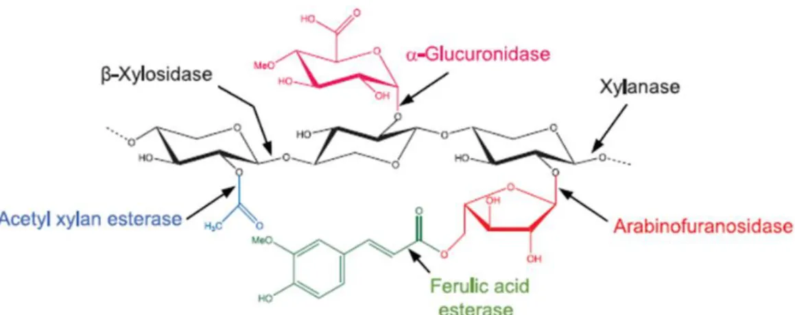
Figure 1.16. Structure of xylan and the xylanolytic enzymes involved in its degradation. ... 23
Figure 1.17. Enzymatic attack on galactomannan structure. ... 23
Figure 1.18. Representative ester linkages between xylans and lignin. ... 24
Figure 1.19. Summary of HG and RG-degrading enzymes. ... 26
Figure 1.20. Schematic representation of cellulosome. ... 29
Figure 1.21. Types of cellulosome systems. ... 30
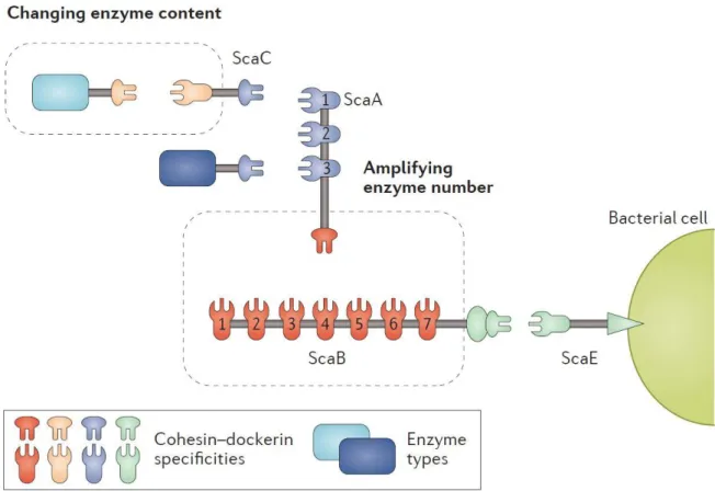
Figure 1.22. The cellulosome of Ruminococcus flavefaciens with alternative roles of adaptor scaffoldins. ... 31
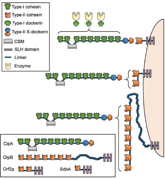
Figure 1.23. Schematic representation of the scaffoldins of simple cellulosome systems. .... 33
Figure 1.24. The cellulosome architecture of C. thermocellum. ... 34
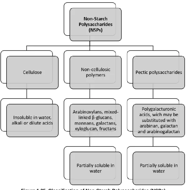
Figure 1.25. Classification of Non-Starch Polysaccharides (NSPs)... 35
Figure 2.1. pH and temperature profiles of CtGlc16A and CtCel5E. ... 47
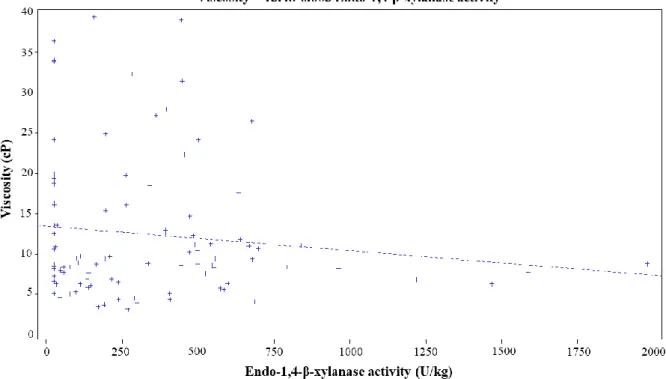
Figure 3.1. Relation between cereal extract viscosity and endogenous endo-1,4-β-xylanase activity in different wheat lots. ... 62
Figure 3.2. Detection of endo-xylanase activity in the gastrointestinal contents of broilers. .. 69
Figure 4.1. MALDI spectra. ... 78
Figure 4.2. Principal Component Analysis (PCA) of bacterial microbiota in the caecum of birds fed on a wheat-based diet. ... 85
Figure 5.1. The distribution of the 1476 enzymes produced in this study by the five CAZy classes. ... 95
Figure 5.2. Protein expression and purity analysis of CAZymes exemplified by a SDS-PAGE gel. 96
Figure 5.3. Efficacy of the recombinant production of CAZymes. ... 98
Figure 5.4. Number of CAZY families and ECs obtained in this study. ... 102
Figure 5.5. Number of CAZy classes and families represented in the current library. ... 102
Figure 6.1. Schematic representation of modular GEs. ... 111
Figure 6.2. Phylogenetic relationships between the 20 GEs used in this study and 3 bacterial GEs which 3D structure were recently determined. ... 112
Figure 6.3. Protein expression and purity analysis of GEs by SDS-PAGE. ... 113
Figure 6.4. pH (a.) and temperature (b.) dependence of the tested GEs’ catalytic activity. . 115
Figure 6.5. Representation of TtCE15A’s structure. ... 117
Figure 6.6. Putative catalytic triad of TtCE15A’s. ... 118
Figure 6.7. Multiple sequence alignment of GE15 members with known structures. ... 119
Figure 7.1. Exemplification of molecular integrity and degree of purity of recombinant FAEs produced in this study... 129
Figure 7.2. Phylogenetic relationships among the 202 active bacterial FAEs characterized in this study and representative fungal FAEs of subfamilies 1 to 13 (SF1-SF13). ... 132
Figure 7.3. Structure of Lactobacillus crispatus feruloyl esterase 1A (LcFAE1A). ... 136
Figure 7.4. Detailed view of LcFAE1A’s putative active center, identified by overlaying its structure with Lactobacillus johnsonii cinnamoyl esterase in complex with ferulic acid. ... 138
LIST OF T ABLES
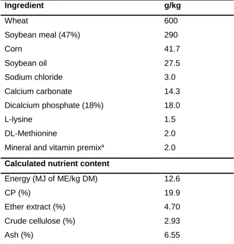
Table 2.1. Ingredient composition and calculated analysis of the barley-based feed. ... 45
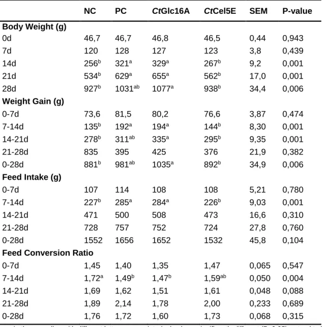
Table 2.2. Growth performance parameters of broilers fed on a barley-based diet ... 48
Table 3.1. Ingredient composition (g/kg) and calculated analysis of the wheat-based diets prepared in this study. ... 56
Table 3.2. Endo-1,4-β-xylanase activity (U/kg of wheat) and viscosity (cP) of different wheat lotsa,b. ... 58
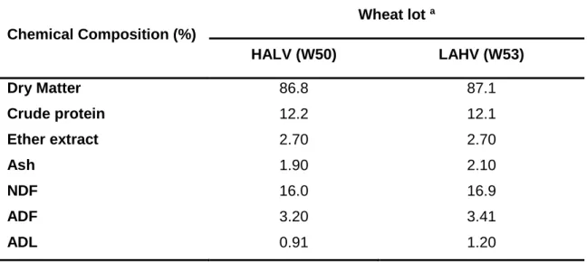
Table 3.3. Nutritional composition of the two different wheat lots used in the animal experiment. ... 64
Table 3.4. Performance of broilers fed with diets prepared with wheat. ... 65
Table 3.5. Relative weight and length of the gastrointestinal tract and viscosity of digesta. .. 67
Table 3.6. Number of birds with enzyme activity in GI tract... 68
Table 4.1. Ingredient composition and calculated analysis of the wheat-based feed of experiments 1 and 2. ... 74
Table 4.2. Performance of broilers fed on the wheat-based diet (Experiment 1). ... 80
Table 4.3. Performance of broilers fed on the wheat-based diet (Experiment 2). ... 82
Table 4.4. Relative weight and length of the GI tract of broilers (Experiment 2). ... 83
Table 4.5. Ingredient composition and calculated analysis of the corn-based feed of experiment 3. ... 87
Table 4.6. Performance of broilers fed on the corn-based diet (Experiment 3). ... 88
Table 5.1. Analysis of the amino acid composition in primary sequences of soluble and insoluble CAZymes produced in this study. ... 100
Table 6.1. Data collection and refinement statistics of TtGE15A. ... 110
Table 6.2. Organism origin, production level, specific activity and optimal conditions of characterized GEs in this study. ... 114
Table 7.1. Data collection and refinement statistics of LcFAE1A. ... 128
Table 7.2. Specific activity of characterized FAEs in this study. ... 133
Table S2.1. Relative weight and length of the gastrointestinal tract and viscosity of digesta.177 Table S4.1. Relative weight and length of the GI tract of broilers (Experiment 1)... 178
Table S4.2. Relative weight and length of the GI tract of broilers (Experiment 3)... 179
Table S5.1. DNA and amino acid sequences, predicted molecular mass, molecular architecture, CAZy family and origin of enzymes produced in this work. ... 180
Table S6.1. Primers used for the PCR amplification of CE15 constructs. ... 181
Table S6.2. Predicted molecular mass, molecular architecture and origin of GEs studied in this work. ... 182
Table S6.3. DNA and amino acid sequences of GEs studied in this work. ... 184
Table S7.2. DNA and amino acid sequences, predicted molecular mass, molecular architecture and origin of FAEs studied in this work. ... 207
LIST OF ABBREVIATION S AND S YMBOLS
% Percentage
[M+Na]+ Sodium adduct ions
Å Angstrom
A. cellulolyticus Acetivibrio cellulolyticus
A550 Absorbance at 550 nanometers
AA Auxiliary Activities
AAO Aryl-alcohol oxidase
ABF α-arabinofuranosidase
AcE Acetyl esterases
AcXEs Acetylxylan esterases
ADF Acid Detergent Fibre
ADL Acid Detergent Lignin
AGA Apiogalacturonan
Ala Alanine (A)
ANOVA Analysis of variance
Araf Arabinofuranose
Arg Arginine (R)
Ash Mineral matter present in feed
Asn Asparagine (N)
Asp Aspartic acid (D)
AXE Acetyl xylan esterase
AXOS Arabinoxylo-oligosaccharides
B. cellulosolvens Bacteroides cellulosolvens
BL Breeding lines
BL21(DE3)
E. coli expression strain containing the DE3 lysogen that carries the gene for T7 RNA polymerase under control of the lacUV5 promoter
BLAST Basic Local Alignment Search Tool
BnzGlcA Benzyl D-glucuronate
BSA Bovine serum albumin
BW Body Weight
BXL 1,4-β-xylosidase
C- Non-supplemented diet
C. acetobutylicum Clostridium acetobutylicum
C. cellulolyticum Clostridium cellulolyticum
C. cellulolyticus Clostridium cellulolyticus
C. cellulovorans Clostridium cellulovorans
C. clariflavum Clostridium clariflavum
C. japonicus Clostridium japonicus
C. josui Clostridium josui
C. papyrosolvens Clostridium papyrosolvens
C. S. usitatus Candidatus Solibacter usitatus
C. thermocellum Clostridium thermocellum
Ca2+ Calcium ion
CaCl2 Calcium chloride
CAI Codon Adaptation Index
CAZyme Carbohydrate-Active enZyme
CBH Cellobiohydrolases
CE Carbohydrate Esterase
CE1 Carbohydrate Esterase family 1
CE15 Carbohydrate Esterase family 15
cm/kg Centimetre per kilogram
coh Cohesin
cP Centipoise
CP Crude Protein
CV Cultivar lines
Cys Cysteine (C)
Dha 2-keto-3-deoxy-D-lyxo heptulosaric acid
DHB 2,5-dihydroxybenzoic acid
DNA Desoxyribonucleic Acid
DNSA 3,5-dinitrosalicylic acid
Doc Dockerin
DyPs Decolorizing peroxidases
E. coli Escherichia coli
EC Enzyme Commission Number
EG Endoglucanase
ES Spanish origin
eV Electron volt
F ratio Variation between sample means / variation within the samples
FA Ferulic acid
FAE Ferulic Acid Esterase
FCR Feed Conversion Ratio
FOS fructo-oligosaccharides
FR French origin
Fuc L-Fucose
G Guaiacyl
g/kg Gram per kilogram
Gal D-Galactose
GalpA Galacturonic acid
GAXs Glucuronoarabinoxylans
GC Gas chromatography
GC–MS Gas chromatography–mass spectrometry
gDNA Genomic desoxyribonucleic Acid
GE Glucuronoyl esterase
GFP Green-fluorescence protein
GH Glycoside Hydrolase
GI Gastrointestinal tract
GlcA Glucuronic acid
Gln Glutamine (Q)
GLOX Glyoxal oxidase
Glu Glutamic acid (E)
Gly Glycine (G)
GOS Galacto-oligosaccharides
GS Gene synthesis
GST Glutathione S-transferase
GT Glycosyl Transferase
GT-B Glycosyltransferase fold B
GUS α-glucuronidase
h Hour
H Hydroxyphenyl
H2O2 Hydrogen peroxide
H2SO4 Sulfuric acid
HA Enzymes with high activity
HALV High endogenous endo-1,4-β-xylanases activity and low viscosity
HCl Hydrogen chloride
Hepes Hydroxyethyl piperazineethanesulfonic acid
Hexn Hexose oligosaccharides
HG Homogalacturonan
His Histidine (H)
His6-tag Six Histidines tag
HTP High-throughput
Ile Isoleucine (I)
IMAC Immobilized metal ion-affinity chromatography IPTG Isopropyl β-D-1-thiogalactopyranoside
IUBMB Union of Biochemistry and Molecular Biology
K2HPO4 Potassium hydrogen phosphate
Kan Kanamycin
kDa Kilodalton
Kdo 2-keto-3-deoxy-D-manno octulosonic acid
Kg Kilogram
KOH Potassium hydroxide
L Litre
LA Enzymes with low activity
Lac Laccases
LAHV Low endogenous endo-1,4-β-xylanases activity and high viscosity
L-Araf L-arabinofuranose
LB Luria Bertani
LCCs Lignin-carbohydrate complexes
Leu Leucine (L)
LiP lignin peroxidases
LPMO Lytic polysaccharide mono-oxygenases
Lys Lysine (K)
M Molar
MALDI Matrix-assisted laser desorption/ionization
MBP Maltose-binding polypeptide
MCA Methyl caffeate
MeGlcA 4-O-methyl-D-glucuronic acid
MES 2-(N-morpholino)ethanesulfonic acid
Met Methionine (M)
MFA Methyl ferulate
mg Milligram
min Minutes
mL Milliliter
Mn2+ Manganese cation (2+)
Mn3+ Manganese cation (3+)
MnP Manganese peroxidases
MOS Mannan-oligosaccharides
MpCA Methyl p-coumarate
MSA Methyl sinapate
MT Enzymes active on methyl ester substrates
MW Molecular Weight
NaCl Sodium chloride
NaOH Sodium hydroxide
NCBI National Center for Biotechnology Information
nd Not detectable
NDF Neutral Detergent Fiber
Ni2+ Nickel ion
nm Nanometer
NRC National Research Council
NSP Non-starch polysaccharide
NZY5α E. coli competent cells with similar properties to DH5α
ºC Celcius degree
OD Optical Density
OSE diet supplemented with xylose (Experiment 1)
OTUs Operational Taxonomic Units
p p-value
p(F) p-value from F-Ratio
PC Phosphate citrate
PCA Principal Component Analysis
PCR Polymerase Chain Reaction
PCW Plant cell wall
PD10 GE Healthcare dessalting columns
PDB Protein data bank
PEG 3350 Polyethylene glycol 3350
Pentn Pentose oligosaccharides
pH Negative decimal logarithm of the hydrogen ion activity in a solution
Phe Phenylalanine (F)
pHTP1 E.coli expression vector containing a N-terminal Histidine tag
pHTP9 E.coli expression vector containing a N-terminal GFP fusion tag
PL Polysaccharide Lyase
pNP P-nitrophenol
pNP-Fe P-nitrophenyl ferulate
pNPP 4-nitrophenol palmitate
PP Enzymes that attack pNP-ferulate but not the methyl substrates
Pro Proline (P)
R. albus Ruminococcus albus
R. flavefaciens Ruminococcus flavefaciens
r.m.s.d Root mean square deviation
RGI Rahmnogalacturonan I
RGII Rahmnogalacturonan II
Rhap Rhamnose
S Syringyl
SAS Statistical Analysis Software
SCFA Short Chain Fatty Acids
SDS-PAGE Sodium dodecyl sulfate-polyacrylamide gel electrophoresis
SEM Standard error of the mean
Ser Serine (S)
SLHs S-layer homology modules
SOS Soybean meal oligosaccharides
T. turnerae Teredinibacter turnerae
TCEP Tris(2-carboxyethyl)phosphine
Thr Threonine (T)
Tris 2-Amino-2-hydroxymethyl-propane-1,3-diol
Trp Tryptophan (W)
TrxA Thioredoxin
Tyr Tyrosine (Y)
U Enzymatic unities
U/g Enzymatic unities per gram
U/kg Enzymatic unities per kilogram
UK United Kingdom origin
Unk Unknown
USA American origin
v/v Volume per volume
Val Valine (V)
VFA Volatile fatty acids
VP Versatile peroxidases
w/v Weight per volume
w/w Weight per weight
×g Ggravity or relative centrifugal force
XGA Xylogalacturonan XLN 1,4-β-endoxylanase XOS Xylo-oligosaccharides XYL 1,4-β-xylanase βG β-Glucosidade μg Microgram μL Microliter μM Micromolar μm Micrometre
1.
BIBLIOGRAPHIC REVIEW AND OBJECTIVES
1.1.
Introduction
Plant cell walls (PCWs) and their carbohydrate components are the most abundant organic compounds found in nature and the most abundant source of organic carbon in the biosphere. These structures are mainly constituted by cellulose, hemicellulose (which include xyloglucans, xylans, mannans, and β-glucans), pectin and lignin. Recycling these highly relevant molecules is a process of growing interest, particularly in the biofuel and bioprocessing sectors. In nature, the recycling of organic carbon stored in the plant cell wall is performed by microbial enzymes that convert the cell wall polysaccharides to monosaccharides and oligosaccharides. These enzymes are generally referred as Carbohydrate-Active Enzymes (CAZymes) and include Glycoside Hydrolases (GHs), Polysaccharide Lyases (PLs), Carbohydrate Esterases (CEs), Glycosyl Transferases (GTs) and Auxiliary Activities (AAs). Additionally, CAZymes are frequently modular, containing one or more catalytic domains connected to non-catalytic carbohydrate-binding modules (CBMs). CAZymes and CBMs have been grouped into sequence-based families on the continuously updated Carbohydrate-Active enZymes database (www.cazy.org). The complete biodegradation of recalcitrant plant cell wall carbohydrates requires the cooperative attack between enzymes that break the polysaccharide backbone, such as cellulase, hemicellulases, pectinases, among others, along with several accessory enzymes, which remove the side chains and break crosslinks between hemicellulosic carbohydrates and other plant polymers, such as lignin. Among the accessory enzymes, feruloyl esterases and glucuronoyl esterases, which belong to CEs enzyme class, play a key role in enhancing the accessibility of backbone enzymes to their target bonds. CAZYmes play not only a relevant role in carbon turnover in nature but also display considerable biotechnological potential, in particular in animal nutrition. It is well established that soluble non-starch polysaccharides, such as arabinoxylans, β-glucans and pectins are considerably anti-nutritive for simple-stomach animals. Particularly, in poultry, elevated levels of PCW Non-Starch Polysaccharides (NSPs) lead to an increase in digesta viscosity with a consequent decreased in nutrient digestion and nutrient absorption. When birds are fed wheat or barley-based diets, the presence of soluble NSPs is considerable. Therefore, it is now well established that the dietary supplementation with exogenous enzymes (such as cellulases and hemicellulases) reduces the degree of polymerization of NSPs, improving the nutritive value of diets and, consequently, increases animal’s performance. However, the biological role of CAZymes in animal nutrition remains poorly understood, in particular in what relates
to the modulation of the gastrointestinal microflora through the production of dietary prebiotics.
This thesis aims to clarify several unresolved hypotheses related with CAZymes and their biotechnological impact in animal nutrition and it is divided in 8 main chapters. The first chapter begins with a general review on plant cell wall structure and composition, followed by a description of the different mechanisms and enzymes that are required for the degradation of structural lignocellulosic material. Then, a brief description of its importance in poultry production is also presented. At the end of the bibliographic review, the main objectives of this work are clearly defined. The following chapters (2 to 7) are organized in papers based on scientific manuscripts, already published, in preparation to submission or submitted to international journals. The 8th chapter aims to provide an integrated discussion and derive
conclusions of all the work presented in this thesis.
1.2.
Plant cell wall
The plant cell wall (PCW) is the most abundant source of terrestrial biomass. It is a rigid and semi-permeable protective layer located outside the cell membrane that also provides support for the cell’s structure. Its main function is to provide cells rigidity, strength and protection, as well as providing a barrier against the environment and the invasion of potentially pathogenic organisms. Thus, PCWs act as an exoskeleton and are crucial for the development and function of plants (Scheller and Ulvskov 2010; Ralet et al. 2016).
1.2.1. Plant cell wall structure
The PCW is multi-layered and it is divided into three sections. These layers are identified as the primary cell wall, the middle lamella and the secondary cell wall. While all plant cells have a middle lamella and a primary cell wall (Figure 1.1), secondary cell walls (Figure 1.2) are characteristic of differentiated cells (Caffall and Mohnen 2009).
The middle lamella is the first synthesized layer of the PCW. It is an outer cell wall layer and contains, predominantly, polysaccharides called pectins (Reiter 2002). These polysaccharides have the function of cementing the cell wall contacts of two adjoining plant cells (Lodish et al. 2000).
When going from the periphery to the center of the cell, the next layer is the primary cell wall. It is formed between the middle lamella and the plasma membrane (Figure 1.2) and it is primarily composed of cellulose microfibrils, hemicellulose and pectin polysaccharides (Figure 1.1). The primary cell wall is classified into Type I and Type II, based on the structure. Type I primary cell walls are present in dicots and to a certain degree in monocots as well (McCann and Carpita 2008). It consists of cellulose microfibrils, which are embedded in a network built of xyloglucans and, to a lesser extent, glucuronoarabinoxylans (GAXs), as
well as pectins such as homogalacturonan and rhamnogalacturonan I (Carpita and Gibeaut 1993; Hoffman et al. 2005). Type II cell walls are present in Poaceae and in related monocots. In these cell walls, cellulose is embedded in a network of highly abundant GAXs and, to a lower extent, pectins, glucomannans, and xyloglucans (Gordon et al. 1985; McCann and Carpita 2008). This layer surrounds growing cells or cells capable of growth and provides the strength and flexibility needed to allow for cell to expand (Lodish et al. 2000; Keegstra 2010).
Figure 1.1. Schematic representation of the cell wall. (Adapted from Sticklen 2008)
The third layer is the secondary cell wall which is formed between the primary cell wall and the plasma membrane, at later stages of plant differentiation (Figure 1.2). Secondary cell walls start developing when primary cell walls stop growing. They correspond to the most rigid and recalcitrant portion of PCWs and provide additional strength and support to the cell (Keegstra 2010). The secondary cell wall comprises cellulose and hemicellulose, such as (acetylated) GAXs in grasses and hard woods or mannans in soft woods (McCann and Carpita 2008). In addition to cellulose and hemicellulose, secondary walls are thickened structures containing lignin and surrounding specialized cells such as vessel elements or fiber cells (Keegstra 2010). Lignin strengthens the cell wall and aids in water conductivity in plant vascular tissue cells (Lodish et al. 2000). Through this mechanism, the cell wall loses its flexibility and becomes thicker to form a consolidated structure (Ralet et al. 2016).
Figure 1.2. Cell wall structure. (Adapted from Sticklen 2008)
1.2.2. Plant cell wall components
The primary cell wall polysaccharides can be divided into three groups: cellulose, hemicellulose and pectin. Cellulose represents the major component in cell wall, hemicellulose is the second most abundant structure and the last one is pectin polysaccharides (Harholt et al. 2010; Scheller and Ulvskov 2010). In the other hand, the secondary cell wall contains, in addition to cellulose and hemicellulose, lignin (Sticklen 2008). Quantitatively, cellulose is the largest fraction in secondary cell wall, followed by lignin and hemicellulose. Other components present in small amount are pectin, fats, oils, proteins and glycoproteins and extractives (Rose and Lee 2010; Shrotri et al. 2017).
1.2.2.1. Cellulose
Cellulose is a polysaccharide composed of 1,4-β-D-glucan chains that interact with each other via hydrogen bonds to form a crystalline microfibril. It is a linear polymer with a chain length of repeating units ranging from 140 or less to 10,000 or more depending on the source (Hallac and Ragauskas 2011). The cellulose chains are parallel and the successive glucose residues are rotated 180º, forming a flat ribbon in which cellobiose is the repeating unit (Figure 1.3) (Lodish et al. 2000; Somerville 2006). The chains pack together to form rodlike microfibrils, which are stabilized by hydrogen bonds between the chains (Lodish et al. 2000).
Cellulose is a polymer that can assume different crystalline structures and, until recently, it was assumed that there were six polymorphic forms of cellulose (O’Sullivan 1997). Cellulose I represents the native material present in plants, and it is this polymorph that is composed of two distinct crystal structures or types called cellulose Iα and Iβ. Cellulose Iα exists as a single-chain triclinic unit cell, whereas cellulose Iβ has a two-chain
monoclinic unit cell (Heiner et al. 1995; Brown Jr 1996; Somerville 2006). Cellulose I allomorphs can be converted to cellulose II as a result of acid regeneration or mercerization, realizing a form that can be more readily hydrolysed. Cellulose IIII and IIIII can be produced
by the ammonium treatment of cellulose I and II, respectively (Marrinan and Mann 1956; Hayashi et al. 1975), whilst these cellulose III allomorphs can be converted to their respective IV allomorphs (IVI and IVII) by heating in glycerol to 206 °C (Gardiner and Sarko
1985). In addition, cellulose chains may align in parallel (Type I) or antiparallel (Type II) orientation to each other (Sugiyama et al. 1991).
Figure 1.3. Cellulose representation. (Adapted from Shrotri et al. 2017)
1.2.2.2. Hemicellulose
Hemicelluloses are heterogeneous polysaccharides that have different backbones and show structural side-chain variations as a result of different types and distributions of substituents along the backbone. In addition, hemicellulosic polysaccharides bind tightly to cellulose microfibrils via hydrogen bonds and most wall models have incorporated this interaction as one important feature of cell wall architecture (Keegstra 2010). These group of polysaccharides include, among other polysaccharides, xylans, xyloglucans, mannans, glucomannans and mixed-linkage glucans (Figure 1.4) (Ebringerová 2006; Scheller and Ulvskov 2010; Zhou et al. 2016).
Xylans represent the largest group of hemicellulose in the PCW of grass plants and can account for up to 50% (w/w) of the hetero-polysaccharides (Ebringerová 2006). Xylans are a diverse group of polysaccharides with the common feature of a backbone of 1,4-β-D- xylan residues (Figure 1.4a). A common modification of xylans is a substitution with 1,2-α-linked glucuronosyl which are often known as glucuronoxylans. Xylans also can contain arabinose attached at positions 2 and/or 3 and are known as arabinoxylans and glucuronoarabinoxylans (Scheller and Ulvskov 2010). Other residues, such as glucuronic acid and ferulic acid esters, are also attached in arabinoxylans, and these are particularly abundant in cereal grasses (Cosgrove 2005).
Xyloglucan is the most abundant hemicellulose in the primary walls of spermatophytes plants with exception of grasses (Scheller and Ulvskov 2010). Xyloglucan
has a backbone structure of 1,4-β-D-glucan residues, like cellulose (Figure 1.4b). However, xyloglucan is heavily decorated with side chains of α-D-xylose residues linked to the C-6 of backbone glucose residues (Caffall and Mohnen 2009). The xylose can also be serially appended with galactose (Gal) and fucose (Fuc) residues (Cosgrove, 2005).
Mannan is the major fraction of gymnosperms (Vries and Visser 2001). Mannan is divided into galactomannan (Figure 1.4c) and glucomannan (Figure 1.4d) based on structure properties (Moreira and Filho 2008) but both mainly consist of (1→4)-β-linked mannosyl units. Galactomannan has a mannose chain decorated with galactose residues connected by 1,6-α-glycosidic linkage (Chaubey and Kapoor 2001; Tamaki et al. 2010), whereas glucomannan possesses both glucose and mannose in the main chain and is decorated with galactose side chain in different degrees (Figure 1.4) (Maeda et al. 1980; Teleman et al. 2003).
Mixed-linkage β-glucans (1,3-1,4-β-glucans) (Figure 1.4e) are mainly present in the primary cell wall of cereal kernels, such as oat and barley (Scheller and Ulvskov 2010). In general, three to four (1→4)-β-linked glycosyl units are linked with each other via (1→3)-β-linkages, but longer (1→4)-β-linked segments have also been reported (Bulone et al. 1995; Fry et al. 2008). They are associated with cellulose microfibrils during cell growth (Ebringerová 2006).
Figure 1.4. Structure of different types of hemicelluloses. (Adapted from Shrotri et al. 2017)
1.2.2.3. Pectin
Pectins are also highly heterogeneous polysaccharides, traditionally characterized by being relatively easily to extract with hot acid or chelators and by containing a backbone 1,4-linked α-D-GalpA residues (Vincken et al. 2003; Scheller and Ulvskov 2010). Various pectic polysaccharides can be detected in plant cell walls, including, homogalacturonans (HG), rhamnogalacturonans I and II (RGI and RGII), and substituted galacturonans (xylogalacturonan (XGA) and apiogalacturonan (AGA)) (Harholt et al. 2010). The ratio between HG, XGA, RGI and RGII is variable but typically HG is the most abundant polysaccharide, constituting about 65% of the pectin, while RGI constitutes 20% to 35% (Mohnen 2008). XGA and RGII are minor components, each constituting less than 10% (Zandleven et al. 2007; Mohnen 2008). The different pectic polysaccharides are not separated molecules but covalently linked domains (as shown in Figure 1.5) (Harholt et al. 2010). The complexity of the pectic polysaccharides and their conservation, to a greater or lesser degree, throughout the plant kingdom, infers specific and important biological functions in plant cell walls (Caffall and Mohnen 2009).
Figure 1.5. Schematic structure of pectin.
Pectin consists of four different types of polysaccharides (RGII, HG, XGA and RGI). (Kdo, 3-Deoxy-D-manno-2-octulosic acid; DHA, 3-deoxy-D-lyxo-2-heptulosaric acid)
(Harholt et al. 2010)
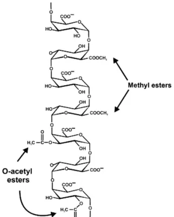
Homogalacturonan (HG) is the simplest and the most abundant pectic polysaccharide and it consists of a linear backbone of 85–320 1,4-linked α-D-GalpA residues (Figure 1.5) (Thibault et al. 1993; Hellín et al. 2005; Round et al. 2010). HG GalpA residues may be methyl-esterified at the C-6 carboxyl or acetylated at the O-2 or O-3 (Figure 1.6) (Ridley et al.
2001). The pattern and degree of methylesterification and acetylation varies from source to source and determines the industrial applicability of pectin (Caffall and Mohnen 2009).
Rhamnogalacturonan I (RGI) has repeating units of [→2)-α-L-Rhap-(1→4)-α-D-GalpA-(1→]n, where n can be larger than 100 (McNeil et al. 1980; Visser and Voragen 1996;
Vincken et al. 2003). Partial acetylation often occurs at the O-2 and/or O-3 positions of the GalpA residues. The rhamnosyl residues can be substituted at O-4 with neutral sugars (McNeil et al. 1980; Lau et al. 1987). Depending on the plant species, the RGI backbone is decorated with galactans (1,4-β-D-galactose units) and/or arabinans (1,5-α-linked L-arabinofuranose units with additional L-arabinofuranose side-chains) (Figure 1.5) (Luis et al. 2018).
Figure 1.6. Homogalacturonan structure and representative sites of methylesterification at the C-6 and O-acetylation at the O-2 or O-3 of the carbohydrate ring.
(Ridley et al. 2001)
The structure of Rhamnogalacturonan II (RGII) is highly complex with 12 different types of glycoside residues, including the rare sugar species 2-O-methyl xylose, 2-O-methyl fucose, aceric acid, lyxo heptulosaric acid (Dha), and 2-keto-3-deoxy-D-manno octulosonic acid (Kdo) (Figure 1.5) (Caffall and Mohnen 2009).
Finally, Xylogalacturonan (XGA) is HG modified by the addition of D-xylose residues at the O-3 of GalpA backbone residues (Figure 1.5) (Cosgrove 2005; Caffall and Mohnen
2009). In addition, in apiogalacturonan (AGA) the galacturonan backbone is decorated with D-apiofuranose at O-2 and/or O-3 (Hart and Kindel 1970; Ovodov et al. 1971; Longland et al. 1989).
1.2.2.4. Lignin
Lignification is the last important process of cell wall development, accounting for increased mechanical strength and protection against pathogen attack (Wei et al. 2009). Lignin has also the function of protecting cellulose and hemicellulose from enzymatic hydrolysis (Schoenherr et al. 2018). Around 25% of all the lignin in wood is found in the middle lamella (Dinwoodie 2000), and the remaining 75% is part of secondary cell walls being deposited following the completion of the cellulosic frame work. Functionally, lignin reinforces plant cell walls through bonding with cellulose. In addition, lignin also enhances the waterproof nature of plant cell walls as a result of its hydrophobicity, while allowing the efficient transport of water in the vascular tissues (Zhou et al. 2016).
Lignin is a highly complex noncrystalline molecule comprised of a large number of phenylpropane monomers (Desch and Dinwoodie 1996). Primarily, three monolignols (monomers) p-coumaryl alcohol, coniferyl alcohol and sinapyl alcohol are present in varying degrees in native lignin. These monolignols are often referred to as phenylpropanoids, which differ in the substitutions at the 3-C and 5-C positions in the aromatic ring (Faix 1991; Wong 2009). Lignin synthesis starts with the random self-replicating radical coupling of phenoxy radical to form an oligomeric product. After polymerization, these polymers are referred as p-hydroxyphenyl (H), guaiacyl (G), and syringyl (S) (from p-coumaryl alcohol, coniferyl alcohol, and sinapyl alcohol, respectively) (Figure 1.7). Monolignols are linked either by C–C bond or C–O–C bond, and more than two third of monolignols are joined by ether linkages (Parthasarathi et al. 2011). The distribution of these monomers varies in different plant species and tissues. Generally, lignin from grasses is a roughly equimolar mixture of G, S and H units, whereas lignin from hardwood contains approximately equal quantities of G and S units but relatively small amounts of the H unit, and lignin from softwood is mainly composed of G units (up to 90%) (Faix 1991).
In the plant cell wall, lignin is built into a network with hemicellulose via ester and ether linkages. These linkages are formed between lignin and residues of hemicellulose, which comprise glucuronic acid or arabinosyl-ferulic acid substituents (Takahashi and Koshijima 1988; Jacquet et al. 1995; Lam et al. 2001).
Figure 1.7. Chemical structure of monolignols and the corresponding units in lignin. (Brown and Chang 2014)
1.2.3. Plant cell wall models
Presently, the organization and interactions established between the repertoire of plant cell wall components remains unclear. Several models have been proposed to represent plant cell wall organization while accounting a dynamic nature appropriate to allow cells to expand and grow. Keegstra and colleagues, in 1973, proposed that the cell wall matrix polymers (xyloglucan, pectin and glycoproteins) are covalently linked to form a giant macromolecular network. In this model, cellulose interacts with the matrix via H-bonding, predominantly to xyloglucan (Figure 1.8-A). More recently, Hayashi (1989) and Fry (1989) proposed that cellulose microfibrils may be tethered together directly via long xyloglucan chains. The cellulose-xyloglucan network is enmeshed in a non-covalently cross-linked pectic network (Figure 1.8-B). This model is currently the most popular one (Carpita and Gibeaut 1993), although, variations to this general organization have been proposed. Talbott and Ray (1992) proposed a model in which each cellulosic microfibril is coated by a series of progressively less-tightly bound polysaccharide layers (Figure 1.8-C) and the linkage between microfibrils is made indirectly via the lateral (non-covalent) associations between the distinctive polysaccharide layers. Ha et al. suggested that xyloglucan molecules are hydrogen bonded to and cross-link cellulose microfibrils and these cellulose-xyloglucan lamellae are separated by strata of pectic polysaccharides (Figure 1.8-D) (Cosgrove 2001).
Figure 1.8. Models of cell wall structure.
A) The covalently cross-linked model; B) The tether model; C) The diffuse layer model, and D) The stratified layer model. (Adapted from Albersheim et al. 2007)
Clearly much research is still required to provide a complete description of the plant cell walls. It is, however, established that primary walls are dynamic structures whose composition and architecture changes during plant growth and development.
1.2.4. Plant cell wall degradation
As referred above, ligno-cellulosic biomass is the most abundant source of organic carbon in the biosphere and the recycling of plant cell wall structures is a general process of considerable biological importance (Hervé et al. 2010). The fixed carbon is recycled by microbial enzymes, generally termed as CAZymes, that convert cell wall polysaccharides to monosaccharides and oligosaccharides. Understanding how microbes deconstruct cell walls is also of growing industrial significance for the biofuel and bioprocessing sectors (Figure 1.9) (Sticklen 2008; Himmel and Bayer 2009; Hervé et al. 2010).
Microbial communities secrete a large repertoire of enzymes that act sequentially and in a synergistic manner to degrade plant cell walls (Wilson 2008; Wei et al. 2009). It is believed that synergy between different types of enzymes produced by bacteria and/or fungi is crucial to effectively degrade plant cell wall components (Wei et al. 2009). The plant cell wall degrading microorganisms have been shown to use two different approaches for cellulose degradation. Aerobic microorganisms secrete large quantities of modular enzymes, usually containing a catalytic module linked to one or more non-catalytic
carbohydrate-binding module (CBM) (Lynd et al. 2002; Hashimoto 2006). In aerobes, enzymes are either secreted into the extracellular milieu or are located on the outer membrane. Although these enzymes do not physically associate, they do display extensive biochemical synergy. In contrast, anaerobic bacteria and fungi organize enzymes in high molecular mass multienzyme complexes, called cellulosomes, which are usually attached to the outer surface of the microorganism (Wilson 2008). It is believed that the anaerobic environment impose a greater selective pressure for the evolution of these highly efficient nanomachines (Bayer et al. 2004). In previous studies, it has been proposed that cellulosomal systems may have greater activity against recalcitrant plant cell walls than non-aggregated cellulase enzymes (Wei et al. 2009).
Figure 1.9. Representation of cell wall deconstruction.
Ligno-cellulosic biomass is the most abundant source of organic carbon in the biosphere. Its degradation is a process of growing interest, particularly in the biofuels and bioprocessing sectors, and
can be performed by microbial enzymes, which are generally referred as Carbohydrate-Active Enzymes.
1.2.4.1. Carbohydrate-Active Enzymes (CAZymes)
Carbohydrate-Active Enzymes (CAZymes) comprise a diversity of enzymes classes including glycoside hydrolases (GHs), polysaccharide lyases (PLs), carbohydrate esterases (CEs) and glycosyl transferases (GTs). While the PLs, CEs, and GHs carry out the breakdown of polysaccharides, the GTs are mainly involved in the formation of the glycosidic
bond and thus in the biosynthesis of carbohydrates (Chakraborty et al. 2017). Recently, the CAZy database incorporated a new category, named Auxiliary Activities (AAs), which covers redox enzymes that act in conjunction with CAZymes. This category groups families of lytic polysaccharide monooxygenases (LPMOs) as well as ligninolytic enzymes (Levasseur et al. 2013).
The enzymes are classified in families according to their amino acid sequence similarities which reflects common structural folds. This classification usually reflects a common evolutionary origin and conservation on the catalytic mechanisms, protein fold and structural features, much better than the Enzyme Commission Number (EC) that is a numerical classification scheme for enzymes, based only on the chemical reactions they catalyse. While the structures of CEs, PLs, and CBMs are dominated by the α/β-hydrolase (Correia et al. 2008), parallel β-helix (Pickersgill et al. 1994), and jelly roll (or β-sandwich) (Czjzek et al. 2001) folds, respectively, there are a large number of different folds within the GHs (Gilbert 2010).
The classification of CAZymes is continuously updated in Carbohydrate-Active EnZymes database (CAZy; www.cazy.org) (Cantarel et al. 2009; Lombard et al. 2014). Currently, the database contains 165 sequence-based families of GHs, 37 families of PLs, 15 families of CEs, 107 families of GTs, 16 families of AAs and finally, 85 families of CBMs (data collected on July 2019).
Figure 1.10. Modular architecture of Carbohydrate-Active enZymes (CAZymes). Examples of modular CAZymes, composed by GHs, PLs, CEs, dockerin (Doc) and CBMs from different CAZy families. a) cellobiohydrolase I from Hypocrea jecorina (SP P00725); b) alginate lyase from Sphingomonas sp. A1 (GB BAB03312.1); c) xylanase from Cellulomonas fimi (GB CAA54145.1);
d) xylanase D/licheninase from Ruminococcus flavefaciens (GB CAB51934.1).