(Annals of the Brazilian Academy of Sciences)
Printed version ISSN 0001-3765 / Online version ISSN 1678-2690
www.scielo.br/aabc
Preparation and evaluation of a biomimetic scaffold
with porosity gradients
in vitro
QIANBIN WANG1,2, QIGUANG WANG2 and CHANGXIU WAN2 1 School of Chemistry and Environment, Beihang University, Beijing 100191, P.R. China 2 College of Polymer Science and Engineering, Sichuan University, Chengdu 610065, P.R. China
Manuscript received on December 21, 2010; accepted for publication on May 30, 2011
ABSTRACT
A novel biodegradable scaffold based on mimetic a natural bone tissue morphology with a porosity gradient structure was prepared in this paper. The result of surface morphology indicated that a graded porous structure was formed in the fabricated scaffold, where the dense layer (0%) was connected with the most porous layer (60%) by a middling porous layer (30%). To evaluate the degradability, graded porous scaffolds compared with homogeneous scaffolds were placed into a Tris-HCl buffer solution (pH=7.4) for 28 days. It was found that both scaffolds presented the same degradation trend, and the graded porous structure did not change the original degradability of the scaffold. Moreover, the compressive strength of the graded porous scaffold was better than that of conventional homogeneous scaffold with the increase of degradation time, and the graded porous
structure can enhanced the mechanical property of the scaffold. These findings suggest that this biodegradable
and porosity-graded scaffold may be a new promising scaffold for loaded bone implant.
Key words: biodegradable, biomimetic, graded porous scaffold, compressive strength.
INTRODUCTION
Bone has a functionally graded structure from the
surface cortical bone towards the inner cancellous
bone (Fig. 1a). Therefore, the biomedical implant
should be designed with a porosity gradient
simulating as much as possible the bimodal structure
of the bone (cortical and cancellous) (Fig. 1a) (Hing
et al. 1999, Castillo et al. 2003). The production of
implant material with graded porosity has been
attempted with hydroxyapatite (HA) by Tampieri
et al., and they found that the high-porosity portion
allows a good and fast bone ingrowth and the
low-porosity side can withstand early physiologic
mechanical stress (Tampieri et al. 2001). Becker
and Pompe reported that functionally graded
material could give the implant a suitable strength
to withstand the physiological loading, and that
the graded porosity structure can optimize the
material’s response to external loading; a similar
feature might prove favorable to an artificial bone
implant (Becker and Bolton 1997, Pompe et al.
2003). Many studies had demonstrated that graded
porous materials provide the advantage to engineer
material with specific structural, morphological
and mechanical properties (Tampieri et al. 2001,
Pompe et al. 2003, Castillo et al. 2003).
Degradability is one of the most concerned
properties in the research field of bone repair
materials because it is crucial for bone induction,
conduction, metabolism and longevity on implants.
According to the research made by Hench LL, the
degradation speed of HA is generally slow, which
is incompatible with bone growth (Hench 1998).
Implants made of alloy or ultra-high molecular
weight polyethylene (UHMWPE) materials
are not degraded either in the body and need to
be removed again (Lieberman and Friedlander
2005). The uncontrollable degradability of
these biomaterials limited them to be used in
bone implants. Calcium polyphosphate (CPP),
a new promising biomaterial for bone tissue
engineering, has been fast developed since its
rediscovery and use in bone regeneration (Pilliar
et al. 2001, Grynpas et al. 2002, El Sayegh et al.
2002, Waldman et al. 2002). CPP has drawn many
researchers’ attention not only for its outstanding
biocompatibility, but also for its controllable
degradability, excellent mechanical property, and
so on (Park et al. 2004, Yang et al. 2004, Ding
et al. 2008, Chen et al. 2008,Wang et al. 2009).
All these studies indicated that CPP is an ideal
bioceramic with excellent osteoinduction and
osteoconduction for bone substitute.
In this paper; we describe a novel calcium
polyphosphate bioceramic scaffold with a graded
pore structure similar to the bimodal structure of
cortical and cancellous bones. Porosity, pore size,
degradability and compressive strength of the
scaffold were investigated
in vitro
. The objective of
this paper is to produce a controllable biodegradable
scaffold with graded and interconnected porosity to
improve its mechanical properties for a possible use
as loaded bone implants.
MATERIALS AND METHODS
The preparation of CPP powder has been described
in details by Qiu (Qiu et al. 2006). Briefly, calcium
phosphate monobasic monohydrate powder was
placed into a 100
cm
3crucible and sintered at 500
◦
C
for 10 h to drive off the crystalline water. Then the
powder was melted at 1200
◦
C for 1 h to further
polymerize. The molten CPP was quenched in
distilled water to obtain CPP amorphous frits. The
frits were milled and screened to yield powders in a
size range of 48-75
μ
m.
The porosity-graded calcium polyphosphate
(PGCPP) scaffold was designed to be composed
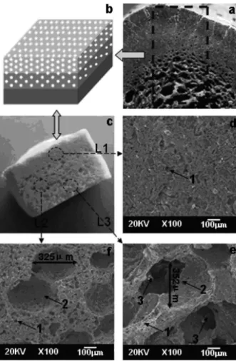
of three functional regions as shown in Figure 1b.
The high porosity region (60%) and denser region
(0%) were connected by a layer with intermediate
Fig. 1 – The porosity-graded calcium polyphosphate scaffold prepared in thispaper. (a) the functionally graded structure of a natural bone, (b) the schematic
representation of an artificial scaffold, (c) the photograph of the scaffold, (d)-(f)
porosity (30%), while the homogeneous calcium
polyphosphate (HCPP) scaffold was designed to
be homogeneous and monolithic. CPP powder and
stearic acid were mixed with paste to fill into a
rectangular stainless-steel mold layer by layer. Then
samples were pressed by a constant compressive
stress between 0.9 to 1 MPa. After drying, these
samples were sintered at 800
◦
C for 3h and then
cooled naturally to room temperature in furnace for
benefiting crystal growth.
The macrostructure of PG-CPP samples was
observed using a digital camera. A JSM-5900LV
scanning electron microscopy (SEM) was used
to evaluate the graded porosity structure of the
samples, and a X’Pert Pro MPD X-ray diffractometer
(Philips, Netherlands) was performed to identify
the crystalline phases of the samples.
The degradation testing was carried out at
37
◦
C for 1, 2, 3, 5, 7, 10, 13, 16, 20, 24 and 28 days
in a Tris-HCl buffer solution (pH=7.4), according
to ISO 10993-14, using triplicate samples. The
quantitative determination of ions released into
the testing solution was preformed using two
different techniques. Calcium ions were measured
by EDTA titration (Dion et al. 2005), whereas
the total orthophosphate ion concentration in
the supernatant was measured by ultraviolet
spectroscopy using the molybdenum blue method
(UV-754, China) (Fiske and Subbarow 1925).
For the compressive strength measurements,
the samples were immersed in a Tris-HCl buffer
solution at 37
◦
C for 0, 14 and 28 days, then taken
out and dried at 80
◦
C for 24 h. The samples were
loaded with a crosshead speed of 1 mm.min−1
using a screw-driven load frame (Instron 4302,
American). The stress and strain responses were
monitored. Five samples in each group were tested
to obtain average values and standard deviations.
The results were presented as arithmetic
means ± standard deviation. The analysis of the
results was carried out using Student’s t-test, with
a significance level of p
<
0.05.
RESULTS AND DISCUSSION
The graded porous structure of calcium polyphosphate
scaffold is presented in Figure 1. Figure 1c shows
the cross-section of the porosity-graded scaffold
with three different porosity layers: L1, L2 and L3.
The surface of the sample becomes coarse and more
porous from L1 to L3. L1 (Fig. 1d) mimicked the
human cortical bone (density), while L3 simulated
the human cancellous bone (porosity). L3 (Fig. 1e)
exhibited a three-dimensionally interconnected pore
structure and connected to the rest of the neighbour
pores by throats (identified by number 3), and the
diameter of the pores was all about 200-400
μ
m
(identified by number 2). When implanted into
the body, the highly interconnected macro-porous
network of the scaffold allows not only cell growth,
but also the flow transport of nutrients and metabolic
waste. L2 (Fig. 1f) was used to connect L1 and L3
and avoided visible layer-layer interface. As shown
in Figure 1c, the contact among various microporous
layers is good. From L3 to L1, the scaffold becomes
denser. These results indicate that a graded porous
structure is formed in the CPP scaffold.
the micro-pores (≤ 50
μ
m) formed from sintering
promote scaffold vascularization (nutrients supplying
for cells, waste products removal).
during the immersion period for both scaffolds were
increased. Two different types of scaffolds presented
the same degradation trend and velocity during the
degradation. The weight loss of H-CPP and PG-CPP
scaffolds were (10.1 ± 0.03)% and (10.4 ± 0.02)%
after 28 days of degradation in a Tris-HCl buffer
solution, respectively. Combined with Table I, the
results indicated that the graded porous structure did
not change the degradability of CPP.
Hench and Polak (2002) postulated the
definition of the third generation of biomaterials,
both bioactive and biodegradable materials (Hench
and Polak 2002). It is known to us that degradability
is a concerned property for implanting biomaterials
because it is crucial for bone induction, conduction,
metabolism and longevity on implants. Nowadays
calcium phosphate-based bioceramics are the
most common biomaterials for bone implant.
However, the degradability of HA and
β
-TCP
was uncontrollable, which limited them to apply
in bone regeneration. Very recently,
β
-CPP, a
novel biomaterial, has drawn many researchers’
attention because of its controllable degradation,
biocompatibility and osteoconduction (Park et al.
2004, Yang et al. 2004, Ding et al. 2008, Chen et
al. 2008,Wang et al. 2009). As shown in Figure
2a, the three char¬acteristic peaks of XRD pattern
indicated that the CPP prepared in this paper was
beta crystalline phase. As a novel biomaterial,
calcium polyphosphate has been wildly developed
in recent years partly due to its controllable
degradability. The crystal structure (Qiu et al.
2005), sintering time and temperature (Wang et al.
2010a, b), polymerization degree (Ding et al. 2008,
Qiu et al. 2005), porosity (Wang et al. 2011), ion
doped (Song et al. 2008) and degradation media
(Wang et al. 2009) can affect the degradation rate
of the CPP bioceramic.
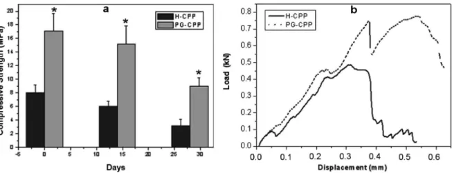
The mean compressive strength for the samples
aged in a Tris-HCl buffer solution (
pH 7.4) for
0, 14 and 28 days is presented in Figure 3a. The
compressive strength of both types of scaffolds
Porosity is defined as the percentage of void
space in a scaffold, and it plays a critical role in
bio-scaffold design
in vitro
and bone formation
in vivo
(Athanasou 1996). It has been demonstrated that
the bio-scaffolds should exhibit pore content and
size. The most porous structures are more likely
to offer good conditions for cell growth and flow
transport of nutrients and metabolic waste because
of their greater specific surface area (Sous et al.
1998). In this article we designed a biomimetic
scaffold with a porosity-graded structure. The most
porosity layer simulated the cancellous bone and
may provide a better condition for cell ingrown,
while the density layer may meet the mechanical
need when implanted in body.
The design of human bone, how it demonstrates
change from a dense stiff external structure of the
cortical bone to the light and porous cancellous
bone on the interior surface, is a perfect example
of porosity-graded material (Fig. 1a). This structure
has been used in scaffolds for bone implant by many
researchers (Becker and Bolton 1997, Castillo et al.
2003, Pompe et al. 2003), and they found that the
porosity-graded structure can optimize the material’s
response to external loading; a similar feature might
prove favorable to an artificial bone implant.
Bio-scaffold requires controllable degradability
and a 3D structure. As shown in Figure 2, the
concentrations of orthophosphate and calcium ions
TABLE I
Porosity of the both types of CPP.
Samples
H-CPP PG-CPP
25 25
32.73 ± 0.71 32.87 ± 0.82 Theoretical porosity
(%)
Fig. 2 – (a) XRD pattern of calcium polyphosphate obtained in this work, and the PO3− ion (b) and Ca2+ ion (c) concentration during the degradation in the Tris-HCl buffer solution (pH 7.4).
Fig. 3 – The mechanical property of H-CPP and PG-CPP: (a) compressive strength of both types of scaffolds before and after degradation. Asterisk (*) means that the compressive strength of PG-CPP has a statistically significant difference compared with the H-CPP, P<0.05, n = 5; (b) the load-displacement curves of both types of scaffolds.
changes significantly (p
<
0.05) within the degradation
test period. For example, as the scaffold immersion in
solution increased from 0 to 14 days, the compressive
strength of H-CPP scaffold decreased 53%, but
the PG-CPP scaffold only decreased 17%. The
compressive strength for H-CPP scaffolds decreased
from 5.16 MPa to 4.48 MPa, while the compressive
strength of PG-CPP scaffolds decreased from
12.57 MPa to 8.98 MPa from 14 to 28 days.
The compressive strength of PG
-
CPP scaffolds
was better than H
-
CPP scaffolds, which was significant
(p
<
0.05) at each time point. It was noted that, after
28 days of degradation, the compressive strength
of PG
-
CPP scaffolds ws still 8.98 MPa, which was
even greater than that of primary H-CPP. The result
indicated that the scaffold with a porosity-graded
structure has a much better mechanical property.
Figure 3b shows the load-displacement
curves of the porosity-graded scaffold and the
conventional homogeneous scaffold, respectively.
It can be seen that the porosity-graded scaffold
exhibits a non-brittle failure, while the conventional
homogeneous scaffold fractures catastrophically.
For the porosity-graded scaffold, after the first
load drop, the load-bearing ability of the testing
bar still retains over 75% of the peak load. Until
totally fractured, the porosity-graded scaffold
gives a prolonged deflection besides the elastic
deformation. This shows that the porosity-graded
scaffold exhibits a different fracture behavior from
that of the conventional homogeneous scaffold.
Hence, it may be inferred that the mechanical
properties of the porosity-graded scaffold can be
substantially improved by a graded porous structure.
Fig. 4 – Schematic diagrams and photographs of scaffolds with a radial gradient porosity: (a) porous scaffold withBiomechanics is one important function of
bone. So, the design of biomaterial scaffold must
have a reasonable mechanical strength. However,
the degradation of the scaffold often leads to a sharp
reduction in its mechanical properties, which limits
the application of these biomaterials. As for H-CPP,
a fast degradation results in a sharp reduction in the
mechanical properties, while for PG-CPP, the density
layer degradation slowly afford the mechanical
property of the scaffold. Hence, as presented in
Figure 3, the mechanical property of PG-CPP was
significantly better than that of H-CPP.
In order to mimic a large segmental bone,
some scaffolds with a radial gradient porosity were
also prepared by using the same method as shown
in Figure 4. The scaffold with a hollow center was
10 mm in diameter, and the diameter of the hollow
center was about 4 mm (Fig. 4a). The porosity of
the outer layer was 15% and could be adjusted
by using different pore-forming agents. Scaffolds
with two and three radial layers were presented in
Figure 4b and 4c. Although the property of these
scaffolds needed further proofs, we believe that
these scaffolds with a radial gradient porosity could
be useful in large segmental bone implants.
CONCLUSION
In this article, a porosity-graded calcium
polyphosphate scaffold was prepared with the
aim of simulating the bone tissue morphology.
Porosity, porosity-graded structure, degradability
and mechanical property of scaffolds were
evaluated
in vitro
for bone implants. The results
indicated that the porosity-graded scaffold showed
a porosity percentage of (32.73 ± 0.71)%, with
a pore size of about 200-400
μ
m in diameter, and
the porosity graded scaffolds are composed of an
inner porous layer and outer compact CPP layers.
In vitro
degradation showed that the degradability
of gradient scaffolds and homogeneous scaffolds
did not have a significant difference due to the same
porosity between them. The compressive strength
test showed that the gradient porosity scaffold was
better than the homogeneous scaffold regarding the
degradation time. These findings may provide an
approach to study and achieve a biomimetic scaffold
with porosity gradients, as well as explore more
biomedical applications.
ACKNOWLEDGMENTS
The authors are grateful to the NSFC for providing
financial support through project 30870614. The
author would also like to thank the professors in the
Centre of Analysis and Testing of Sichuan University
who provided the SEM measurement.
RESUMO
Um novo esqueleto mimetizando a morfologia de tecido ósseo e com uma estrutura de porosidade gradiente
foi preparado e é descrito neste artigo. O resultado
da avaliação da morfologia da superfície indicou que uma estrutura porosa gradiente se formou no esqueleto fabricado no qual uma camada densa (0%) foi conectada com a camada mais porosa (60%) por uma camada porosa média (30%). Para avaliar a degradabilidade, esqueletos de porosidade gradiente e esqueletos homogêneos foram colocados em uma solução tampão
Tris-HCL (pH = 7,4) durante 28 dias. Observou-se que
ambos os esqueletos apresentaram a mesma tendência de degradação e a estrutura de porosidade gradiente não
modificou a degradabilidade original do esqueleto. Além
disso, a força compressiva do esqueleto de porosidade gradiente foi melhor do que aquela do esqueleto homogêneo convencional, com aumento do tempo de degradação, e que a estrutura porosa em gradiente pode acentuar a propriedade mecânica do esqueleto. Estas observações sugerem que este esqueleto biodegradável de porosidade gradiente pode ser promissor para implantes ósseos.
REFERENCES
ATHANASOU N. 1996. Current Concepts Review – Cellular
Biology of Bone – Resorbing Cells. J Bone Joint Surg 78: 1096.
BECKER B AND BOLTON J. 1997. Corrosion behaviour and mechanical properties of functionally gradient materials developed for possible hard-tissue applications. J Mater Sci-Mater M 8: 793–797.
CASTILLO M, MOORE J, SCHOWENGERDT F, AYERS R, ZHANG
X, UMAKOSHI M, YI H AND GUIGNE J. 2003. Effects of gravity on combustion synthesis of functionally graded biomaterials. Adv Space Res 32: 265–270.
CHEN F, WANG K AND LIU C. 2008. Crystalline structure and its effects on the degradation of linear calcium polyphosphate bone substitute. Appl Surf Sci 255: 270–272. DING Y, CHEN Y, QIN Y, SHI G, YU X AND WAN C. 2008. Effect of of polymerization degree of calcium polyphosphate on its microstructure and in vitro degradation performance. J Mater Sci-Mater M 19: 1291–1295.
DION A, LANGMAN M, HALL G AND FILIAGGI M. 2005. Vancomycin release behaviour from amorphous calcium polyphosphate matrices intended for osteomyelitis treatment. Biomaterials 26: 7276 –7285.
EL SAYEGH T, PILLIAR R AND MCCULLOCH C. 2002. Attachment,
spreading, and matrix formation by human gingival
fibroblasts on porous-structured titanium alloy and
calcium polyphosphate substrates. J Biomed Mater Res Part A 61: 482–492.
FISKE C AND SUBBAROW Y. 1925. The colorimetric determination of phosphorus. J Biol Chem 66: 375.
GRYNPAS M, PILLIAR R, KANDEL R, RENLUND R, FILIAGGI M
AND DUMITRIU M. 2002. Porous calcium polyphosphate scaffolds for bone substitute applications in vivo
studies. Biomaterials 23: 2063–2070.
HENCH L AND POLAK J. 2002. Third-generation biomedical
materials. Science’s STKE 295: 1014.
HENCH LL. 1998. Bioceramics. J Am Ceram Soc 81: 1705–1728.
HING K, BEST S AND BONFIELD W. 1999. Characterization of porous hydroxyapatite. J Mater Sci-Mater M 10: 135–145.
LIEBERMAN JR AND FRIEDLANDER GE. 2005. Humana Press, Totowa, New Jersey, Ch. 2 and Ch. 8.
PARK E, LEE Y, CHOI J, OH S, SHIN H, KIM K, KIM SYAND
KIM S. 2004. Cellular biocompatibility and stimulatory effects of calcium metaphosphate on osteoblastic differentiation of human bone marrow-derived stromal cells. Biomaterials 25: 3403–3411.
PILLIAR R, FILIAGGI M, WELLS J, GRYNPAS M AND KANDEL
R. 2001. Porous calcium polyphosphate scaffolds for bone substitute applications – in vitro characterization. Biomaterials 22: 963–972.
POMPE W, WORCH H, EPPLE M, FRIESS W, GELINSKY M,
GREIL P, HEMPEL U, SCHARNWEBER D AND SCHULTE
K. 2003. Functionally graded materials for biomedical applications* 1. Mater Sci Eng A 362: 40–60.
QIU K, WAN CX, CHEN X, ZHANG Q AND SU HF. 2005. In vitro
degradation studies of calcium polyphosphate ceramics prepared by controlled degree of polymerization and crystallization. Asbm6: Advanced Biomaterials Vi 288-289: 553–556.
QIU K, ZHAO X, WAN C, ZHAO C AND CHENY. 2006. Effect
of strontium ions on the growth of ROS17/2.8 cells on
porous calcium polyphosphate scaffolds. Biomaterials 27: 1277–1286.
SONG W, TIAN M, CHEN F, TIANY, WAN C ANDYU X. 2008. The study on the degradation and mineralization mechanism of ion-doped calcium polyphosphate in vitro. J Biomed Mater Res Part B: Applied Biomaterials 89: 430–438. SOUS M, BAREILLE R, ROUAIS F, CLEMENT D, AMEDEE J,
DUPUY B AND BAQUEY C. 1998. Cellular biocompatibility
and resistance to compression of macroporous [beta]-tricalcium phosphate ceramics. Biomaterials 19: 2147–2153.
TAMPIERI A, CELOTTI G, SPRIO S, DELCOGLIANO A AND
FRANZESE S. 2001. Porosity-graded hydroxyapatite ceramics to replace natural bone. Biomaterials 22: 1365–1370.
WALDMAN S, GRYNPAS M, PILLIAR R AND KANDEL R. 2002. Characterization of cartilagenous tissue formed on calcium polyphosphate substrates in vitro. J Biomed Mater Res (USA) 62: 323–330.
WANG Q, WANG J, ZHANG X, YU X AND WAN C. 2009. Degradation kinetics of calcium polyphosphate bioceramic: an experimental and theoretical study. Mater Res 12: 495–501.
WANG QB, WANG QG AND WAN CX. 2010a. The Effect of sintering time on the microstructure and properties of inorganic polyphosphate bioceramics. Science of Sintering 42(3): 337–343.
WANG QB, WANG QG AND WAN CX. 2011. The effect of porosity on the structure and properties of calcium polyphosphate bioceramics. Ceram-Silikaty 55(1): 43–48. WANG QB, WANG QG, ZHANG XH, YU XX AND WAN
CX. 2010B. The Effect of Sintering Temperature on the Structure and Degradability of Strontium-Doped Calcium Polyphosphate Bioceramics. Ceram-Silikaty 54: 97–102.
YANG SM, KIM SY, LEE SJ, LEEYK, LEEYM, KU Y, CHUNG
CP, HAN SB AND RHYU IC. 2004. Tissue Response of Calcium Polyphosphate in Beagle Dog. Part II:
1
2 Month Result. Key Eng Mat, p. 245–248.YOSHIKAWA H AND MYOUI A. 2005. Bone tissue engineering with