REVIEW
Carbohydrate/Glycan-Binding Specificity of Legume Lectins
in Respect to Their Proposed Biological Functions
Márcio Viana Ramos
1*, Thalles Barbosa Grangeiro
1, Benildo Sousa Cavada
1, Iain
Shepherd
2, Roberval Oliveira de Melo Lopes
1and Alexandre Holanda Sampaio
11
Laboratório de Moléculas Biologicamente Ativas (BioMol-Lab). Universidade Federal do Ceará, Campus do Pici, Caixa Postal 6033-CEP 60451-970. Fortaleza-Ce, Brazi; 2Department of Biochemistry, Irvine Building, University of St. Andrews, St. Andrews, Fife, Scotland
ABSTRACT
The lectins, proteins which specifically recognize carbohydrate moieties, have been extensively studied in many biochemical and structural aspects in order to establish the molecular basis of this non-catalytic event. On the other hand, their clinical and agricultural potentials have been growing fast. Although lectins, mainly those from legume plants, had been investigated for biological properties, studies about the physiological functions of lectins are scarce in literature. Therefore, despite the accumulated data on lectins (as proteins), the role played by these signalizing molecules is poorly discussed. In the light of our accumulated results on legume lectins, specially those obtained from plants belonging to the Diocleinae sub-tribe and available data in literature, we discuss here the main hypothesis of their functions according to their carbohydrate/glycan-binding specificity.
Key Words: Binding-site; biological functions; fine sugar-specificity; legume lectins
*
Author for correspondence
INTRODUCTION
Lectins are generically defined as proteins which interact non-covalently with carbohydrate moieties, displaying high affinity and specificity for their ligands. At the present, a large number of lectins have been isolated and their biochemical characteristics established. Recently, an increasing number of three-dimensional structures of plant lectins have been solved from experimental analysis by x-ray diffraction (Bourne et al., 1990a; Hamelryck et al., 1999; Chandra et al., 1999) and others are in progress (Calvete et al., 1999). Furthermore, the co-crystallization of some of these proteins with specific ligands has also given information towards the understanding of the interaction between lectins with simple and complex carbohydrates, revealing which amino acids residues are involved in the interaction between molecular partners. Therefore more properly establishing the monosaccharide binding-sites of these proteins (Bourne et al., 1990b; Shaanan et
al., 1991; Delbaere et al., 1993; Naismith et al., 1996).
According to their specificity, lectins have been classified as glucose/mannose, N-acetylglucosamine, galactose/N-acetylgalactosamine and fucose-binding specific. Recently, a new group of highly mannose specific lectins was described. The mannose specific lectins constitute a growing group of proteins from Monocotyledoneae plants, whose interaction is specially towards mannose, a fact not observed in glucose/mannose lectins (Chandra et al., 1999). In addition, lectins have been demonstrated to interact with syalic acid and some derivatives, although glucose and galactose specific lectins also show ability to interact with this acid (Konami et al., 1994).
the possible involvement of some lectins as molecules of recognition in leguminous plants, displayed in the complex establishment of symbiosis with bacteria rhizobia (Diaz et al., 1989). The second describes lectins as defense proteins protecting plants against infections or physical attack caused by microrganisms and predators (Cavada et al., 1993). Although these hypothesis have received great adhesion by lectinologists, many questions remain to be solved. How could legume lectins play similar roles when they show broad monosaccharide-binding specificities and thus, how can specificity and functions be correlated? On the other hand, some non legume lectins which are theoretically involved in plant defense usually have similar specificities and related structural domains which provide strong relationships between carbohydrate specificity and function (Cavada et al., 1993). It had also been suggested that lectins are proteins able to recognize foreign molecules or organisms (Ayouba et al., 1992). Although no conclusive data has been obtained in this way, a consensus is accepted independently of the specificity expressed by the lectin that their functions would be in accordance to their ability to interact with specific ligands.
It is now clear that the similarities observed in the tertiary structures of legume lectins are larger than the homology in their amino acid sequences. In fact, legume lectins seem to form an homo- or polyfunctional family of proteins sharing a common three-dimensional folding of their functional subunit, although their quaternary arrangement to form dimmer and tetramer is quit different. Thus, new evidence suggests that conserving the general features of their 3-D structures, legume lectins may be mainly evolved and diverged in the combining-site region to exhibit different specificities, therefore, play specific role in plant metabolism.
In the light of the recent discussion of the lectin definition and new insight achieved by structural and functional studies on the legume lectin monosaccharide/glycan binding-site, we discuss some aspects of lectin functions comparing the amino acid composition of their monosaccharide-binding site and the recent concept of extended binding site with the main hypothesis pointing to their functions.
Lectins in the life cycle of leguminous plant Although relevant information about plant lectins is available, the function of lectins in plants remains without a conclusive statement. Some functions were earlier suggested and investigated. The role of storage or transport proteins was firstly proposed as many legume plants investigated were shown to possess high levels of lectins located in the protein bodies in the seeds, on one hand, and these proteins interacted with sugars, on the other (Etzler, 1986). However no further evidence supported this hypothesis. Einhoff and colleagues proposed that lectins should be involved in the packaging of storage proteins, since various lectins from Leguminosae tested, were able to interact with storage proteins belonging to their own plants (Einhoff et al., 1986). Recent results from Wenzel and co-workers (Wenzel & Rudiger, 1995) showed that, pea lectin does interact with vicilin and legumin fraction from the protein bodies of pea seeds, suggesting that the protein body membranes might be a further candidate for lectin interaction (Gers-Barlag et al., 1993). Similar observations were previously obtained from soybean lectins and their storage proteins (Rudiger & Schecher, 1993). Since, up to date, no significant data is available about glycosilation of storage proteins in plants any specific ligand could be attributed to these lectins in seeds. Although it was not emphasized by these authors, a possible role in the maintenance of the protein body structure could be suggested. The search to determine the exactly moment of appearance of the lectin in protein bodies during the development of seeds should be appreciated in the attempt to establish if a parallelism exists between storage protein deposition and lectin appearance in the establishment of protein bodies.
seedlings (Cavada et al.,1990). During maturation of Canavalia brasiliensis seeds, the lectin was detected only after high molecular weight protein appearance (Moreira et al., 1993). A lectin precursor that bound to polydextran was detected previously, displaying extremely weak hemagglutinating activity. If the lectin is synthesized concomitantly to the storage proteins, perhaps it is accumulated as a pre-protein, not completely active and could become active in a specific stage of seed development. It should be taken in account that many of these lectins investigated are from a closely related group of plants and one could reasonable consider these results are of low significance. However, recently a lectin from a distantly related taxonomic group of Leguminosae belonging from the Erythrinae tribe, in which all lectins investigated are galactose-specific, showed similar behavior when seeds were challenged to germinate in absence of light (Oliveira et al., 1998).
Considering all these results, legume lectins may be involved in the protein body structure, but it does not implicate that they are storage proteins. Indeed, results of these two different approaches are in agreement that lectins are unlikely to be storage proteins although a correlation between an endogenous receptor, specificity and functions could not be evaluated for them. If legume lectins may support protein body membranes, the lectins from these two groups which exhibit differences in their monosaccharide-specificity should share some other molecular ability to act in a similar form. In fact, pioneer studies on the complex binding-specificity of lectins, elegantly showed that lectins possessing differences in monosaccharide-specificity could interact with the same complex glycan which will be discussed later (Debray et al., 1981).
Lectins as a recognition factor in leguminous plant
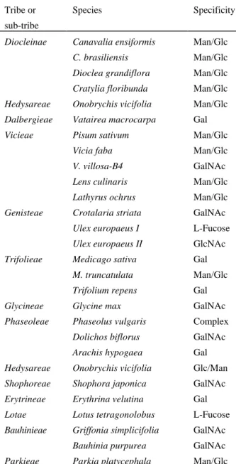
Leguminoseae is the main source of all lectins isolated and characterized so far (Van Damme et al., 1998). Also, within this family are the greater number of the lectins for which three-dimensional structures have been described (Mourey et al., 1998) (Table I). Although these proteins belong to the same taxon and present many common biochemical characteristics, they exhibit different monosaccharide/glycan binding specificities and
quaternary arrangement (Debray et al., 1981). A proposed role for Leguminoseae lectins as molecules of recognition in the initial events of symbiosis between the Leguminoseae-Rhizobium interaction arose from the observation of the strong specificity expressed between plant and microrganism to establish functional symbiotic nodules and the detection of lectins in root hair of various leguminous plants (Kijne et al.,1992). Rhijn et al. (1998) using trangenic Lotus corniculatus plants expressing soybean lectin gene observed the involvement of legume lectins in the symbiont process. This hypothesis became more attractive after the discovery that Rhizobium bacteria produced an extra-cellular factor known to be involved in the initial events of symbiosis, which always contained a carbohydrate moiety in its structure and could eventually interact with root hair lectin (Lerouge et al.,1990). Indeed, these lipo-oligosaccharide signalizing molecules, named NOD factors, synthesized as a result of the expression of rhizobial nod genes, a key factor hosts in the initial events of symbiosis (Rhijn et al., 1998). Therefore, the ability of a NOD factor to induce root-hair curling and infection, leading to nodule formation, have been demonstrated (Lerouge et al., 1990).
Table 1. Lectins from Leguminoseae family distributed in various tribes showing specificities for different monosaccharides.
Tribe or
sub-tribe
Species Specificity
Diocleinae Canavalia ensiformis Man/Glc
C. brasiliensis Man/Glc
Dioclea grandiflora Man/Glc
Cratylia floribunda Man/Glc
Hedysareae Onobrychis vicifolia Man/Glc
Dalbergieae Vatairea macrocarpa Gal
Vicieae Pisum sativum Man/Glc
Vicia faba Man/Glc
V. villosa-B4 GalNAc
Lens culinaris Man/Glc
Lathyrus ochrus Man/Glc
Genisteae Crotalaria striata GalNAc
Ulex europaeus I L-Fucose
Ulex europaeus II GlcNAc
Trifolieae Medicago sativa Gal
M. truncatulata Man/Glc
Trifolium repens Gal
Glycineae Glycine max GalNAc
Phaseoleae Phaseolus vulgaris Complex
Dolichos biflorus GalNAc
Arachis hypogaea Gal
Hedysareae Onobrychis vicifolia Glc/Man
Shophoreae Shophora japonica GalNAc
Erytrineae Erythrina velutina Gal
Lotae Lotus tetragonolobus L-Fucose
Bauhinieae Griffonia simplicifolia GalNAc
Bauhinia purpurea GalNAc
Parkieae Parkia platycephala Man/Glc
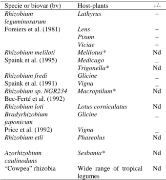
The NOD factors are lipo-oligosaccharide signal molecules structurally organized as tri-, tetra- or pentasaccharides of N-acetylglucosamine, generally sulfated at C-6 position in the reducible termini and modified by the presence of an acylated N-acetylglucosamine termini (Lerouge et al., 1990). Recently, different NOD factors of other symbiotic bacteria have been isolated and characterized. These compounds show similar structural features and only few changes in general structure are observed (Spaink et al., 1995). Some symbiotic bacteria, which have had NOD factors isolated and their structure determined, are listed in Table 2, associated to the specific host-plant.
Although different pieces of evidence pointed some legume lectins as biological agents to interact with NOD factors, how could the role of its specific recognition be attributed to these proteins; whether Leguminoseae family possesses a large number of lectins exhibiting a broad sugar specificity on one hand, and NOD factor studied until now, exhibit only a N-acetylglucosamine backbone in its structure? As non interaction apparently occurs between research groups investigating lectins and NOD factor, this question remains obscure.
Few lectins used to test this hypothesis were from the group glucose/mannose specific, which also interact with N-acetylglucosamine. Indeed, although some legume lectins such as Ulex
europaeus lectin-I, Arachis hypogaea and
Erythrina corallodendron are well studied, their physiological functions have not been frequently questioned and there are no comments present to relate these lectins to NOD factor. These lectins have fucose, galactose and galactose specificity, respectively. On the other hand, the symbiosis process was extensively studied in Medicago sativa and Glycine max, which expressed galactose specific lectins. They are noduled by R. melioti and R. fredii, respectively, which produce NOD factors without galactose or N-acetyl galactosamine units (Kijne et al., 1992). Besides this, no effort has been made to provide the lectin interaction with NOD factor, expressed by their respective symbiotic bacteria.
In Bradyrhizobium japonicum - Glycine max
NOD-factor receptor in the host plant. Although the involvement of legume lectin in symbioses remains in evidence, this possible biological activity still requires more detailed investigations as at the moment the sugar binding specificity of lectins seems to be not related to this specific function.
Table 2. Specificity of nodulation expressed by host-plants and symbiotic bacteria of known NOD factor structure.
Specie or biovar (bv) Host-plants
+/-Rhizobium leguminosarum
Lathyrus + Foreiers et al. (1981) Lens +
Pisum +
Viciae +
Rhizobium meliloti Melilotus* Nd Spaink et al. (1995) Medicago _
Trigonella* Nd
Rhizobium fredi Glicine _ Spaink et al. (1991) Vigna _
Rhizobium sp. NGR234 Macroptilum* Nd Bec-Ferté et al. (1992)
Rhizobium loti Lotus corniculatus Nd
Bradyrhizobium japonicum
Glicine _ Price et al. (1992) Vigna _
Rhizobium etli Phaseolus Nd
Azorhizobium caulinodans
Sesbania* Nd “Cowpea” rhizobia Wide range of tropical
legumes
Nd
*- Plants which lectins have not been isolated, nd - not determined (+/-) the monosaccharide inhibitor of the lectin is present or absent in the structure of NOD factor
Assuming that the ability to specifically recognize a receptor moiety is related to function, only limited replacement of amino acids in the binding-site of legume lectins was permitted during evolution to conserve the same specificity. A good example is ConA and DGL lectins, compared to
Vicieae lectins. Phenylalanine and glycine
residues, completely conserved within Vicieae lectins, correspond to arginine and tyrosine, respectively in Diocleinae lectins although the same specificity was conserved. On the other hand, regarding LOL and EcorL, the changing of three amino acids in the binding-site led to a new specificity, although the two lectins showed very similar three-dimensional folding and the spatial arrangement of the amino acids in the binding-site were almost identical. In the case of LOL and PSL, which exhibited the same amino acids in the binding-site and have the same specificity, the
monosaccharide, mannose, was directly bound by a network of hydrogen bonds in different forms (Eijsden et al., 1994). Thus, one may presume that apparently major changes of features in amino acid composition of the carbohydrate binding-site may lead to different specificity and consequently to differential recognition too. Considering all these facts, what could be the role played by fucose or Gal/GalNAc specific legume lectins in their native plants and what is the actual significance of a particular lectin within its plant? Whether legume lectins play a role of recognition in the initial phase of symbiosis process, perhaps different specificity represent a strategy to interact with specific symbionts in a specific way, mediated or not by NOD factor, thus assuring a highly specific recognition. Presently, this hypothesis constitutes a challenge to be demonstrated. The evidence presented by Ho and Kijne (Kijne et al., 1992) in soybean-Bradyrhizobium system and pea-Rhizobium leguminosarum system pointed out this and an increasing number of results showed the involvement of lectins in the symbiosis process, although their role had been almost undetermined. Spaink et al. (1995) have recently suggested that the recognition of NOD factors by the host legume could be related to the different hydrophobicities exhibited by fatty acids present in NOD factor structure, thus opening the question if the chitin unit from NOD factors plays same role in the recognition process. Presently there are only a few available structures of NOD factors produced by symbiotic bacteria, although a new NOD factor showing an additional O-metyl-fucoside moiety in its structure was reported (Sanjuan et al., 1992; Cohn et al., 1998). Many others could be isolated and their structures established, especially in bacteria which interact with legume plants which do not express glucose/mannose lectins and the actual interaction between lectin and NOD factor (if possible) should be evaluated.
determined by a loop length and conformation corresponding to Thr-97 to Glu-102 in ConA (Thr216 to Glu 224 in EcorL and Thr-28α to Ala-33α in the lectins from pea, lentil and Lathyrus) (Sharma & Surolia, 1997; Loris et al., 1998). The results suggested that both length and conformation of this loop defined the monosaccharide-specificity of legume lectins. In this view, differences in monosaccharide specificity became more interesting and attractive. Studies have been shown that affinity of these lectins for oligosacchaides was still stronger Brewer & Gupta, 1994). Debray (1981) interpreted these data structurally. The complexity of lectin-glycan interaction have been attributed to the existence of an extended-binding site surrounding the monosaccharide site, including few additional residues on the lectin that formed addition hydrogen bounds directly or via water with the sugar units in the glycan structure (Imberty & Perez, 1994; Bourne et al., 1994; Dessan et al., 1995). Although the molecular basis of this phenomena has been investigated, its biological relevance has not been considered.
Legume lectins as defense proteins
The genetic program of plant defence against a broad wild spread predator has been investigated by many different groups. Not surprisingly, almost of these genetic programs seem to be built up of a highly integrated system involving closely related proteins possessing very precise function whose sometimes work on a cooperative or complementary way. It is now understood that some plants synthesizes defense proteins only under adverse conditions as salt or water stress and fungi or insect attack.
Within legume lectins already studied, ConA and other ConA-like lectins were shown to protect artificial seeds against the beetle Callosobruchus maculatus (Gatehouse et al., 1995). However, it is not certain that the intrinsic toxic activity which leads insect to die or to a delay in its development is due the carbohydrate-binding activity of the lectins. The lectins from seeds of Canavalia brasiliensis, Dioclea grandiflora, D. rostrata and
Cratylia floribunda which have identical
monosaccharide specificity (Glc/Man) exhibit differences when tested to C. maculatus (Gatehouse et al., 1995). The former being totally ineffective and the two latter being toxic. Although at present any structural basis could
explain the ineffectiveness of two of these lectins, the toxic effects of the other have been credited to the ability of the lectins to binding to membrane glycoproteins in mouth and/or epithelial cells in the mid-gut inducing changes in performance of feed. Recently, Zhu-Salzman and colleagues (1998) reported the carbohydrate binding and resistance to proteolysis control insecticidal activity of Griffonia simplicifolia lectin II in order to investigate its toxic activity. In the case of ConA which does not appear to be toxic to mammals, at least at low levels of dietary inclusion, its gene could be used in an transgenic plant project against C. maculatus. However, the first lectin reported to possess insecticidal activity was PHA. It was attributed the inability of the cowpea bruchid beetle, C.maculatus, to attack the seeds of Phaseolus vulgaris due the presence of PHA, the haemagglutinating lectin present in the seeds. Nevertheless, many years latter, the toxic effects were more attributed to the isecticidal protein arcelin and to α-amylase inhibitor, two lectin-like proteins occurring in Phaseolus vulgaris seeds exhibiting sequence homology to PHA (Mirkov et al., 1994; Schroeder et al., 1995; Fabre et al., 1998). The PHA protein family seems to be a classical example of a integrated defense system which each protein has a precise role to protect seeds, although undefined yet. Structural studies of arcelin, PHA and α-amylase inhibitor have proved that these proteins share very similar three-dimensional folding but differing in quaternary structure and in their active binding site whose are ready distinct (Bompard-Gilles et al., 1996; Hamelryck et al., 1999; Mourey et al., 1998). Once, the activity and function do seem to be defined by the combine site-region. Apart of present status of the investigation about the lectins as tools in crop biotechnology, a condition sine qua non for their use is the test to engineered plant for resistance in field. In this way, some non legume plants have already been used (Rao et al., 1998).
A set of amino acid to define the monosaccharide specificity
present some interesting features. Although the well conserved triad Asp-Asn-Gly support the saccharide in the site, the other residues have been not strongly conserved during evolution. The replacement of amino acid residues is unlikely to have generated different specificities among the lectins as the contribution of these semi-conserved and non conserved residues is made by hydrogen bonds established with the sugars via the CO and NH groups or architecture well conserved (Loris et al., 1998). Nevertheless, minor differences may change the affinity of lectins by structurally related ligands. Lectins from the same group (Gal/GalNAc) can interact
more strongly with a sugar derivative than with the simple monosaccharide as demonstrated by, EcorL and DBL both specific for galactose but DBL binds more tightly to N-acetyl-galactosamine. This phenomena is also true to the lectins from
Diocleinae which are similar over 90% by
pairwise alignment (Ramos et al., 1996). Although the lectins are gifted of at least a monosacharide-binding site, it is reasonable to suppose that their biological ligand are larger than a single monosaccharide. Indeed, lectins not only bind to oligosacchaides, but this interaction is tighter than that to their monosacchaide inhibitor (Konami et al., 1994; Do & Lee, 1998; Dam et al., 1998). Although the ability to interact with oligosacchaide had been proved early, the first complex of a lectin with a complex glycan was established later by crystallographic studies (Bourne et al., 1990b, 1994; Naismith et al., 1996).
These crystallography works clearly identified the involvement of additional amino acid residues in the vicinity of the monosaccharide-binding site, with the glycans complexes. The hydrogen bounds occurring between the glycans and other amino
acid residues (also mediated by water) are responsible for the correct anchorage of the larger receptor and by the magnitude of the affinity constant determined to be so far strong than to that for monosaccharides.
The new set of amino acid composing these sites in legume lectins has been named extended binding-site and has been proved to occur in all lectins which structure has been investigated in complex with oligosaccharides (Imberty & Pérez, 1994; Naismith & Field, 1996).
Although the completeness of the complexes between legume lectins and complex glycans is of an extraordinary elegance, of most intrinsic and biological relevance seems to be the ability of lectins possessing different monosaccharide specificity to binding the same receptor, through attachment in different epitope present in the same glycan structure (for review see Debray et al., 1981). This very interesting biological event puts under suspect the actual role of the monosacchaide-binding site. According to these results, we could speculate that despite the monosaccharide specificity, legume lectins (and probably non legume lectins) could display similar roles as they can interact with complex glycans broadly distributed in naure. In this context, the monosacchaide-binding site would serve essentially to attach the ligand, being the specificity of the interaction defined by different epitope in the structure. Although the available results allow speculations on basis of the molecular structure of legume lectins and their specificity, identification of endogenous receptors remains obscure and up to date, our attempt to identify endogenous receptor on free-lectin fractions from seed lectins of some members of Diocleinae lectins has failed.
Table 3. Comparison of the carbohydrate specificity and amino acid composition of the monosaccharide-binding site of legume lectins of known structure.
Specificity
Strongly conserved Amino acids
Semi-conserved Amino acids
Not conserved Amino acids
ConAa Man/Glc N-14 D-208 R-288 Y-12 L-99 Y-100
DGLb Man/Glc N-14 D-208 R-228 Y-12 L-99 Y-100
CFLb Man/Glc N-14 D-208 R-228 Y-12 L-99 Y-100
OVLc Man/Glc N-131 D-89 G-107 F-129 D-214 L-215
AHLb Gal N-127 D-83 G-104 Y-125 L-212 G-213
VFLa Man/Glc N-126 D-82 G-100 F-124 A-211 E-212
PSLa Man/Glc N-125 D-81 G-99 F-123 A-210 E-211
UEL-Ic L-Fucose N-135 D-87 G-105 F-127 T-219 Y-220 UEL-IIc GlucNAc N-139 D-88 S-106 F-130 V-222 G-223
LCLc Man/Glc N-127 D-82 G-99 F-123 A-189 E-190
LOLa Man/Glc N-125 D-81 G-99 F-123 A-210 E-211
GSL-Iva GalNAc N-135 D-89 G-107 W-133 V-221 G-222
ECLa Gal N-133 D-89 G-107 F-131 A-218 Q-219
VMLd Gal N-129 D-87 G-105 F-127 L-213 S-214
DBLa GalNAc N-129 D-85 G-103 L-127 L-214 S-215
GMLa GalNAc N-130 D-88 G-106 F-128 G-218 E-219
a - determined by three-dimensional structure, b and c predicted by alignment to ConA and GML, respectively. d-predicted by alignment to ECL. ConA (Naismith et al., 1996), DGL (Cavada et al. 1993), CFL (Cavada et al. 1999), OVL (Sharon & Lis, 1990), AHL (Sharma & Surolia, 1997), VFL (Reeke et al., 1986), PSL (Bourne et al., 1990b), UEL-I, II (Sharma & Surolia, 1997), LCL (Sharon & Lis, 1990), LOL (Bourne et al., 1990b), GSL-IV (Delbaere et al., 1993), ECL (Shaanan et al., 1991), DBL (Imberty et al., 1994), GML (Dessan et al., 1995). VML (Calvete et al. 1998; Ramos et al. 1999) Lectins are designed by initial letter of the plant names.
CONCLUSIONS
Legume plants, are the most rich source of lectins and the continuous investigation of new lectins in members of this group may give new insight on the lectin function in nature. Although investigations under the physiological role of lectins in plant have received few attention, results from biochemical and structural features and mainly the understanding of the molecular basis of the specificity of new lectins may help the discussion. The hypothesis discussed here focused only the interaction of lectins with carbohydrate/glycan. It should be take in account that some legume lectins have been shown to possess a hydrofobic cavity forming a combining site other than that to carbohydrate/glycan, which has a high affinity to adenine and some adenine-related compounds, including plant hormones substances (Loris et al., 1998). In fact the legume
Dolichos biflorus lectin has recently been
successfully crystallized and its complex with adenine solved (Hamelryck et al., 1999). The capability to bind adenine was earlier showed (Gegg & Etzler, 1994; Puri & Surolia, 1994). It should be expected that a new field to discuss the
polyfunctional features of these proteins will be initiated fast, adding new data to the present discussion.
Acknowledgements. This work was supported by CNPq, BNB, FINEP, PADCT, FUNCAP, CAPES/COFECUB and BioTools Ecological Foundation.
RESUMO
moléculas de sinalização é pobremente discutido. Valendo-se de nossos estudos sobre lectinas de leguminosas, principalmente da sub-tribo Diocleinae, e outros dados presentes na literatura, discutimos aqui, as principais hipóteses de suas funções com base na especificidade por carboidratos e glicanos complexos.
REFERENCES
Ayouba, A.; Martin, D. & Rougé, P. (1992), Recognition of muramic acid and N-acetylmuramic acid by Leguminosae Lectins: possible role in plant-bacteria interactions. FEMS, 92:41-46
Bompard-Gilles, C. Rouseau, P.; Rougé, P. & Payan, F. (1996), Substrate mimicry in the active center of a mammalian amylase: structural analysis of an enzyme-inhibitor complex. Structure, 4:1441-1452 Bourne, Y.; Abergel, C.; Cambillau, C.; Frey, M.;
Rougé, P. & Fontecilla-Camps, J-C. (1990a), X-ray crystal structure determination and refinement at 1.9 Ao resolution of isolectin I from the seeds of Lathyrus ochrus. J. Mol. Biol., 214: 571-584
Bourne, Y.; Rougé, P. & Cambillau, C. (1990b), X-ray structure of a (α-Man (1-3)β-Man(1-4)GlcNac)-lectin complex at 2.1Ao resolution. J. Biol. Chem.265 (30): 18161-18165
Bourne, Y.; Mazurier, J.; Legrand, D.; Rougé, P.; Montreuil, J.; Spik, G. & Cambillau, C. (1994), Structures of a legume lectin complexed with the human lactotranferrin N2 fragment, and with an isolated biantennary glycopeptide: role of the fucose moiety. Structure, 2:209-219
Brewer, C.F. & Gupta, D. (1994), Mutivalent carbohydrate-protein interactions. A new dimension of binding specificity. Current Topics in Peptide & Prot. Res., 1:177-191
Calvete, J.J., Santos, C.F., Mann, K., Grangeiro, T.B., Himtz, M., Urbanke, C. & Cavada, B.S. (1998), Amino acid sequence, glycan structure, and proteolytic processing of the lectin of Vatairea macrocarpa seeds. FEBS Letters, 425(2): 286-292.
Calvete, J.J., Thole, H.H., Raida, M., Urbanke, C., Romero, A., Grangeiro, T.B., Ramos, M.V., Rocha, I.M.A., Guimarães, F.N. & Cavada, B.S. (1999), .Molecular characterization and crystallization of
Diocleinae lectins. Biochem. Biophys. Acta,
1430:367-375
Cavada, B.S.; Vieira, C.C.; Silva, L.M.A.; Oliveira, J.T.A. & Moreira, R.A. (1990), Comportamento da lectina de sementes de Canavalia brasiliensis Mart.
durante a germinação em presença de luz. Acta Bot. Bras. 4 (2):13-20
Cavada, S.B.; Moreira, R.A.; Oliveira, J.T.A. & Granjeiro, T.B. (1993). Primary structures and functions of plan lectins. R. Bras. Fisiol. Veg. 5 (2): 193-201
Cavada, B.S.; Grangeiro, T.B.; Ramos, M.V.; Crisostomo, C.V.; Silva, L.M.A.; Moreira, R.A. and Oliveira, J.T.A. (1994), Lectin from Dioclea guianensis var. lasiophylla Duke seeds mobilization during germination and seedling growth in the dark.
R. Bras. Fisiol. Veg.6 (1): 21-25
Cavada, B.S., Nogueira, N.A.P., Farias, C.M.S.A., Grangeiro, T.B., Ramos, M.V., Thole, H.H., Raida, M., Rougé, P. & Calvete, J.J. (1999), Primary structure and kinetic interaction with glycoproteins of the lectin from seeds of Cratylia floribunda. Prot. Peptide Letters6(1): 27-34
Chandra, N.R., Ramachandraiah, G., Bachhawat, K., Dam, T.K., Surolia, A. & Vijayan, M. (1999), Crystal structure of a dimeric mannose-specific agglutinin from Garlic: quaternay association and Carbohydrate specificity. J. Mol. Biol.285(3):1157-1168
Cohn, J., Bradley, D.R. & Stacey, G. (1998), Legume nodule organogenesis. Trends in Plant Science,
3:105-110
Dam, T.K.; Cavada, B.S.; Grangeiro, T.B.; Santos, C.F.; Sousa, F.A.M.; Osacarson, S. & Brewer, C.F. (1998), Diocleinae lectins are a group of proteins with conserved binding sites for the core trimannoside of asparagine-linked oligosaccharide and differential specificities for complex carbohydrates. J. Biol. Chem., 273(20):12082-12088
Debray, H., Decout, D., Strecker, G., Spik, G. & Montreuil, J. (1981), Specificity of twelve lectins towards oligosaccharides and glycoproteins related to
N-glycoproteins. Eur. J. Biochem., 117:41-55
Delbaere, L.T.J.; Vandonselaar, M.; Prasad, L.; Quail, J.W.; Wilson, K.S.; & Dauter, Z. (1993), Structures of the lectin IV of Griffonia simplicifolia and its complex with the Lewis b human blood group determinant at 2.0 Ao resolution. J. Mol. Biol. 230: 950-965
Dessan, A.; Gupta, D.; Sabesan, S.; Brewer, C.F. & Sacchettini, J.C. (1995), X-ray crystal structure of the soybean agglutinin cross-linked with a biantennary analog of blood group I carbohydrate antigen.
Biochem., 34: 4933-4942
Diaz, C.L.; Melchers, L.S.; Hooykaass, P.J.J.; Lugtenberg, B.J.J. & Kijne, J.W. (1989), Root lectin as a determinant of host-plant specificity in the
Rhizibium-legume symbiosis. Nature338: 579-581 Do, Su-Il & Lee, K.Y. (1998), Jacalin interacts with
Asn-linked glycopeptides containing multi-antennary oligosaccharide structure with terminal α-linked galactose. FEBS Letters, 421:169-173
seed lectins with seed proteins - lectins as packing aids of storage proteins. In: Lectins: Biol., Biochem., Clinical Biochem., ed. Driessche, E.V.; Bog-Hansen, T.C., Textop Denmark, Textop, Denmark, 5:45-52 Eijsden, R.R.V., Pater, B.S. & Kijne, J.W. (1994)
Mutational analysis of the sugar-binding site of pea lectin. Glycoconjugate J., 11:375-380.
Etzler, M.E. (1986), Distribution and functions of plant lectins. In: The lectins: Properties, Functions, and applications in Biology and Medicine, eds Leiner, I.E., Sharon, N. & Goldstein, I.G. New York, Academic Press, 600pp
Fabre, C, Barre, A., Demont, N., Promé J.C. & Rougé, P. Do Leguminosae lectins interact with nod factors? (1994). In: Lectins, Biol. Biochem. Clinical Biochem. ed. Driessche E.van, Fischer, J., Beeckmans, S., Bøg-Hansen, T.C. Textop Denmark, Textop, Denmark,
10:142-148.
Fabre, C., Causse, H., Mourey, L., Koninkx, J., Riviere, M., Hendriks, H., Puzo, G., Samama, J.P. & Rougé, P. (1998), Characterization and sugar-binding properties of arcelin-1, na insecticidal lectin-like protein isolated from kidney bean (Phaseolus vulgaris
L. cv. RAZ-2) seeds. Biochem. J., 329:551-560 Gatehouse, A.M.R., Powell, K.S., Peumans, W.J., Van
Damme, E.J.M. & Gatehouse, J.A. (1995), Insecticidal properties of plant lectins: their potential in plant protection. Pp 35-58. In: Lectins: Biomedical Perspectives, ed. Pusztai, A. & Bardocz, S. Taylor e Francis., London, 331 pp
Gegg, C.V. & Etzler, M.E. (1994), Photoaffinity labeling of the adenine binding sites of two Dolichos biflorus lectins. J. Biol. Chem.269:5687-5692 Gers-barlag, H.; Schecher, S.; Kumar, N.S. & Rudiger,
H. (1993), Protein body membranes as a binding partners of lectins In: Lectins: Biol. Biochem., Clinical Biochem. ed. Driessche, E.V., Franz, H.; Beeckmans, S.; Pfuller, U., Kallikorm & Bog-Hansen, T.C. Textop, Denmark, 8:97-100.
Hamelryck, T.W., Loris, R., Bouckaert, J., Dao-Thi, M.H., Wyns, L. & Etzler, M. (1999) Carbohydrate Binding, Quaternary Structure and a Novel Hydrophobic Binding Site in Two Legume Lectin Oligomers from Dolichos biflorus. J Mol Biol. 286(4):1161-1177.
Imberty, A. & Perez, S. (1994), Molecular modelling of protein-carbohydrate interactions. Understanding the specificities of two legume lectins towards oligosaccharides. Glycobiology, 4(3): 351-366. Kijne,, J; Diaz, C.; Sylvia de Pater & Lugtenberg, B.
(1992), Lectins in the symbiosis between rhizobia andd leguminous plants. In: Advances in Lectin Research ed. Franz, H. 5:15-50.
Konami, Y., Yamamoto, K., Osawa, T. & Irimura, T. (1994), Strong affinity of Maackia amurensis
hemagglutinin (MAH) for silaic acid acid-containing Ser/Thr-linked carbohydrate chains of N-terminal
octapeptides from human glycophorin A. FEBS Letters, 342:334-338.
Lerouge, P., Roche, P., Faucher, C., Truchet, G., Promé, J.C. & Dénarié, J. (1990). Symbiotic host-specificity of Rhyzobium meliloti is determined by a sulphated and acylated glucosamine oligosaccharide signal.
Nature, 344:781-784.
Loris, R. Hamelryck, T., Bouckaert J. & Wyns, L. (1998). Legume lectin structure. Biochem. Bioph. Acta, 1383:9-36.
Mirkov, T.E.; Waslstrom, J.M.; Hagiwara, K.; Finardi-Filho, F.; Kjemtrup, S. & Chrisppels, M.J. (1994), Evolutionary relationships among proteins in the phytohemagglutinin-arcelin-α-amylase inhibitor family of the common bean and its relatives. Plant Mol. Biol., 26:1103-1113.
Moreira, R.A. & Cavada, B.S. (1984), Lectin from
Canavalia brasilienses. Isolation, characterization and Behavior during germination. Biol. Plant., 26 (2):113-120.
Moreira, R.A.; Silva, L.M.A.; Horta, A.C.G.; Oliveira, J.T.A. & Cavada, B.S. (1993), Lectin from Canavalia brasiliensis Mart. Behaviour during maturation and detection of a lectin precursor. R. Bras. Fisiol. Veg.5 (2): 133-138.
Mourey, L., Pédelacq, J.D., Birck, C., Fabre, C., Rougé, P. & Samama, J.P. (1998), Crystal structure of the arcelin-1 dimer from Phaseolus vulgaris at 1.9 A° resolution. J. Biol. Chem., 273(21): 12914-12922. Naismith, J.H. & Field, R.A (1996),. Structural basis of
trimannoside recognition by Concanavalin A. J. Biol. Chem., 271(2): 972-976.
Oliveira, J.T.A., Moraes, S.M.D., Cavada, B.S., Moreira, B.S. & Vasconcelos, I.M. (1998), Protein and lectin mobilization during Erythina velutina
forma aurantica seed germination and seedling growth in the Dark. R. Bras. Fisiol. Veg.10(1):25-30. Puri, K.D. & Surolia, A. (1994), Amino acid sequence
of the winged bean (Psophocarpus tetragonolobus) basic lectin. Adenine binding and identification of the active-site tryptophan residue. J. Biol. Chem.,
269(49):30917-26.
Ramos, M.V., Moreira, R.A., Oliveira, J.T.A., Cavada, B.S. & Rougé, P. (1996), The carbohydrate-binding specificity and molecular modelling of Canavalia maritima and Dioclea grandiflora lectins. Mem. Inst. Oswaldo Cruz,91(6):761-766.
Ramos, M.V. Cavada, B.S., Calvete, J.C., Sampaio, A.H., Mazard, A.M., Barre, A., Grangeiro, T.B., Freitas, B.T., Leite, K.B. & Rougé, P. (1999). Specificity of the Vataireamacrocarpa lectin towards glycans exhibiting exposed Gal/GalNAc residues.
Protein and Peptide Letters6(3):163-172
M., Bown, D.P., Powell, K.S., Spence, J., Gatehouse, A.M.R. & Gatehouse,.A. (1998), Expression of snowdrop lectin (GNA) in transgenic rice plants confers resistence to rice brown plant hopper. The Plant Journal, 15(4):469-477.
Reeke, G.N.,Jr., & Becker, J.W. (1986), Three-dimentional structure of favin: saccharide-binding cyclic permutation in leguminous lectins. Science 234:1108-1111.
Rhijn, P.V., Goldberg, R.B. & Hirsch, A.M. (1998),
Lotus corniculatus nodulataion specificity is changed by the presence of a soybean lectin gene. Plant Cell 10:1233-1250.
Rudiger, H. & Schecher, G. (1993), The protein body membrane of soybean seeds as a possible lectin-biding component In: Lectins: Biol. Biochem., Clinical Biochem, ed. Driessche, E.V., Franz, H.; Beeckmans, S.; Pfuller, U., Kallikorm and Bog-Hansen, T.C. Textop, Denmark, 8:101-104.
Sanjuan, J. Carlson, R.W., Spaink, H.P., Bhat, U.R., Barbour, W.M., Glushka, J. & Stacey, G. (1992). A 2
O-methylfucose moiety is present in the lipo-oligosaccharide nodulation signal of Bradrhyzobium japonicum, Proc. Natl. Acad. Sci. USA, 89 :8789-8793.
Schroeder, H.E.; Gollasch, S.; Moore, A.; Tabe, L.M.; Craig, S.; Hardie, D.C.; Chrispeels, M.J.; Spencer, D. & Higgins, T.J.V. (1995), Bean α-amylase inhibitor confers resistance to the pea weevil (Bruchus pisorum) in transgenic peas (Pisum sativum L.) Plant Physiol.107:1233-1239.
Shaanan, B.; Lis, H. & Sharon, N. (1991), Structure of a legume lectin with an ordered N-linked carbohydrate in complex with lactose. Science254: 862-866. Sharma, V. & Surolia, A. (1997), Analysis of the
carbohydrate recognition by legume lectin: size of the
combining site loops and their primary specificity. J. Mol. Biol., 267:433-445.
Sharon, N. & Lis, H. (1990). Legume lectins - a large family of homologous proteins. The FASEB Journal 4: 3198-3207
Spaink, H.P., Bloemberg, G.V., Brussel, A.A.N.V., Lugtemberg, B.J.J., Drift, K.M.G.V.D., Havwerkamp, J. & Oates, J.E.T. (1995). Host specificity of
Rhyzobium leguminosarum is determined by the hydrophobicity of highly unsaturated fatty acyl moieties of the nodulation factors. Molecular Plant-Microbe interaction, 8:155-154.
Van Damme, E.J.M., Peumans, W.J., Barre, A. & Rougé, P. (1998), Plant lectins: A composite of several distinct families of structurally and evolutionary related proteins with diverse biological roles. Critical Reviews in Plant Science 17(6) :575-692.
Wenzel, M. & Rudger, H. (1995), Interaction of pea (Pisum sativum) lectin with pea storage proteins. J. Plant Physiol. 145: 191-194
Zhu-Salzman, K., Shade, R.E., Koiwa, H. Salzman, R.A., Narasimhan, M., Bressan, R.A., Haseggawa, P.M. & Murdock, L.L. (1998), Carbohydrate binding and resistance to proteolysis control insecticidal activity of Griffonia simplicifolia lectin II. Proc. Natl. Acad. Sci.USA, 95(25):15123-15128.