Correlações entre
straylight
, aberrometria, opacidade e
densitometria do cristalino em pacientes com catarata
Correlations of Straylight, Aberrometry, and
Lens Scattering in Cataract Patients
Bruno Freitas Valbon
1,3, Milton Ruiz Alves
2, Renato Ambrósio Jr
31Ph.D. Student in Ophthalmology, São Paulo University (USP), São Paulo/SP, Brazil.
2Ph.D., Adjunct Professor, Medical School of the São Paulo University (USP), São Paulo/SP, Brazil.
3Corneal Tomography and Biomechanics Study Group - Rio de Janeiro/RJ, Brazil.
The authors declare no conflict of interest.
Received for publication: 18/3/2013 - Accepted for publication: 26/4/2013
RESUMO
Objetivo: Correlacionar os níveis de straylight, de scattering, de opacidade do cristalino (LOCS III) e das aberrações ópticas intraoculares em pacientes com catarata nuclear. Métodos: Estudo observacional, prospectivo envolvendo 30 olhos de 30 pacientes com diagnóstico de catarata nuclear. A avaliação do straylight (espalhamento de luz) através do Log(s) foi realizada pelo C-Quant (Oculus, Wetzlar, Germany). A densitometria do cristalino (scattering) através do PNS(Pentacam Nucleus Staging) foi realizada pela tomografia de córnea e segmento anterior (Pentacam® - Oculus, Wetzlar, Germany) e a análise do tipo e graduação da opacidade do cristalino foi feito pelo Lens Opacities Classification System (LOCS III). O Ray tracing (iTrace, Vista, Tracy Technologies) foi utilizado para avaliação da aberrometria total e intraocular onde foi calculado com a integração da Topografia de Plácido. Os critérios de exclusão foram: doença corneana, doenças da retina e/ou nervo óptico e cirurgia ocular prévia. O Teste de Kolmogorov-Smirnov foi utilizado para avaliar a distribuição normal e o Teste de Spearman (não paramétrico) utilizado para as correlações entre as variáveis Log(s) x PNS; Log(s) x LOCS III; LOCS III x PNS; RMS IO x PNS; RMS IO x LOCS III e RMS IO x Log(s). Foi considerado como estatisticamente significante p≤ 0,05%. Resultados: A média de idade dos pacientes foi de 72,3 anos (±8,9). As médias do PNS, do LOCS III, do Log(s) e da RMS IO, foram 2,7 (±1,14), 3,7 (±1,15), 1,49 (±0,38) e 1,23 (±0,5), respectivamente. As correlações de Spearman e o seu coeficiente de correlação (rho) foram p=0,270 (rho 0,20) entre Log(s) x PNS, um p=0,717 (rho 0,06) entre Log(s) x LOCS III, um p=0 (rho 0,76) entre LOCS III x PNS, um p=0,41 (rho -0,15) entre RMS IO x PNS, um p=0,39 (rho -0,15) entre RMS IO x LOCS III e um p=0,83 (rho 0.03) entre RMS IO x Log(s). Conclusão: A perda da qualidade de visão não se restringe somente aos resultados da acuidade visual, logo novos métodos objetivos e qualitativos do desempenho visual têm sido introduzidos. Os estudos do espalhamento de luz na retina, da sensibilidade ao contraste e da análise da óptica intraocular através da aberrometria são novos parâmetros funcionais que associados a métodos objetivos de diagnóstico como o PNS vão nos permitir o entendimento e o conhecimento da qualidade de visão destes pacientes e o seu prognóstico visual.
Descritores: Catarata; Cristalino/fisiopatologia; Acuidade visual; Técnicas de diagnóstico em oftalmologia
ABSTRACT
Purpose: To correlate straylight levels, lens scattering, lens opacity and intraocular aberrometry in patients with nuclear cataract. Methods: This is a prospective study. Thirty consecutive patients (30 eyes) were including. Ocular Straylight was evaluated using the C-Quant. Lens scattering was evaluated using Scheimpflug anterior segment tomography, using the new software PNS. Dilated slip lamp evaluation was performed and lens opacity was classified according to LOCS III scale. Total wavefront was measured by ray tracing (iTrace); intraocular aberrations were calculated by the integration of the Placido topography (Vista, Tracy Technologies. Exclusion criteria: corneal disease, retinal or optic nerves disease or previous ocular surgery. This is study analyzed was Kolmogorov-Smirnov for Normality. Spearman correlation test was performed. Results: The mean age from patients was 72,3 y (±8,9). The mean PNS was 2,7 (±1,14), the mean LOCS III was 3,7 (±1,15) and the mean Log(s) was 1,49(±0,38). The correlations between Log(s) x PNS, Log(s) x LOCS III and LOCS III x PNS obtained respectively p=0,270 (rho 0,20), p=0,717 (rho 0,06) e p=0 (rho 0,76). The mean RMS IO was 1,23 (+/- 0,5). The correlations between RMS IO x PNS was p=0, 41 (rho -0,15) and RMS IO x LOCS III was p=0,39 (rho -0.15) and between RMS IO x Log(s) was p=0,83 (rho 0,03). Conclusion: There is an association between the straylight level and cataract development, causing glare disability and visual performance. Quality of vision loss because of eye media disturbances is limited not only to visual acuity effects, but also to other effects such as caused by straylight and intraocular aberrations.
I
NTRODUCTIONC
ataract is the leading cause of visual decline in elderly patients(1). Of the many types of lens opacities, nuclearcataract has a strong relationship with the aging process, with patients experiencing a decline of visual function with the progression of nuclear sclerosis. This, in turn, leads to changes in refractive indices, resulting in refractive errors. However, the deterioration of visual function in these patients can not be fully explained by spherical or cylindrical errors(2-4). Thus, the quality
of vision loss in eye disorders due to changes in lens opacity can not be due solely to the effects of visual acuity(5).
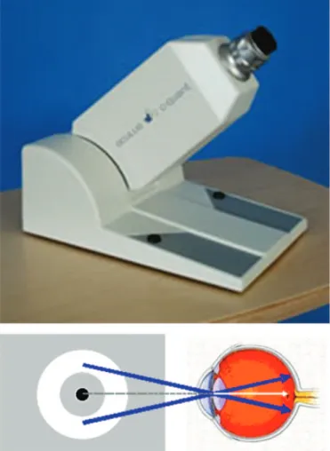
Since cataract surgery has been increasingly indicated earlier, even before the reduction in high contrast visual acuity, it is interesting to assess functional visual performance using more advanced approaches. The objective study of optical aberrations (wavefront analysis), contrast sensitivity, and light scattering (straylight) is important and should be associated with optical densitometry (scattering) of the lens. Light scattering can be assessed using the C-Quant (Cataract-Quantifier, Oculus) device developed by van den Berg(6). The method uses an objective
measure — log(s) — that corresponds to the degree of light scattering in the eye. Contrast sensitivity testing can be done using optotypes (e.g., Snellen chart, Landolt ring) or Ginsburg’s sine-wave gratings(7).
Such an approach is essential for observing and confirming cataract-related changes and helps inform decisions on surgical indication and visual prognosis(8).
In this study, we evaluated changes in density and the optical properties of the lens in eyes with nuclear cataract, establishing correlations between qualitative functional measures and subjective (LOCS III) and objective (PNS) diagnostic methods for the altered lens.
M
ETHODSProspective observational study on 30 eyes of 30 patients with nuclear cataract using the Lens Opacification Classification System III.
The study was conducted in compliance with the standards and guidelines of the Declaration of Helsinki and the Research Ethics Committee of Fluminense Federal University’s Medical School.
The inclusion criterion was a diagnosis of nuclear cataract according to the LOCS III classification.
Exclusion criteria were corneal disorders or opacities, retinal or optic nerve disorders, previous ocular surgery, and no informed consent.
After a routine eye examination, all patients included in the study were assessed with the following methods: ray tracing associated with Placido topography (iTrace, Vista, Tracy Technologies, Houston, USA), used to assess total and intraocular aberrometry (IO RMS [intraocular root-mean-square]); C-Quant, to assess light scattering using log(s); and cornea and anterior segment tomography (Pentacam™, Oculus, Wetzlar, Germany), which uses a Scheimpflug image to determi-ne PNS (Pentacam Nucleus Staging), classifying lens densitometry in a scale from 1 to 5.
Aberrometry testing was performed in dynamic conditions of accommodation and pupillary reflex using the fogging strategy, by tracing individual laser beams projected on the retina
associated with Placido topography (Figure 1). Two sequences of 128 beams, totalling 256 projected beams were used in the pupillary aperture for each test. The position of each beam on the retina is identified to reconstruct the Retinal Spot Diagram (RSD), providing a visualisation of the point spread function. In an eye without optical aberrations, all points would converge to the centre of the RSD. Each point’s deviation is calculated to reconstruct the wavefront according to Zernike polynomials in the form of RMS (Root Mean Square) wave height (microns). The 4-mm optical zone is typically used to calculate the autorefraction of an aberrometre(4,72). We calculated the following
RMS values for 4mm: total, corneal, intraocular (IO RMS), low-order, high-low-order, unfocused, astigmatism (and its axis), higher-order aberrations, spherical aberration, coma (and its axis), and trefoil (and its axis).
C-Quant was used to assess intraocular light scattering. The overall strength of the scattering is calculated as a measure of dispersion using log(s). Higher log(s) values mean greater light scattering, with worse visual quality. The result of this calculation also includes a parameter representing the accuracy of the respective measure (Esd). The normal accuracy of C-Quant (Figure 2) is Esd 0.07 log units. Two measures were taken in each eye without mydriasis, and measures with Esd e” 0.08 were excluded.
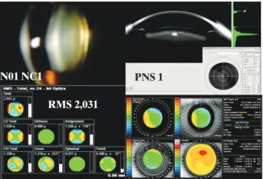
Tomography of the cornea and anterior segment employs a 360-degree rotating camera and a monochromatic light source (475 nm blue LED), using the Scheimpflug principle to obtain 25-100 slit images of the anterior segment in approximately 2 seconds, thus building 3-D images of the cornea and anterior segment. After adequate face alignment, the patient is instructed to look at the fixation target. The examiner observes a real-time
Figure 2:C-Quant was used to assess light scattering using log(s).
image of the patient’s eye on a computer screen; the image is focused and centred automatically. The PNS parameter is provided by the instrument’s software, classifying lens densitometry in a scale from 1 to 5 (Figure 3).
The Kolmogorov-Smirnov test was used to assess the nor-mal distribution, and the Spearman nonparametric test was used for correlations between variables LOCS III vs. PNS; LOCS III vs. log(s); log(s) vs. PNS; IO RMS vs. PNS; IO RMS vs. LOCS III; and IO RMS vs. log(s). Statistical analysis was done using Bioestat (Version 5.0; Belém/PA, Brazil) software. p-values d”0.05 % were considered statistically significant.
RESULTS
The sample included 30 eyes of 30 patients, with 16 males and 14 females. Mean age was 72.3 years (± 8.9).
Mean LOCS III between NO1 and NO6 was 3.7 (±1.15). Mean PNS was 2.7 (±1.14).
Mean log(s) was 1.49 (±0.38), with a mean Esd of 0.05 (±0.02).
Mean IO RMS was 1.23 (±0.5).
The correlation between variables LOCS III and PNS was statistically significant, with p = 0 and a correlation coefficient rho = 0.76 — a strong correlation, as shown in Chart 1.
The correlation between variables log(s) and LOCS III was not statistically significant, with p = 0.71 and a correlation coefficient rho = 0.06 (Chart 2). Although the correlation
Figure 3: Scheimpflug images and classification of lens densitometry — PNS 1 to 3, from left to right, respectively (above) and PNS 4 and 5, from left to right, respectively (below).
between variables log(s) and PNS was not statistically significant, with p = 0.27 and rho = 0.20 (Figure 3), they were positively associated, with a stronger correlation than between log(s) and LOCS III.
The correlations between variables IO RMS vs. PNS, IO RMS vs. LOCS III, and IO RMS vs. log(s) were not statistically significant, with p = 0.41 (rho = -0.15), p = 0.39 (rho = -0.15, and p = 0.83 (rho = 0.03), respectively.
D
ISCUSSIONThe advent of cataract surgery has brought the need for advanced diagnostic and prognostic methods of visual performance. Traditionally, the method used to classify cataracts has been the LOCS III system(9), which is based on slit-lamp
biomicroscopy; however, this is a subjective method and its interpretation by different observers is variable. Thus, considering medical-legal aspects and the tendency to perform surgery earlier, even before a reduction in visual acuity, objective, reproducible, scientifically-validated methods to confirm the diagnosis and classification of cataract become critically important.
Lens densitometry through Scheimpflug imaging is computed tomographically using the PNS(10) (Pentacam Nucleus
Staging) system. This is an objective method which, when associated with the study of wavefront analysis to detect intraocular optical aberrations, assessment of light scattering(11),
and objective tests of contrast sensitivity(7), can help us
understand the actual state of functional changes in each cataract patient. The variables generated by such more advanced tests not only provide useful diagnostic and prognostic information, but also are very helpful for femtolaser surgery and for choosing premium intraocular lenses.
Our study supports findings in the literature, such as Ortiz et al.(12), who found a decrease in optical quality with increasing
morphological changes detected by lens densitometry using PNS. Datiles et al.(13) showed that lens densitometry using PNS is more
sensitive than LOCS III to evaluate the progression of lens opacity, detecting earlier changes. Other studies, such as Pei et al.(14) and Magalhães et al.(15), show a linear correlation between
PNS and the LOCS III in nuclear cataracts.
Chart 2
Correlation between log(s) and LOCS.
Chart 3
Correlation between log(s) and PNS. Chart 1
Correlation between LOCS and PNS.
shows that IO RMS for cataract with PNS 1 is 2.031, while in Figure 5 PNS was classified as 4, and IO RMS was 0.729. Lee et al.(16) found
that in eyes with nuclear cataract, most higher-order aberrations came from intraocular optical defects (IO RMS), and the degree of opalescence of nuclear cataract was negatively correlated with spherical aberration, in agreement with our study. These findings may explain the “subjective” symptoms in these patients.
In conclusion, assessment of the lens should go beyond the traditional and essential tests of biomicroscopy and Snellen visual acuity, especially in the early stages of cataract. Thus, the introduction of new technologies is critical to provide actual qualitative information on changes in visual performance in eyes with cataract.
R
EFERENCES1. Kuroda T, Fujikado T, Ninomiya S, Maeda N, Hirohara Y, Mihashi T. Effect of aging on ocular light scatter and higher order aberra-tions. J Refract Surg. 2002;18(5):S598-602
2. Porter J, Guirao A, Cox IG, Williams DR. Monochromatic aberra-tions of the human eye in a large population. J Opt Soc Am A Opt Image Sci Vis. 2001;18(8):1793-803.
stronger correlation between PNS vs. log(s) than between LOCS III vs. log(s), showing that light scattering is more closely associated with the objective than the subjective method. Furthermore, even though the correlations between IO RMS and other variables such as LOCS III and PNS were not statistically significant, they are negative correlated, showing that greater degrees of lens opacity are associated with fewer aberrations. However, our study may have limitations, because ray tracing analysis can be difficult in more advanced cataracts, as not all laser beams reach the retina beyond the 4 mm optic zone assessed by the study. Thus, aberrometry is more important in early cataracts. This can be seen in Figures 4 and 5, which show biomicroscopy images with the LOCS III classification, the Scheimpflug image with PNS, and aberrometry maps. Figure 4
Figure 4: Schematic analysis of the subjective, objective and qualitative assessment of lens N01NC1 using the LOCS III classification. log(s) in this patient was 1.16.
Figure 5: Schematic analysis of the subjective (LOCS III), objective (PNS) and qualitative assessment of lens N04NC4 using the LOCS III classification. log(s) in this patient was 1,85.
N01 NC1
RMS 2,031
PNS 1
PNS 4
N04 NC4
Corresponding author:
Bruno de Freitas Valbon
Av. Marechal M. de Moraes, 2767/Apto 102 - Bento Ferreira CEP: 29052-121 - Vitória (ES), Brazil.
Telephone: +5521 8103 7117 E-mail: [email protected]
3. Applegate RA, Marsack JD, Ramos R, Sarver EJ. Interaction between aberrations to improve or reduce visual performance. J Cataract Refract Surg. 2003;29(8):1487-95.
4. Donnelly WJ 3rd, Pesudovs K, Marsack JD, Sarver EJ, Applegate RA. Quantifying scatter in Shack-Hartmann images to evaluate nuclear cataract. J Refract Surg. 2004;20(5):S515-22.
5. Van Den Berg TJ, Van Rijn LJ, Michael R, Heine C, Coeckelbergh T, Nischler C, et al. Straylight effects with aging and lens extrac-tion. Am J Ophthalmol. 2007;144(3):358-63.
6. Van den Berg TJ. Depth-dependent forward light scattering by donor lenses. Invest Ophthalmol Vis Sci. 1996;37(6):1157-66. 7. Ginsburg AP, Cannon MW. Comparison of three methods for rapid
determination of threshold contrast sensitivity. Invest Ophthalmol Vis Sci. 1983;24(6):798-802.
8. Ambrosio Jr R, Chalita MR, Netto MV, Schor P, Chamon W, Fontes BM, editores. Wavefront e topografia, tomografia e biomecânica da córnea. Propedêutica complementar em cirurgia refrativa. 2a ed. Rio de Janeiro: Guanabara Koogan; 2012. 9. Chylack LT Jr, Wolfe JK, Singer DM, Leske MC, Bullimore MA,
Bailey IL, et al. The Lens Opacities Classification System III. The Longitudinal Study of Cataract Study Group. Arch Ophthalmol. 1993;111(6):831-6.
10. Nixon DR. Preoperative cataract grading by Scheimpflug imag-ing and effect on operative fluidics and phacoemulsification en-ergy. J Cataract Refract Surg. 2010;36(2):242-6.
11. Van den Berg TJ, Coppens JE. Method and device for measuring retinal straylight. Patent W02005023103, NL1024232C. 2005. Avail-able from: http://patentscope.wipo.int/search/en/WO2005023103 12. Ortiz D, Alió JL, Ruiz-Colechá J, Oser U. Grading nuclear
cata-ract opacity by densitometry and objective optical analysis. J Cataract Refract Surg. 2008;34(8):1345-52.
13. Datiles MB 3rd, Magno BV, Freidlin V. Study of nuclear cataract progression using the National Eye Institute Scheimpflug sys-tem. Br J Ophthalmol. 1995;79(6):527-34.
14. Pei X, Bao Y, Chen Y, Li X. Correlation of lens density measured using the Pentacam Scheimpflug system with the Lens Opacities Classification System III grading score and visual acuity in age-related nuclear cataract. Br J Ophthalmol. 2008;92(11):1471-5. 15. Magalhães FP, Costa EF, Carrielo AJ, Rodrigues EB, Hofling-Lima AL. Comparative analysis of the nuclear lens opalescence by the Lens Opacities Classification System III with nuclear den-sity values provided by Oculus Pentacam: a cross-section study using Pentacam Nucleus Staging software. Arq Bras Oftalmol. 2011;74(2):110-3.