This in vitro study evaluated the effect of sodium bicarbonate and sodium ascorbate on the microtensile bond strength of an etch-and-rinse system to bleached bovine enamel. Sixty bovine enamel blocks (4x4 mm) were flattened and randomly allocated into 5 groups: G1 (negative control): without treatment; G2 (positive control): bleached with 35% hydrogen peroxide (HP); G3: bleached and stored for 7 days in artificial saliva before restorative procedures; G4: bleached and treated with 10% sodium bicarbonate solution for 5 min; G5: bleached and treated with 10% sodium ascorbate hydrogel for 15 min. HP gel was applied twice (20 min each, except in G1) and the adhesive restorations were performed. After 24 h, the specimens were sectioned into sticks and submitted to microtensile bond strength testing with a crosshead speed of 0.5 mm/min (n=12). As a complementary visual observation, the enamel surfaces of the G1 and G2 specimens were evaluated with scanning electron microscopy. Data were analyzed by one-way ANOVA (p<0.05). The means (standard deviation) were: G1: 24.22±7.74; G2: 18.29±5.88; G3: 40.88±7.95; G4: 19.95±5.67 and G5: 24.43±6.43. Adhesive failures were predominant in all groups. The comparison between the treatments indicates that waiting 7 days after bleaching is still the most effective approach. When this waiting period is not possible, application of sodium ascorbate or sodium bicarbonate seems to be a good alternative. Therefore, the practicality of obtaining sodium bicarbonate in the bleaching kits and its higher stability enables its clinical use.
E f f e c t o f T w o A n t i o x i d a n t s
A g e n t s o n M i c r o t e n s i l e B o n d
S t r e n g t h t o B l e a c h e d E n a m e l
Marina Studart Alencar, Juliana Fraga Soares Bombonatti, Rafael Massunari Maenosono, Ana Flávia Soares, Linda Wang, Rafael Francisco Lia Mondelli
Department of Operative Dentistry, Endodontics and Dental Materials, Bauru School of Dentistry, USP - University of São Paulo, Bauru, SP, Brazil.
Correspondence: Rafael Francisco Lia Mondelli, Alameda Dr. Octávio Pinheiro Brisolla, 9-75, 17012-901, Bauru, SP, Brasil. Tel: +55-14-3235-8265, +55-14-98156-4686. e-mail: [email protected]
Key Words: tooth bleaching, hydrogen peroxide, antioxidants, sodium bicarbonate.
Introduction
Pigmentation of dental structures is one of the most common clinical issues related to aesthetics in dentistry. Dental bleaching could be a conservative and simple option to reverse this condition (1). Moreover, after the bleaching procedure, many situations call for adhesive restorations (1), such as remodeling the anatomical form, closure of diastema or replacing anterior restorations.
Even though the literature presents consistent results regarding the stability of adhesive restorations placed in dental enamel, there are some clinical situations in which this adhesion is questioned, such as when the restorations are performed immediately after bleaching (2,3).
After bleaching, the residual oxygen remains among the enamel prisms and the polymerization of the resin monomers can be inhibited by its presence, damaging the marginal sealing, promoting an early microleakage process and reducing the bond strength of the restorations (1,4,5). However, the residual oxygen slowly dissipates to the extent that the oxidation reaction ends over time. The general approach is to defer any adhesive restorative procedure for a time interval ranging from 24 h to 4 weeks after bleaching (1,4-7), because reduction of the bond strength
is temporary.
Aiming to reduce this time interval, the antioxidant products were introduced as an interesting strategy since they can remove or reduce the residual oxygen (8,9). Sodium ascorbate is one of the most studied antioxidants, however, despite its efficiency (7,8,10-12), there is still controversy regarding its action mechanisms (9). In addition, Ozelin et. al (11) concluded that this substance must to be applied for at least 60 min to improve bond strength values, which hinders its clinical practice.
Another substance that has been recently tested is 10% sodium bicarbonate solution. It is usually indicated to neutralize the adverse effects of bleaching gel in soft tissue (13). However, Tostes et al. (14) noted its use as a possible alternative treatment prior to restoring bleached teeth. In addition, this solution accompanies most bleaching kits (14), which eliminates the need of manipulation in specialized pharmacies. It is important to mention that sodium bicarbonate used as a chemical activator associated with a high concentration of hydrogen peroxide, this association is able to protect the organic enamel content (15).
Effect of antio
xidants on bleached enamel.
the use of antioxidants and lack of evidence for the use of 10% sodium bicarbonate solution for this purpose, the present study aimed to evaluate its effect after bleaching in comparison to 10% sodium ascorbate hydrogel. The null hypothesis was that the bond strength of an adhesive restorative system to bleached bovine enamel is not influenced by the treatment protocol after bleaching.
Material and Methods
Experimental Design
The present in vitro study involved one factor: treatment protocol (in five levels: no bleaching, bleaching, bleaching followed by a waiting time of 7 days, 10% sodium bicarbonate solution or 10% sodium ascorbate hydrogel). The microtensile bond strength was assessed quantitatively and expressed in MPa. Optical microscopy of failure mode was performed in order to complement the microtensile bond strength values.
Preparation of Specimens
Sixty bovine incisors were collected from a local slaughterhouse and stored in 0.1% supersaturated thymol solution at 4 °C, renewed weekly. A previous analysis of the mesiodistal dimensions and the original length was made to select similar units. Teeth with fissures or cracks on the crown surface were excluded from the sample.
The teeth were sectioned at the mesiodistal and incisal-cervical regions with a low speed saw (Isomet; Buehler, Lake Bluff, IL, USA) under running water to obtain 4x4 mm fragments . At this point, the fragments that presented cracks and structural defects were discarded and the palatal side was excluded. The buccal face of each fragment was ground flat in a polishing machine (Politriz Metalográfica; Aropol 2V, Cotia, SP, Brazil) with a series of silicon carbide papers (320-, 400-, 600- and 1200-grit) under continuous irrigation. This procedure was performed carefully in order to avoid dentin exposure, which was controlled with a stereomicroscopic lens with 40x magnification (Carl Zeiss Microimaging GMBH; Göttingen, Germany). The specimens were randomly assigned to 5 groups (n=12): G1 (negative control): Not bleached and restored; G2 (positive control): 35% HP bleaching and immediately restored; G3: 35% HP bleaching, 7-day storage in distilled water and restored; G4: 35% HP bleaching, 10% sodium bicarbonate solution for min and restored; G5: 35% HP bleaching, 10% sodium ascorbate hydrogel for 15 min and restored.
Bleaching Procedure
The 35% hydrogen peroxide bleaching gel (Total Blanc Office; Nova DFL, Rio de Janeiro, RJ, Brazil) was applied according to the manufacturer’s instructions: a
1 mm thick layer was applied for 20 min on the enamel surfaces of all groups, except for G1. After this period, the gel was removed and a new layer of gel was applied for an additional 20 min. Next, the specimens were washed and immersed in distilled water for 5 min.
Application of Antioxidant
A drop of the 10% sodium bicarbonate solution from the bleaching kit, was passively applied with a microbrush on the enamel surfaces of the previously bleached G4 group. The application was performed for 5 min according to the Tostes et al. (14) study. The bleached enamel specimens of G5 received a uniform layer of 10% sodium ascorbate hydrogel passively applied with a microbrush for 15 min (7).
Restorative Procedure
The enamel surfaces were etched for 30 s with 37% phosphoric acid (Alpha Etch; Nova DFL, Rio de Janeiro, RJ, Brazil), rinsed for the same time and air-dried. For adhesion, a two-step etch-and-rinse system (Natural Bond DE; Nova DFL, Rio de Janeiro, RJ, Brazil) was applied according to the manufacturer’s instructions: the first coat was placed and scrubbed for 5 s, and after a 20-s interval, an air jet was performed for 5 s to facilitate the evaporation of the solvent. A second coat was applied and, after 20 s, the adhesive system was light cured (Radi Cal, SID, Victoria, Australia - 1000mW/cm2) for 20
s. To build up the restoration, a microhybrid composite resin (Natural Look; Nova DFL) was placed on the enamel surface in 1 mm increments and light cured for 20 s to obtain a 4 mm crown.
Microtensile Bond Strength Test
After 24 h in artificial saliva at 100% humidity at 37 °C, the specimens were cut in parallel sections measuring approximately 1 mm, made from the mesial to the distal surface and from the cervical to the incisal surface with a low speed saw (Isomet; Buehler) under running water, obtaining an average of 4 sticks for each specimen (the lateral sticks were discarded in order to obtain more standardized specimens).
M.S. Alencar et al.
(Messen Sensor Technology Co., Guangdong, China). The load required for the fracture of the specimens was converted to MPa by dividing the fracture load (kgf) by the cross-sectional area of the specimen (mm2).
Failure Mode Analysis
The failure modes were evaluated with a digital stereomicroscope at 200× magnification (DINO-LITEplus, Anmo Electronics Corporation, Hsinchu, China). Failures were classified as: adhesive (when the failure occurred at the substrate/restorative material interface); cohesive in enamel (when the failure occurred in the enamel); cohesive in resin (when the failure occurred in the restorative material); mixed (when there was a combination of adhesive and cohesive failure). The percentage of each type of failure within each group was calculated.
SEM Analysis
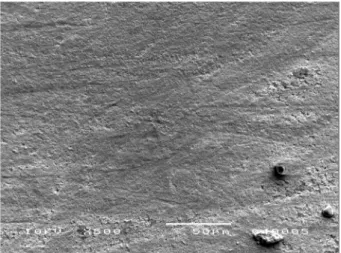
As a complementary visual observation, the enamel surfaces of the G1 and G2 specimens were evaluated with scanning electron microscopy in order to observe the surface characteristics of these samples (i.e. porosities and surface roughness), solely for observational reasons.
Statistical Analysis
The data were evaluated on the treatment protocol variable. Under the normal distribution, the data was analyzed by one-way ANOVA (α=0.05).
Results
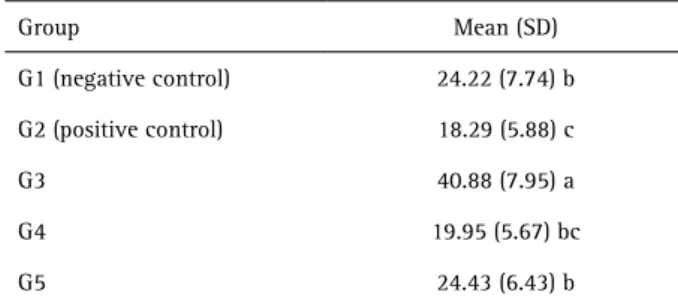
The mean microtensile bond strength values and standard deviations after different treatments are shown in Table 1. The factor treatment protocol was statistically significant (p<0.05).
Comparing the treatment protocols, waiting 7 days after bleaching to perform the adhesive procedures provided G3 significant higher values (p<0.0001) of
microtensile bond strength. The application of 10% sodium bicarbonate solution (G4) and 10% sodium ascorbate hydrogel (G5) recovered the microtensile bond strength values after bleaching since the mean values were similar to the negative control group G1 (p>0.05), which showed an intermediate performance. The positive control (G2 – bleached and immediately restored) showed the lowest values and G4 behaved statistically similar to this group as well.
Optical microscopy analysis revealed predominance of adhesive failures followed by the cohesive in enamel fractures. The percent distribution of failure pattern was as follows: adhesive (65.26%), cohesive in enamel (14.74%), cohesive in resin (1.58%) and mixed (18.42%).
The scanning electron microscopy showed that the enamel surface of G1 (specimen polished and etched with 37% phosphoric acid for 30 s) appeared less porous compared to the enamel surface of G2 (specimens bleached with 35% hydrogen peroxide for 40 min and etched with 37% phosphoric acid for 30 s). G2 presented a highly porous and rough surface.
Discussion
Based on the results, the null hypothesis was rejected since the treatment protocol was able to affect positively or negatively the bond strength values of an adhesive restorative system to bovine bleached enamel.
There was a reduction in the bond strength of G2 (positive control) compared to G1 (negative control). This decrease in the bond strength may be attributed to changes in the enamel surface, such as precipitation that may negatively influence the adhesive process (16) or perhaps due to the presence of residual oxygen which inhibits the full polymerization and proper penetration of the resin tags (1,4,5). In addition, the bond strength values shown in G1 group had a mean value consistent with the mean values found for other microtensile bond strength test studies performed in the same conditions (17-20).
Among the evaluated treatments, the only one that showed a statistically significant difference compared to G1 was G3, with higher mean bond strength values. The storage in distilled water for 7 days after bleaching seems to be enough to eliminate the residual oxygen (1,4-7). Furthermore, the significant increase in the mean bond strength values after exposure to hydrogen peroxide may be associated with an improvement in the etch pattern of the enamel surface (21). Phosphoric acid etching may remove calcium and phosphate from the enamel, increasing the roughness and surface area to be restored (22), however, it is not ble to alter the protein layer, which may adversely affect the adhesive bond with the composite resin. Prolonged exposure of Table 1. Microtensile bond strength mean values (MPa) and standard
deviations
Group Mean (SD)
G1 (negative control) 24.22 (7.74) b
G2 (positive control) 18.29 (5.88) c
G3 40.88 (7.95) a
G4 19.95 (5.67) bc
G5 24.43 (6.43) b
Effect of antio
xidants on bleached enamel.
enamel to higher concentrations of hydrogen peroxide makes this protein layer more susceptible to dissolution by rinsing and drying (23). Prolonged exposure to hydrogen peroxide followed by etching with phosphoric acid may increase the porosity of enamel, resulting in improved retention of the restoration and higher bond strength values (16,21).
Evaluating the specimens with scanning electron microscopy, which was performed for complementary visual observation, it was found that the enamel surface was modified due to the different treatments, corroborating the studies of Titley et al. (16) and Torneck et al. (21). The specimens polished and etched with 37% phosphoric acid (Fig. 1) showed less porous and rough surface areas compared to the specimen exposed to 35% hydrogen peroxide for 40 min, followed by etching with 37% phosphoric acid (Fig. 2).
Analyzing the results of G5, it was noted that 10% sodium ascorbate hydrogel was able to recover the bond strength values, probably due to its capacity to neutralize and reverse the oxidative effects of bleaching agents. It contradicts the results of Sasaki et al. (9) who stated that this substance was not effective for this purpose and the results of Ozlein et al. (11) that affirmed that effective action of this substance required at least a 60-minute application.
Evaluating the sodium bicarbonate solution, the main purpose of the present study, it was observed that G4 presented a mean bond strength value not statistically different from G1 and G2. In addition, Lima et al. (15) noted a great advantage in associating this substance with hydrogen peroxide, because when used as chemical activator, it protected the organic content of enamel.
The results of G4 and G5 could possibly be different if
the application time of antioxidants were standardized. This was not possible because there was lack of papers in the literature confirming the application time of these two substances. Due to reduced availability of researches about the application of sodium bicarbonate at the time of this study, it was decided to follow the protocol suggested by Tostes et al. (14), which was used as a reference. There is no consensus in the literature regarding the application time of sodium ascorbate, which may vary from 10 min to 8 h (7,8,10-12,24). Further studies assessing the long-term effects of antioxidants (applied for a standardized time) are required for comparison with the present study, and thus make them possible to apply them safely in clinical practice.
It should be mentioned that in addition to the bond strength values, evaluating the failure modes with a digital stereomicroscope at 200× magnification, there was prevailance of adhesive failures in all groups. According to Sano et al. (25), due to the small surface area of the specimen, the highest stresses occurred at the periphery of the adhesive interface, increasing the probability of defects that lead to the increase of adhesive failures.
Although the application of antioxidants in G4 and G5 reflected on the immediate satisfactory results, statistically similar to G1, these values are reduced compared to G3. Therefore, the safest strategy is waiting 7 days after bleaching to perform the adhesive restorative procedure.
Only in very specific situations, when it is not possible to wait for a week, dentists may use 10% sodium bicarbonate solution or 10% sodium ascorbate hydrogel on the bleached enamel, since these substances have shown performance similar to the negative control
Figure 1. SEM micrograph (500×) of the specimen polished and etched with 37% phosphoric acid for 30 s. Appears less porous compared to the previously bleached specimen.
M.S. Alencar et al.
group. Although both substances were able to recover the bond strength values after bleaching, the practicality of obtaining sodium bicarbonate solution in bleaching kits enables its clinical use.
Resumo
Este estudo in vitro avaliou o efeito do bicarbonato de sódio e do ascorbato de sódio na resistência de união de um sistema adesivo convencional unido ao esmalte bovino clareado. Sessenta blocos de esmalte bovino (4x4 mm) foram planificados e distribuídos aleatoriamente em 5 grupos: G1: (controle negativo); G2 (controle positivo): clareamento com peróxido de hidrogênio 35% (HP); G3: clareamento com HP seguido de armazenamento por 7 dias em saliva artificial antes do procedimento restaurador; G4: clareamento com HP seguido de tratamento com a solução de bicarbonato de sódio 10% por 5 min; G5: clareamento com HP seguido de tratamento com hidrogel de ascorbato de sódio 10% por 15 min. O HP foi aplicado duas vezes (20 min cada, com exceção do grupo G1) e então as restaurações adesivas foram realizadas. Após 24 h, os espécimes foram seccionados em palitos e submetidos ao teste de resistência de união a uma velocidade de 0,5 mm/min (n=12). As superfícies de esmalte de G1 e G2 foram avaliadas com microscopia eletrônica de varredura para fins de análise visual complementar. Os dados foram analisados por ANOVA a um critério (p<0,05). As medias (desvio-padrão) foram: G1: 24,22±7,74; G2: 18,29±5,88; G3: 40,88±7,95; G4: 19,95±5,67 e G5: 24,43±6,43. Falhas adesivas foram predominantes em todos os grupos. A comparação entre os diferentes tratamentos indica que esperar 7 dias após o clareamento é ainda a abordagem mais eficaz. Nos casos em que este período de espera não é possível, a aplicação do ascorbato de sódio e do bicarbonate de sódio parecem ser boas alternativas. Entretanto, a praticidade na obtenção da solução de bicarbonato de sódio nos kits de clareamento e sua maior estabilidade favorecem o seu uso clínico.
Acknowledgements
This study was performed by M.S.A. as fulfillment of her graduation research that was supported by grant 134179/2011-0 from CNPq (Conselho Nacional de Desenvolvimento Científico e Tecnológico), Brazil. The authors wish to thank Nova DFL for donation of the bleaching gel kit and restorative materials.
References
1. Bittencourt ME, Trentin MS, Linden MS, de Oliveira Lima Arsati YB, França FM, Flório FM, et al.. Influence of in situ postbleaching times on shear bond strength of resin-based composite restorations. J Am Dent Assoc 2010;141:300-306.
2. Briso AL, Toseto RM, Rahal V, dos Santos PH, Ambrosano GM. Effect of sodium ascorbate on tag formation in bleached enamel. J Adhes Dent 2012;14:19-23.
3. Haywood VB, Heymann HO. Nightguard vital bleaching: how safe is it? Quintessence Int 1991;22:515-523.
4. Dishman MV, Covey DA, Baughan LW. The effects of peroxide bleaching on composite to enamel bond strength. Dent Mater 1994;10:33-36. 5. Muraguchi K, Shigenobu S, Suzuki S, Tanaka T. Improvement of bonding
to bleached bovine tooth surfaces by ascorbic acid treatment. Dent Mater J 2007;26:875-881.
6. Cavalli V, Reis AF, Giannini M, Ambrosano GM. The effect of elapsed time following bleaching on enamel bond strength of resin composite. Oper Dent 2001;26:597-602.
7. Gökce B, Comlekoglu ME, Ozpinar B, Turkun M, Kaya AD. Effect of antioxidant treatment on bond strength of a luting resin to bleached enamel. J Dent 2008;36:780-785.
8. Lai SC, Mak YF, Cheung GS, Osorio R, Toledano M, Carvalho RM, et al.. Reversal of compromised bonding to oxidized etched dentin. J Dent Res 2001;80:1919-1924.
9. Sasaki RT, Florio FM, Basting RT. Effect of 10% sodium ascorbate and 10% alpha-tocopherol in different formulations on the shear bond strength of enamel and dentin submitted to a home-use bleaching treatment. Oper Dent 2009;34:746-752.
10. Kaya AD, Türkün M, Arici M. Reversal of compromised bonding in bleached enamel using antioxidant gel. Oper Dent 2008;33:441-447. 11. Ozelin AA, Guiraldo RD, Carvalho RV, Lopes MB, Berger SB. Effects of
green tea application time on bond strength after enamel bleaching. Braz Dent J 2014;25:399-403.
12. Kavitha M, Selvaraj S, Khetarpal A, Raj A, Pasupathy S, Shekar S. Comparative evaluation of superoxide dismutase, alpha-tocopherol, and 10% sodium ascorbate on reversal of shear bond strength of bleached enamel: An in vitro study. Eur J Dent 2016;10:109-115. 13. Torres CRG, Koga AF, Borges AB. The effects of anti-oxidant agents as
neutralizers of bleaching agents on enamel bond strength. Braz J Oral Sci 2006;5:971-976.
14. Tostes BO, Mondelli RFL, Lima-Arsati YB, Rodrigues JA, Costa LC. The effect of baking soda when applied to bleached enamel prior to restorative treatment. Gen Dent 2013;61:5-9.
15. Lima DA, Aguiar FH, Pini NI, Soares LE, Martin AA, Liporoni PC, et al.. In vitro effects of hydrogen peroxide combined with different activators for the in-office bleaching technique on enamel. Acta Odontol Scand 2015;73:516-521.
16. Titley K, Torneck CD, Smith D. The effect of concentrated hydrogen peroxide solutions on the surface morphology of human tooth enamel. J Endod 1988;14:69-74.
17. Barcellos DC, Benetti P, Fernandes VV Jr, Valera MC. Effect of carbamide peroxide bleaching gel concentration on the bond strength of dental substrates and resin composite. Oper Dent 2010;35:463-469. 18. Lago AD, de Freitas PM, Netto NG. Evaluation of the bond strength
between a composite resin and enamel submitted to bleaching treatment and etched with Er:YAG laser. Photomed Laser Surg 2011;29:91-95.
19. Reis AF, Giannini M, Kavaguchi A, Soares CJ, Line SR. Comparison of microtensile bond strength to enamel and dentin of human, bovine, and porcine teeth. J Adhes Dent 2004;6:117-121.
20. Sen Tunc E, Bayrak S, Tuloglu N, Ertas E. Evaluation of microtensile bond strength of different fissure sealants to bovine enamel. Aust Dent J 2012;57:79-84.
21. Torneck CD, Titley KC, Smith DO, Adibfar A. Effect of water leaching on the adhesion of composite resin to bleached and unbleached bovine enamel. J Endod 1991;17:156-160.
22. Silverstone LM, Saxton CA, Dogon IL, Fejerskov O. Variation in the pattern of acid etching of human dental enamel examined by scanning electron microscopy. Caries Res 1975;9:373-387.
23. Wolff SP, Dean RT. Fragmentation of proteins by free radicals and its effect on their susceptibility to enzymic hydrolysis. Biochem J 1986;234:399-403.
24. Lai SC, Tay FR, Cheung GS, Mak YF, Carvalho RM, Wei SH, et al. Reversal of compromised bonding in bleached enamel. J Dent Res 2002;81:477-481.
25. Sano H, Shono T, Sonoda H, Takatsu T, Ciucchi B, Carvalho R, et al.. Relationship between surface area for adhesion and tensile bond strength - evaluation of a micro-tensile bond test. Dent Mater 1994;10:236-240.