1. PhD in Cardiovascular Surgery; Pediatric Surgeon of INCL. 2. Master’s Degree in Cardiology; Head of the Cardiopediatric Service
of the INCL.
3. Cardiovascular Surgery Specialist; Head of the Cardiovascular Surgery Service of the INCL.
This study was carried out at the National Institute of Cardiology. Cardiovascular Surgery Service. Child and Adolescent Cardiology Service, Rio de Janeiro, RJ, Brazil.
José Alberto CALIANI1,Luiz Carlos SIMÕES2,Odilon Nogueira BARBOSA3
RBCCV 44205-996
Modificação técnica para correção de coarctação aórtica com hipoplasia do arco aórtico
Technical modification for correction of aortic
coarctation using hypoplastic arch
Abstract
Objectives: To study technical feasibility and early results
of the technical modification suggested by Caliani et al. for correction of aortic coarctation and aortic arc.
Methods: Between January 2005 and July 2006, nine
neonatal patients with aortic coarctation and significant aortic hypoplasia were selected, and underwent a new surgical approach in order to correct this aortic defect. The definition of aortic arch hypoplasia according to Moulaert’s criteria is an aortic arch with a diameter that is less than 50% of the diameter of the ascending aorta. In this study, only patients with proximal and distal aortic hypoplasia were selected. Many techniques were previously used, but significant residual gradients were observed, as well as the incovenience of definitive occlusion of the left subclavian artery. The aim of this study is to describe a new surgical technique that includes left posterolateral thoracotomy, wide mobilization of descending aorta with occlusion of the first two intercostal branches, transection of the left subclavian artery at its base, wide resection of the hypoplastic area and the surronding regions of the ductus arteriosus; end-to-end anastomosis between the aortic arch and descending aorta, with 7-0 PDS thread and reimplantation of the subclavian artery into the left carotid artery with side-to-end anastomosis.
Results: There were no perioperative or late deaths. The
mean residual gradient was 5 mmHg. Up to now, there were no cases of recoarctation or medullary neurological lesions.
Conclusion: Despite the small number of cases and the
short follow-up, this technique modification may be an excellent option for the treatment of this complex situation.
Descriptors: Aortic coarctation/surgery. Heart defects,
congenital. Cardiac surgical procedures/methods. Infant, newborn.
Resumo
Objetivo: Estudar a viabilidade técnica e resultados imediatos
da modificação técnica proposta por Caliani et al. para correção da coarctação aórtica com hipoplasia do arco aórtico.
Métodos: Entre janeiro de 2005 e julho de 2006, nove
neonatos com coarctação aórtica e hipoplasia do arco aórtico foram submetidos a uma nova abordagem cirúrgica para correção do defeito. A definição de hipoplasia do arco aórtico seguiu os critérios de Moulaert, segundo os quais o arco aórtico é considerado hipoplásico quando seu diâmetro atinge 50% do diâmetro da aorta ascendente. Nesta série, foram selecionados apenas pacientes com hipoplasia proximal e distal do arco aórtico. Várias técnicas foram propostas anteriormente, mas gradientes residuais importantes foram observados, assim como há o inconveniente da ligadura definitiva da artéria subclávia esquerda. A modificação técnica consiste em: toracotomia póstero-lateral esquerda, ampla mobilização da aorta descendente, com ligadura dos dois primeiros ramos intercostais, transecção da artéria subclávia esquerda em sua base, ressecção ampla de toda zona hipoplásica e adjacências do ducto arterioso, anastomose término-terminal entre o arco aórtico e aorta descendente com fio de PDS 7-0 e reimplante da artéria subclávia sobre a artéria carótida esquerda com anastomose látero-terminal.
Resultados: Não houve óbito per-operatório ou tardio, o
gradiente residual médio foi de 5 mmHg. Até o presente, não observamos nenhum caso de recoarctação ou de lesão neurológica medular.
Conclusão: A despeito do reduzido número de casos e do
seguimento curto, esta modificação técnica pode representar uma excelente opção para tratamento deste complexo grupo de pacientes.
Descritores: Coartação aórtica/cirurgia. Procedimentos
cirúrgicos cardíacos/métodos. Cardiopatias congênitas. Recém-nascido.
Correspondence address:
José Alberto Caliani. Instituto Nacional de Cardiologia – Rua das Laranjeiras, 374 4° andar - Rio de Janeiro, RJ, Brasil - CEP 22240-005 E-mail address: jcaliani@incl.rj.saude.gov.br
INTRODUCTION
Aortic coarctation is defined as the narrowing of the descending aorta just below the origin of the left subclavian artery in a region called the isthmus, where the ductus arteriosus is connected by proximity. The disease may develop alone, or it may be associated with intracardiac anomalies or aortic arch hypoplasia.
The surgical basics of the first correction of aortic coarctation in newborns were presented by Mustard et al. in 1953 [1]. Since then, we have seen a gradual improvement in outcomes. However, the correction of coarctation with aortic arch hypoplasia still represents a challenge for surgeons [2].
The definition of aortic arch hypoplasia was proposed by Moulaert et al. [3]. The aortic arch is considered hypoplastic when its diameter reaches 50% of the diameter of the ascending aorta. Hypoplasia can be distal (isthmian - type A) or proximal/distal (transverse arch - Type B - Figure 1). When the aortic coarctation is associated with the arch hypoplasia, we can conclude that it deals with duct-dependent heart disease, where, during fetal development, there was less infusion in this region caused by left-right blood flow, which resulted in hypodevelopment of the arc and the consequent migration of ductal cells in the isthmian region [3].
consensus on which surgical technique to use for this expansion [2,4,5].
Recent progress in neonatal intensive care and in two-dimensional Echo-Doppler techniques have allowed for the correction of complex intracardiac malformations associated with aortic arch abnormalities in a single stage, and with satisfactory results [6-8]. In these cases, aortic coarctation is surgically approached as an interruption of the aortic arch, whether associated with arch hypoplasia or not, and surgeons use cardiopulmonary bypass with deep hypothermia and circulatory arrest.
Other techniques have been described for correction of this malformation [4,9,10], but significant residual gradients were observed, frequent recoarctations and growth deficit in the left superior limb, significantly when the procedure of occlusion or patch of the subclavian artery is used [4, 11]. In addition to this is the common denominator of these techniques: the preservation of the hypoplastic zone, in which growth is restricted in long-term [12], which may cause retraction.
The aim of this study is to report a new technique for correction of coarctation with aortic arch hypoplasia type B described by Moulaert. This technique preserves the vessels of base, resects the hypoplastic zone while providing a good hemodynamic profile. Cases of isolated isthmian coarctation or with hypoplasia type A were not included in this study because these pathological entities can be treated with conventional techniques of resection and end-to-end anastomosis with good outcomes. In our view, the real challenge for the surgeon is type B, which is a more complex type of hypoplasia.
METHODS
Patients
Between January 2005 and July 2006, nine newborns with aortic coarctation and aortic arch hypoplasia type B (Moulaert) were admitted to the ICU (during use of prostaglandin in continuous infusion aiming to maintain the permeability of the ductus arteriosus, developing into presentation of heart failure, arterial hypertension and metabolic acidosis). They underwent surgical correction with the technique to be described. Three patients presented small interventricular communication. The patients’ages ranged from 3 to 20 days, with a mean age of 7 days. All patients underwent transthoracic echocardiogram prior to the operation. No hemodynamic studies were performed.
Technique
The surgical approach for all patients was the left posterolateral thoracotomy. The left lung is pulled away, and the descending aorta, ductus arteriosus and vessels of the base are widely dissected up to the ascending aorta Current studies show that surgeons typically agree on
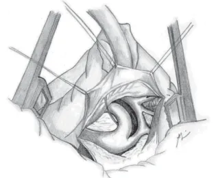
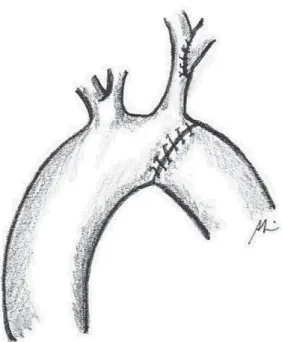
(Figure 2). The ductus arteriosus and the first two or three intercostal branches are connected with 6-0 polypropylene for wide mobility of the descending aorta (Figures 3 and 4). A 0.5 mg/kg dose of heparin is administered. Aortic clamping is performed immediately after the brachiocephalic trunk and in descending aorta, after emergence of the second or third intercostal branch (previously connected). The left carotid arteries and subclavian are clamped. The subclavian artery is then sectioned at the base of its implantation on the aorta (Figure 5). The distal segment remains clamped. The coarctation area and, specifically, the hypoplastic segment, located between the subclavian artery and the left carotid artery, are widely resected (Figure 6). A small opening in the concavity of the arc toward the ascending aorta is performed in order to enlarge the maximum area of anastomosis. Subsequently, an end-to-end anastomosis is performed between the aortic arch and the descending aorta with continuous 7-0 PDS suture (Figure 7).
Fig.2 – Surgical anatomy of aortic coarctation with arch hypoplasia
Fig. 3 – Intercostal arteries occlusion
Fig.4 – Ductus arteriosus occlusion
Fig. 5 – Section of the left subclavian artery
Fig. 7 – End-to-end anastomosis between the crest and the decending aorta
Fig. 8 – Lateroterminal reimplantation of the subclavian artery in the left carotid artery
The forceps are removed and the anastomosis is revised. Partial clamping of the left carotid artery is then performed in its medium third, where surgeons perform a vertical incision in its wall. The subclavian artery is reimplanted in the carotid artery through lateroterminal anastomosis with continuous 7-0 PDS suture (Figure 8).
Fig. 9 – Control Echo-Doppler in operated patient, showing aortic arch after surgical correction without residual stenosis
RESULTS
There were no surgical mortalities, medullary ischemic accidents or neurological sequels caused by the partial clamping of the left carotid artery. The aortic clamping time was 25 + 11 minutes.
The postoperative hospital stay ranged from 7 to 19 days, with mean of 11.7 + 1.1 days.
Control Doppler echocardiogram performed just before discharge and at 3 and 6 months after the operation showed no gradient in six patients (Figure 9) and residual gradient (10mmHg) in three.
All patients received clinical follow-ups, and at the time of this article, there have been no deaths or late complications.
DISCUSSION
The incidence of the aortic arch hypoplasia in coarctation ranges from 65% to 81% [13]. The introduction of therapy with prostaglandins in the neonatal period changed the history of this anomaly [9] with significant improvement in the results. Anatomopathological [14] and surgical [2,4,15] studies reported the clear obstruction created by aortic arch hypoplasia.
A high percentage of recoarctation in newborns was reported after correction with end-to-end anastomosis [16],
flapping of the subclavian artery [11] and a patch made of
end-to-end anastomosis [18-20] requires recoarctation in more than 10% of cases.
Histological studies confirm the abnormal structure of the hypoplastic aortic arch. It is characterized by high percentage of collagen fibers in relation to the aorta’s diameter and the limited presence of alpha-actin positive cells, which are responsible for vessel growth [12]. This reinforces the theory that, to achieve a satisfactory result, the surgical technique should include total resection of the hypoplastic area and ductus arteriosus.
Other techniques have been described [9,10] with apparently good immediate results. However, both techniques preserve the structure of the hypoplastic aortic arch, which may result in limited growth or retraction of the area around the anastomosis,causing recoarctation [12], or which may permanently sacrifice the left subclavian artery with the consequent growth deficit in the superior left limb and the consequences that this may cause [13]. Other authors prefer to treat this anomaly as an interruption of the aortic arch, using sternotomy, cardiopulmonary bypass and deep hypothermia [8]. In our view, this procedure is not necessary, except in the case of associated intracardiac abnormality, which may require single stage correction.After approaching the coarctation with aortic arch hypoplasia in all its aspects, we propose a technique without use of cardiopulmonary bypass. This technique takes into account the factors that are directly responsible for the high occurrence of recoarctations. This technique is based on the following principles: 1) it completely removes the ductal tissue; 2) it ressects the hypoplastic area of the arch and its pathological tissues with low growth potential; 3) it preserves the vessels at the basis through the left subclavian artery reimplantation; and 4) it does not generate gradient during the anastomosis.
CONCLUSION
From a technical point of view, we believe that the surgery proposed herein is feasible if there is good mobilization of the descending aorta. This can be achieved through wide dissection and occlusion of the first intercostal branches. Otherwise, it seems difficult to lead the distal segment toward the implantation base of the left carotid artery and to perform a tension-free end-to-end anastomosis. In this series, we did not have accidents or ischemic medullary sequels; however, this is always a possibility, and additional clinical observations are still needed.
Despite the small number of cases and short follow-up, the technique without use of cardiopulmonary bypass and without use of synthetic patch or prosthesis can be an excellent option for the treatment of this complex group of patients and can contribute to the improvement of immediate and mid-term clinical outcomes.
REFERENCES
1. Mustard WT, Rowe RD, Keith JD, Sirek A. Coarctation of the aorta with special reference to the first year of life. Ann Surg. 1955;141(4):429-36.
2. Karl TR, Sano S, Brawn W, Mee RB. Repair of hypoplastic or interrupted aortic arch via sternotomy. J Thorac Cardiovasc Surg. 1992;104(3):688-95.
3. Moulaert AJ, Bruins CC, Oppenheimer-Dekker A. Anomalies of the aortic arch and ventricular septal defects. Circulation. 1976;53(6):1011-5.
4. Hart JC, Waldhausen JA. Reversed subclavian flap angioplasty for arch coarctation of the aorta. Ann Thorac Surg. 1983;36(6):715-7.
5. Jahangiri M, Shinebourne EA, Zurakowski D, Rigby ML, Redington AN, Lincoln C. Subclavian flap angioplasty: does the arch look after itself? J Thorac Cardiovasc Surg. 2000;120(2):224-9.
6. Haas F, Goldberg CS, Ohye RG, Mosca RS, Bove EL. Primary repair of aortic arch obstruction with ventricular septal defect in preterm and low birth weight infants. Eur J Cardiothorac Surg. 2000;17(6):643-7.
7. Gaynor JW, Wernovsky G, Rychic J, Rome JJ, DeCampli WM, Spray TL. Outcome following single-stage repair of coarctation with ventricular septal defect. Eur J Cardiothorac Surg. 2000;18(1):62-7.
8. Elgamal MA, McKenzie ED, Fraser CD Jr. Aortic arch advancement: the optimal one-stage approach for surgical management of neonatal coarctation with arch hypoplasia. Ann Thorac Surg. 2002;73(4):1267-73.
9. Lacour-Gayet F, Bruniaux J, Serraf A, Chambran P, Blaysat G, Losay J, et al. Hypoplastic transverse arch and coarctation in neonates. Surgical reconstruction of the aortic arch: a study of sixty-six patients. J Thorac Cardiovasc Surg. 1990;100(6):808-16.
10. Rajasinghe HA, Reddy VM, van Son JA, Black MD, McElhinney DB, Brook MM, et al. Coarctation repair using end-to-side anastomosis of descending aorta to proximal aortic arch. Ann Thorac Surg. 1996;61(3):840-4.
11. Metzdorff MT, Cobanoglu A, Grunkemeier GL, Sunderland CO, Starr A. Influence of age at operation on late results with subclavian flap aortoplasty. J Thorac Cardiovasc Surg. 1985;89(2):235-41.
12. Machii M, Becker AE. Hypoplastic aortic arch morphology pertinent to growth after surgical correction of aortic coarctation. Ann Thorac Surg. 1997;64(2):516-20.
Touati G, et al. Aortic coarctation with hypoplastic aortic arch: Results of extended end-to-end aortic arch anastomosis. J Thorac Cardiovasc Surg. 1988;96(4):557-63.
14. Bharati S, Lev M. The surgical anatomy of the heart in tubular hypoplasia of the transverse aorta (preductal coarctation). Thorac Cardiovasc Surg. 1986;91(1):79-85.
15. Myers JL, McConnell BA, Waldhausen JA. Coarctation of the aorta in infants: does the aortic arch grow after repair? Ann Thorac Surg. 1992;54(5):869-75.
16. Tawes RL, Aberdeen E, Waterston DJ, Carter REB. Coarctation of the aorta in infants and children: a review of 333 operative cases including 170 infants. Circulation. 1969;39(suppl I):173-84.
17. Hesslein PS, McNamara DG, Morriss MJ, Hallman GL,
Cooley DA. Comparison of resection versus patch aortoplasty for repair of coarctation in infants and children. Circulation. 1981;64(1):164-8.
18. Conte S, Lacour-Gayet F, Serraf A, Sousa-Uva M, Bruniaux J, Touchot A, et al. Surgical management of neonatal coarctation. J Thorac Cardiovasc Surg. 1995;109(4):663-75.
19. Van Heurn LW, Wong CM, Spiegelhalter DJ, Sorensen K, de Leval MR, Stark J, et al. Surgical treatment of aortic coarctation in infants younger than three months: 1985 to 1990. Success of extended end-to-end arch aortoplasty. J Thorac Cardiovasc Surg. 1994;107(1):74-86.