Myocyte necrosis is the basis
for fibrosis in renovascular
hypertensive rats
1Departamento de Clínica Médica and 2Departamento de Patologia,
Faculdade de Medicina de Botucatu, Universidade Estadual Paulista, Botucatu, SP, Brasil
M.P. Okoshi1,
L.S. Matsubara1, M. Franco2,
A.C. Cicogna1 and
B.B. Matsubara1
Abstract
The pathogenesis of fibrosis and the functional features of pressure overload myocardial hypertrophy are still controversial. The objec-tives of the present study were to evaluate the function and morphol-ogy of the hypertrophied myocardium in renovascular hypertensive (RHT) rats. Male Wistar rats were sacrificed at week 4 (RHT4) and 8 (RHT8) after unilateral renal ischemia (Goldblatt II hypertension model). Normotensive rats were used as controls. Myocardial function was analyzed in isolated papillary muscle preparations, morphological features were defined by light microscopy, and myocardial hydroxy-proline concentration (HOP) was determined by spectrophotometry. Renal artery clipping resulted in elevated systolic arterial pressure (RHT4: 178 ± 19 mmHg and RHT8: 194 ± 24 mmHg, P<0.05 vs
control: 123 ± 7 mmHg). Myocardial hypertrophy was observed in both renovascular hypertensive groups. The myocardial HOP concen-tration was increased in the RHT8 group (control: 2.93 ± 0.38 µg/mg; RHT4: 3.02 ± 0.40 µg/mg; RHT8: 3.44 ± 0.45 µg/mg of dry tissue, P<0.05 vs control and RHT4 groups). The morphological study dem-onstrated myocyte necrosis, vascular damage and cellular inflamma-tory response throughout the experimental period. The increased cellularity was more intense in the adventitia of the arterioles. As a consequence of myocyte necrosis, there was an early, local, conjunc-tive stroma collapse with disarray and thickening of the argyrophilic interstitial fibers, followed by scarring. The functional data showed an increased passive myocardial stiffness in the RHT4 group. We con-clude that renovascular hypertension induces myocyte and arteriole necrosis. Reparative fibrosis occurred as a consequence of the inflam-matory response to necrosis. The mechanical behavior of the isolated papillary muscle was normal, except for an early increased myocardial passive stiffness.
Correspondence
M.P. Okoshi
Departamento de Clínica Médica Faculdade de Medicina de Botucatu UNESP
18618-000 Botucatu, SP Brasil
Fax: 55 (014) 822-2238
Publication supported by FAPESP.
Received November 27, 1996 Accepted July 8, 1997
Key words
•Coronary vascular remodeling
•Myocardial function
•Pressure overload hypertrophy
•Papillary muscle
Introduction
Myocardial hypertrophy is an adaptive re-sponse to ventricular overload. Although there has been considerable investigation on myo-cardial function with pressure overload hyper-trophy, the functional features of the myocar-dium remain controversial; mechanical activ-ity has been reported to be increased (1,2), unchanged (3-5), or depressed (6,7). Changes in myocyte and/or interstitial components of the myocardium have been pointed out as the causes of cardiac dysfunction (8-10). Intersti-tial collagen accumulation occurs in various experimental models of pressure overload myocardial hypertrophy (10-14) and has been associated with myocardial dysfunction by many authors (8,10-13). However, others have failed to demonstrate an association between fibrosis and cardiac muscle dysfunction (15). Another matter of disagreement among the studies is about the pathogenetic origin of myocardial fibrosis. Weber et al. (8,16) have hypothesized that the fibrosis occur-ring in pressure overload hypertrophy might be both reparative and reactive. In the former case, myocyte necrosis may initiate collagen deposition and lead to replacement scarring. Studies (17-19) have demonstrated that ei-ther renovascular or angiotensin II (AII)-induced hypertension causes cardiomyocyte necrosis followed by fibrosis. In addition, vascular injury is the basis of patchy multifo-cal myocardial necrosis and scarring in hy-pertensive rats with aortic banding (20). On the other hand, reactive fibrosis is defined as the result of an increased collagen synthesis by the AII-stimulated interstitial fibroblasts, with no cell loss (8). Accordingly, an exces-sive accumulation of collagen in the hyper-trophied myocardium of rats with renovas-cular hypertension or aortic constriction has been described with no evidence of myocyte necrosis (12,21). Under some experimental conditions, both types of fibrosis might oc-cur sequentially as reported for unilateral renal ischemia (22).
To further address this issue, we ana-lyzed the pathogenesis of myocardial fibro-sis in renovascular hypertensive rats and evaluated the influence of fibrosis on me-chanical performance of papillary muscles. We demonstrated that the myocardial fibro-sis was always related to myocyte necrofibro-sis, suggesting rather a reparative than a reactive fibrosis. There were an early collapse and disarray of the reticular stroma in the areas of myocyte loss and a later replacement by fibrosis. Regardless of the remarkable mor-phological changes, the functional behavior of the papillary muscles was close to normal, except for an early transitory increase in myocardial stiffness.
Material and Methods
Animal and groups
Eight-week old male Wistar rats weigh-ing 130 to 170 g were anesthetized ip with thiopental sodium (50 mg/kg). Renovascular hypertension (RHT) was induced by con-stricting the left renal artery to an outer diameter of 0.25 mm with the aid of a silver clip. The contralateral kidney was untouched (Goldblatt II hypertension model). All ani-mals were housed in a temperature-controlled room (24oC) on a 12-h light/dark cycle, and
food and water were supplied ad libitum. The rats were sacrificed 4 weeks (RHT4 group, N = 15) or 8 weeks (RHT8 group, N = 15) after surgery. Results were compared to sex-matched 14-week old rats (control group, N = 15). Before sacrifice, systolic arterial pressure (SAP) was measured in all animals using a tail cuff.
Functional study
were dissected from the left ventricle (LV), mounted between two spring clips, and placed vertically in a chamber containing Krebs-Henseleit solution at 28oC and gassed with
95% O2 and 5% CO2. The composition of the
Krebs-Henseleit solution was as follows: 118.5 mM NaCl, 4.69 mM KCl, 2.52 mM CaCl2, 1.16 mM MgSO4, 1.18 mM KH2PO4,
5.50 mM glucose, and 25.88 mM NaHCO3.
The lower spring clip was attached to a Kyowa model 12OT-2OB force transducer by a thin steel wire (1/15,000 inch) which passed through a mercury seal at the bottom of the chamber. The upper spring clip was connected by a thin steel wire to a rigid lever arm above which a micrometer stop was mounted for the adjustment of muscle length. The lever arm was made of magnesium with a ball-bearing fulcrum and a lever arm ratio of 4:1. A displacement transducer (Hewlett-Packard, 7 DCDT-50) was mounted above the short end of the lever arm. Preparations were stimulated 12 times/min with 5-ms square wave pulses through parallel plati-num electrodes, at voltages 10% greater than the minimum required to produce a maximal mechanical response.
The muscles were kept contracting iso-tonically with light loads for 60 min and then loaded to contract isometrically and stretched to the maximum of their length-tension curves.
After a 5-min period in afterloaded iso-tonic contractions, muscles were again placed under isometric conditions, and the peak of the length-tension curve (Lmax) was carefully
determined. A 15-min period of stable iso-metric contraction was imposed prior to the experimental period and one isometric traction was then recorded. The isotonic con-traction parameters were obtained using the lightest preload able to maintain Lmax. The
passive length-tension curves were derived from data obtained at lengths of 90%, 92%, 94%, 96%, 98% and 100% of Lmax.
The following parameters were meas-ured from isometric contractions: peak
de-veloped tension (DT; g/mm2), resting
ten-sion (RT; g/mm2), time to peak tension (TPT;
ms), maximum rate of tension development (+dT/dt; g/mm2/s), maximum rate of tension
decline (-dTdt; g/mm2/s) and time for
ten-sion to fall from peak to 50% of peak tenten-sion (RT50; ms). For isotonic contractions, the
following parameters were assessed: peak shortening (PS; mm), time to peak shorten-ing (TPS; ms), maximum velocity of iso-tonic shortening (+dL/dt; muscle length/s), maximum velocity of isotonic relengthening (-dL/dt; muscle length/s) and relative varia-tion of length [(Lmax - PS)/Lmax].
At the end of each experiment, the muscle length at Lmax was measured and the muscle
between the two clips was blotted dry and weighed. Cross-sectional areas were calcu-lated from the muscle weight and length by assuming cylindrical uniformity and a spe-cific gravity of 1.0. All force data were nor-malized for the muscle cross-sectional area and length data were normalized by the muscle length (Lmax).
Biochemical study
Hydroxyproline (HOP) was measured in tissue obtained from the LV apex according to the method described by Switzer (23). Briefly, the tissue was dried for 4 h using an SC 100 SpeedVac Concentrator attached to a TR 100 refrigerated condensation trap and a VP 100 vacuum pump (Savant Instruments Inc., Farmingdale, NY). Tissue dry weight was determined and the samples were hy-drolyzed overnight at 110oC with 6 N HCl (1
min. The addition of 1 ml of 3.6 M sodium thiosulfate with thorough mixing for 10 s stopped the oxidation process. The solution was saturated with 1.5 g KCl and the tubes were capped and heated in boiling water for 20 min. After cooling to room temperature, the aqueous layer was extracted with 2.5 ml toluene. One ml of toluene extract was trans-ferred to a 12 x 75-mm test tube and 0.4 ml of Ehrlich’s reagent was added to allow the color to develop for 30 min. Absorbances were read at 565 nm against a reagent blank. Deionized water and 20 µg/ml HOP were used as blank and standard, respectively.
Morphological study
Five animals from each group were sacri-ficed for morphological study. Hearts were excised and the ventricles separated, weighed and fixed in 10% buffered formalin for 24 h. Formalin-fixed tissue was serially dehydrated and embedded in paraffin; serial 5-µm thick sections were stained with hematoxylin and eosin, Sirius Red F3BA or silver for reticular fibers. Histological indicators of myocardial necrosis were the presence of muscle cells with no nuclei or cross striation, alongside with local accumulation of inflammatory cells and confluence of fibrotic tissue.
Statistical analysis
All grouped data are reported as means ± SD and compared by one-way analysis of variance and the post hoc Tukey test. The normalized LV and right ventricular (RV) weights were analyzed by the nonparametric Kruskal-Wallis test.
Prior to comparing the diastolic length-tension relationship for the three groups, the resting tension at L90 was subtracted from all
subsequent tension data in each experiment in order to have all length-tension curves intercepting the Y axis origin at L90. The
diastolic length (L)-tension (RT) curves for the 3 groups were fitted to
mono-exponen-tial relations of the form RT = A[eB(L-L0) -1],
where A and B are fitting parameters, and L0
is the muscle length corresponding to zero resting tension. These nonlinear relations were compared by constructing an F ratio from the residual sum of squares (24). This test determines whether separate fits to data from two groups are significantly better than the combined fit to all data from two groups. Accordingly, a significant F ratio indicates that the two sets of data being compared were significantly different from each other. For all comparisons, statistical significance was taken to be P<0.05/k, where k is the number of comparison.
Results
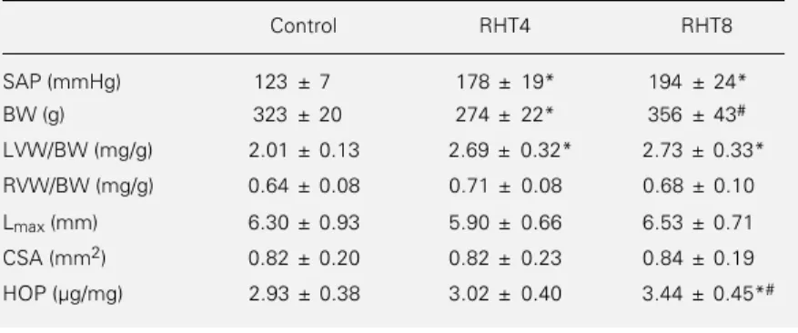
The mean values obtained for the mor-phometric parameters, SAP and myocardial HOP concentration for all groups are sum-marized in Table 1.
Renal artery clipping resulted in an in-creased SAP (RHT4: 178 ± 19 mmHg and RHT8: 194 ± 24 mmHg, P<0.05 vs control: 123 ± 7 mmHg). Analysis of the hypertrophy index, expressed by left or right ventricular/ body weight ratio, showed a significant LV hypertrophy after 4 weeks of RHT, which remained unchanged after 8 weeks. There was no RV hypertrophy in the RHT groups. Myocardial HOP concentration was signifi-cantly increased only after 8 weeks of RHT (control: 2.93 ± 0.38 µg/mg; RHT4: 3.02 ± 0.40 µg/mg; RHT8: 3.44 ± 0.45 µg/mg dry tissue, P<0.05 vs control and RHT4).
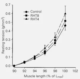
Tables 2 and 3 respectively show the isometric and isotonic contraction data of the papillary muscles. There was no signifi-cant difference among groups. Figure 1 shows the mean curves for the passive length-ten-sion relations for all groups. Statistical anal-ysis demonstrated a significant shift to the left of the relations in the RHT4 group, indicating increased passive myocardial stiff-ness compared to controls.
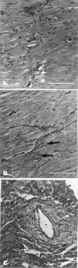
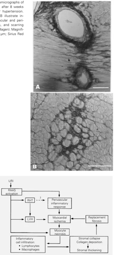
func-tional behavior, the histological study showed striking arterial and myocardial lesions in both ventricles in the RHT groups (Figures 2 and 3). Myocardial histology was normal in control animals. After 4 weeks of renal is-chemia, there were scattered foci of myocyte necrosis of different sizes. The necrotic pro-cess comprised one single cell or groups of myocytes (Figure 2A). The cell necrosis in-duced a predominantly lymphomononuclear inflammatory response with few polymor-phonuclear neutrophils. Collapse of the re-ticular framework occurred at the sites of myocyte loss as demonstrated by silver stain-ing (Figure 2B), as well as accumulation of inflammatory cells. In addition, segmental medial hypertrophy, hyalinization and fi-brinoid necrosis of the intramural coronary arteries were observed. Many arteries pre-sented adventitial lymphomononuclear in-flammatory exudate and edema (Figure 2C). In the RHT8 group, the areas of myocardial necrosis were more frequent and larger. Re-cent myocyte necrosis was found in combi-nation with foci of reparative fibrosis and healing. After eight weeks of renal ischemia, the changes of the intramural coronary arter-ies were more striking. The adventitial in-flammatory process involved the adjacent myocytes and was accompanied by increased perivascular and interstitial fibrosis and scar-ring (Figure 3A,B).
Discussion
This study demonstrated that rats with left ventricular hypertrophy due to renovas-cular hypertension presented marked mor-phological alterations, such as myocyte ne-crosis, vascular damage and interstitial fi-brosis.
The presence of cardiomyocyte necrosis has been previously described in the present experimental model (19). In rats with RHT, the myocyte injury and necrosis have been attributed to the resulting abnormal increase of the renin-angiotensin system activity,
sys-Table 1 - Group comparisons of systolic arterial pressure (SAP), morphometric param-eters and myocardial hydroxyproline concentration (HOP).
Data are reported as means ± SD for N = 15. BW, Body weight; LVW, left ventricle weight; RVW, right ventricle weight; Lmax, muscle length at peak of developed
tension; CSA, cross-sectional area; RHT4, renovascular hypertension for 4 weeks; RHT8, renovascular hypertension for 8 weeks. *P<0.05 vs control group; #P<0.05 vs
RHT4 group (one-way analysis of variance and post hoc Tukey test).
Control RHT4 RHT8
SAP (mmHg) 123 ± 7 178 ± 19* 194 ± 24*
BW (g) 323 ± 20 274 ± 22* 356 ± 43#
LVW/BW (mg/g) 2.01 ± 0.13 2.69 ± 0.32* 2.73 ± 0.33*
RVW/BW (mg/g) 0.64 ± 0.08 0.71 ± 0.08 0.68 ± 0.10
Lmax (mm) 6.30 ± 0.93 5.90 ± 0.66 6.53 ± 0.71
CSA (mm2) 0.82 ± 0.20 0.82 ± 0.23 0.84 ± 0.19
HOP (µg/mg) 2.93 ± 0.38 3.02 ± 0.40 3.44 ± 0.45*#
Table 2 - Isometric contraction data.
Data are reported as means ± SD for N = 10. DT, Peak developed tension; RT, resting tension; TPT, time to peak tension; +dT/dt, maximum rate of tension development; -dT/dt, maximum rate of tension decline; RT50, time for tension to fall from peak to
50% of peak tension; RHT4, renovascular hypertension for 4 weeks; RHT8, renovascu-lar hypertension for 8 weeks (one-way analysis of variance and post hoc Tukey test).
Control RHT4 RHT8
DT (g/mm2) 8.59 ± 2.12 8.95 ± 2.50 8.24 ± 1.92
RT (g/mm2) 0.80 ± 0.24 0.87 ± 0.30 0.77 ± 0.33
TPT (ms) 201 ± 16 201 ± 10 205 ± 15
+dT/dt (g/mm2/s) 76 ± 21 82 ± 29 74 ± 20
-dT/dt (g/mm2/s) 19 ± 5.6 21 ± 5.2 20 ± 4.1
RT50 (ms) 310 ± 57 278 ± 39 276 ± 36
Table 3 - Isotonic contraction data.
Data are reported as means ± SD for N = 10. PS, Peak shortening; TPE, time to peak shortening; +dL/dt, maximum velocity of isotonic shortening; -dL/dt, maximum ve-locity of isotonic relengthening; ML, muscle length; (Lmax - PS)/Lmax, relative variation
of length; Lmax, muscle length at peak of developed tension; RHT4, renovascular
hypertension for 4 weeks; RHT8, renovascular hypertension for 8 weeks (one-way analysis of variance and post hoc Tukey test).
Control RHT4 RHT8
PS (mm) 1.70 ± 0.46 1.53 ± 0.18 1.73 ± 0.41
TPE (ms) 215 ± 18 218 ± 14 223 ± 21
+dL/dt (ML/s) 2.45 ± 0.52 2.28 ± 0.41 2.46 ± 0.72
-dL/dt (ML/s) 4.36 ± 1.24 3.97 ± 1.08 3.92 ± 1.35
temic or local (19,25), and to the coronary vascular damage (20). The toxic effect of AII on the myocardium is thought to be due to its direct action or to the AII-stimulated catecholamine release (19,26).
Our findings of lymphomononuclear in-flammatory accumulation around the foci of necrosis and around the arteries in both the RHT4 and RHT8 groups suggest a sustained injury to the myocardium throughout the follow-up period. This is not in accordance with other investigators (17,18), who showed that the necrotic damage after a chronic in-crease in plasma AII levels was limited to the first two days, despite a continued increase in plasma AII levels. Kabour et al. (18,19) showed that the AII-induced myocyte necro-sis is mediated by the angiotensin type 1 receptor and speculated that a chronic in-crease in circulating AII may cause receptor downregulation protecting the myocardium against further AII-mediated necrosis (18). The different experimental approaches, RHT and AII infusion, may explain the discrepant results and further studies are needed for a better understanding of this point.
Another possible mechanism for multi-focal myocyte necrosis is myocardial is-chemia due to a coronary artery abnormality. In hypertensive rats with aortic banding, myo-cyte and arterial necrosis and scarring have
been reported up to the sixth week of follow-up (20). There was a close association be-tween arterial and myocardial lesions, sug-gesting that muscle necrosis and scars were due to ischemia. Our findings of arteriole damage, perivascular edema and cellular in-flammatory response may support this hy-pothesis. The presence of inflammatory cells within the adventitia of intramyocardial cor-onary vessels has been reported by others (27). It was suggested that the production of mediators by those cells might contribute to the fibrous tissue response. Besides the vas-cular wall injury and the inflammatory re-sponse, other mechanisms might be involved in the perivascular edema and fibrosis. It has been reported that activation of the renin-angiotensin system is related to increased coronary vascular permeability (28-31). This vascular dysfunction may be the result of increased release of nitric oxide (30), brady-kinin and prostaglandins (31). Our study indicates that, in addition to changes in arte-riolar permeability, cell necrosis and the sub-sequent inflammatory response are associ-ated with perivascular fibrosis.
In the present study, since myocyte ne-crosis was found to be so variable, restricted to one single cell or involving a large group of cells, the pattern of the subsequent fibro-sis also presented variable characteristics. Collagen accumulation was observed in deli-cate septa between muscle cells or in large scars. In our interpretation, the late perivas-cular and interstitial myocardial fibrosis was reparative. The fibrosis replaced areas of myocyte loss or arterial smooth muscle cell damage. The stromal collapse reinforced this interpretation. This suggestion is in disagree-ment with the hypothesis of reactive fibrosis occurring in RHT (8,16), which was based on the absence of inflammatory cells near the sites of myocardial fibrosis (12,21). As observed in the present study, Hinglais et al. (14) found increased myocardial fibrosis in hypertensive rats closely related to the pres-ence of lymphocytes and macrophages
Figure 1 - Passive length-tension relations for renovascular hyper-tension at 4 (RHT4 group) and 8 weeks (RHT8 group) and for con-trol rats (concon-trol group). Results are reported as means ± SD. The curve for the RHT4 group was significantly different from the curve for the control group (P<0.05) (nonlinear regression modeling comparing parameters estimated by the residual sum of squares, F ratio test). The shift to the left indicates increased myocardial passive stiffness. Lmax: muscle length at peak of
developed tension.
Resting tension (g/mm
2) 0.7
0.6
0.5
0.4
0.3
0.2
0.1
0.0
90 92 94 96 98 100 102
Muscle length (% of Lmax) Control
around degenerative, myocytolytic muscle cells, indicating that the myocardial fibrosis was a reparative process. In contrast, Campbell et al. (22) described inflammatory cells in the cardiac tissue only during the few days following the induction of unilateral renal ischemia in rats. Nevertheless, they observed progressive fibrosis until the eighth week of RHT, which suggested that a mech-anism other than cell necrosis was the agent of abnormal collagen accumulation.
These different results in morphological patterns might have been due to variations introduced in the classical Goldblatt model of RHT. In order to produce renal ischemia, the authors promoted aortic constriction in-cluding the right renal artery (21,22), or both renal arteries (20). These technical varia-tions may induce different degrees of hor-monal release and consequently different degrees of heart damage.
Our hypothesis for myocardial fibrosis is summarized in Figure 4. The activation of the renin-angiotensin system by unilateral renal ischemia causes arterial hypertension, myocardial hypertrophy, myocyte necrosis and a perivascular inflammatory response. Myocardial hypertrophy is further stimulat-ed by pressure overload and increases myo-cardial oxygen consumption, with oxygen delivery possibly being impaired by the perivascular inflammatory process. All of these factors, taken together, result in myo-cardial hypoxemia and further cell necrosis. Myocyte necrosis initiates the healing pro-cess, which ends with fibrosis replacement. The results about the mechanical func-tion of isolated papillary muscles did not show a depressed systolic function in spite of the remarkable morphological changes. In fact, there was a disagreement between myo-cardial function and the intensity of morpho-logical damage. Other authors have observed the same behavior. Conrad et al. (32) found significant functional changes in the papil-lary muscles of spontaneously hypertensive rats only when there was heart failure in rats.
Before this, the myocardial function was normal, even in the presence of significant morphological changes. Accordingly, Cicogna et al. (15) induced expressive myo-cardial fibrosis in normotensive rats by chronic administration of colchicine and found unimpaired myocardial function, as analyzed in the isolated papillary muscle preparation. These results suggest that, in the experimental models of chronic heart damage, morphological changes precede myocardial dysfunction. However, it is nec-essary to remember that studies of systolic function in renovascular (33) or spontane-ously hypertensive rats (34) have revealed that the myocardium can present a better systolic function than in normotensive con-trol rats. Thus, the apparent normal results obtained in this study may in fact indicate impaired systolic function.
In the pressure overload hypertrophy, the relation between myocardial fibrosis and di-astolic dysfunction has been previously de-scribed (11,12,35). In the present study, it was interesting to find increased myocardial passive stiffness in the RHT4 group, which had normal HOP concentration, and normal passive stiffness in the RHT8 group, which had increased myocardial HOP concentra-tion. One possible explanation for this ob-servation may be the qualitative changes of the myocardial collagen content. The RHT4 group had an increased accumulation of ar-gyrophilic reticular fibers in areas of myo-cyte loss rather than replacement collagen fibrosis. The reticular fibers consist mainly of collagen type III, fibronectin and one or more non-collagenous glycoproteins (36), whereas replacement fibrosis consists mainly of type I collagen (37). Therefore, one would expect a decreased type I:III ratio early after myocyte necrosis. This imbalance may pro-mote the diastolic dysfunction in the RHT4 group. In fact, Mukherjee and Sen (38) hy-pothesized that the type of collagen, rather than the total amount, might influence myo-cardial function.
Figure 3 - Photomicrographs of rat myocardium after 8 weeks of renovascular hypertension. Panels A and B illustrate in-creased perivascular and peri-cellular fibrosis, and scarring (dark-stained collagen). Magnifi-cation bar: 500 µm; Sirius Red F3BA staining.
Figure 4 - Diagrammatic representation of a hypothesis for the physiopathol-ogy of myocardial fibrosis in renovascular hypertension. URI, Unilateral renal ischemia; RAAS, renin-angiotensin-aldosterone system; RHT, renovascular hypertension; LVH, left ventricular hypertrophy.
Perivascular inflammatory response
Myocardial ischemia
Replacement fibrosis RAAS
activation
RHT
LVH
Myocyte necrosis Inflammatory
cell infiltration: · Lymphocytes · Macrophages
Stromal collapse Collagen deposition
Stromal thickening URI
In conclusion, the Goldblatt II model of renovascular hypertension in rats caused myocyte necrosis and arteriolar remodeling followed by fibrosis. Despite the extensive morphological changes, myocardial function remained close to normal.
Acknowledgments
The authors thank José Carlos Georgette and Vitor de Souza for technical assistance and Mario Augusto Dallaqua for secretarial assistance.
References
1. Capasso JM, Strobeck JE & Sonnenblick EH (1981). Myocardial mechanical alter-ations during gradual onset long-term hy-pertension in rats. American Journal of Physiology, 241: H435-H441.
2. Bing OHL, Wiegner AW, Brooks WW, Fishbein MC & Pfeffer JM (1988). Papil-lary muscle structure-function relations in the aging spontaneously hypertensive rat. Clinical and Experimental Hypertension, 10: 37-58.
3. Wisenbaugh T, Allen P, Cooper IV G, Holzgrefe H, Beller G & Carabello B (1983). Contractile function, myosin ATPase activity and isozymes in the hy-pertrophied pig left ventricle after a chronic progressive pressure overload. Circulation Research, 53: 332-341. 4. Capasso JM, Malhotra A, Scheuer J &
Sonnenblick EH (1986). Myocardial, bio-chemical, contractile, and electrical per-formance after imposition of hypertension in young and old rats. Circulation Re-search, 58: 445-460.
5. Cohen ME & Bing OHL (1987). Perfor-mance of papillary muscles from the ag-ing spontaneously hypertensive rat: tem-poral changes in isometric contraction pa-rameters. Proceedings of the Society for Experimental Biology and Medicine, 185: 318-324.
6. Williams Jr JF, Mathew B, Hern DL & Potter RD (1983). Myocardial hydroxyline and mechanical response to pro-longed pressure loading followed by un-loading in the cat. Journal of Clinical In-vestigation, 72: 1910-1917.
7. Lecarpentier Y, Bugaisky LB, Chemla D, Mercadier JJ, Schwartz K, Whalen RG & Martin JL (1987). Coordinated changes in contractility, energetics, and isomyosins after aortic stenosis. American Journal of Physiology, 252: H275-H282.
8. Weber KT, Pick R, Jalil JE, Janicki JS & Carrol EP (1989). Patterns of myocardial fibrosis. Journal of Molecular and Cellular Cardiology, 21 (Suppl V): 121-131. 9. Conrad CH, Brooks WW, Robinson KG &
Bing OHL (1991). Impaired myocardial function in spontaneously hypertensive rats with heart failure. American Journal of Physiology, 260: H136-H145. 10. Bing OHL, Brooks WW, Robinson KG,
Slawsky MT, Hayes JA, Litwin SE, Sen S & Conrad CH (1995). The spontaneously hypertensive rat as a model of the transi-tion from compensated left ventricular hy-pertrophy to failure. Journal of Molecular and Cellular Cardiology, 27: 383-396. 11. Thiedemann K-U, Holubarsch Ch,
Medugorac I & Jacob R (1983). Connec-tive tissue content and myocardial stiff-ness in pressure overload hypertrophy. A combined study of morphologic, morpho-metric, biochemical, and mechanical pa-rameters. Basic Research in Cardiology, 78: 140-155.
12. Doering CW, Jalil JE, Janicki JS, Pick R, Aghili S, Abrahams G & Weber KT (1988). Collagen network remodelling and dia-stolic stiffness of the rat left ventricle with pressure overload hypertrophy. Cardiovas-cular Research, 22: 686-695.
13. Jalil JE, Janicki JS, Pick R & Weber KT (1991). Coronary vascular remodeling and myocardial fibrosis in the rat with reno-vascular hypertension. Response to captopril. American Journal of Hyperten-sion, 4: 51-55.
14. Hinglais N, Heudes D, Nicoletti A, Mandet C, Laurent M, Bariéty J & Michel J-B (1994). Colocalization of myocardial fibro-sis and inflammatory cells in rats. Labora-tory Investigation, 70: 286-294.
15. Cicogna AC, Brooks WW, Hayes JA, Robinson KG, Sen S, Conrad CH & Bing OHL (1997). Effect of chronic colchicine administration on the myocardium of the aging spontaneously hypertensive rat. Molecular and Cellular Biochemistry, 166: 45-54.
16. Weber KT, Janicki JS, Pick R, Capasso J & Anversa P (1990). Myocardial fibrosis and pathologic hypertrophy in the rat with re-novascular hypertension. American Jour-nal of Cardiology, 65: 1G-7G.
17. Tan LB, Jalil JE, Pick R, Janicki JS & We-ber KT (1991). Cardiac myocyte necrosis induced by angiotensin II. Circulation Re-search, 69: 1185-1195.
18. Kabour A, Henegar JR & Janicki JS (1994). Angiotensin II (AII)-induced myocyte ne-crosis: role of the AII receptor. Journal of Cardiovascular Pharmacology, 23: 547-553.
19. Kabour A, Henegar JR, Devineni VR & Janicki JS (1995). Prevention of angio-tensin II induced myocyte necrosis and coronary vascular damage by lisinopril and losartan in the rat. Cardiovascular Re-search, 29: 543-548.
20. Rodrigues MAM, Bregagnollo EA, Montenegro MR & Tucci PJF (1992). Cor-onary vascular and myocardial lesions due to experimental constriction of the ab-dominal aorta. International Journal of Car-diology, 35: 333-341.
21. Jalil JE, Doering CW, Janicki JS, Pick R, Shroff SG & Weber KT (1989). Fibrillar collagen and myocardial stiffness in the intact hypertrophied rat left ventricle. Cir-culation Research, 64: 1041-1050. 22. Campbell SE, Janicki JS & Weber KT
23. Switzer BR (1991). Determination of hy-droxyproline in tissue. Journal of Nutri-tional Biochemistry, 2: 229-321. 24. Ratkowsky DA (Editor) (1983). Comparing
parameter estimates from more than one data set. In: Nonlinear Regression Model-ling; a Unified and Practical Approach. Dekker, New York, 135-152.
25. Hirsch AT, Talsnec CE, Schunkert H, Paul M & Dzau VT (1991). Tissue specific acti-vation of cardiac angiotensin converting enzyme in experimental heart failure. Cir-culation Research, 69: 475-482. 26. Ratajska A, Campbell SE, Sun Y & Weber
KT (1994). Angiotensin II-associated car-diac myocyte necrosis: role of adrenal cat-echolamines. Cardiovascular Research, 28: 684-690.
27. Ratajska A, Campbell SE, Cleutjens JPM & Weber KT (1994). Angiotensin II and structural remodeling of coronary vessels in rats. Journal of Laboratory and Clinical Medicine, 124: 408-415.
28. Giacomelli F, Anversa P & Wiener J (1976). Effect of angiotensin-induced hy-pertension on rat coronary arteries and myocardium. American Journal of Pathol-ogy, 84: 111-125.
29. Laine GA & Allen SJ (1991). Left ventricu-lar myocardial edema: lymph flow, inter-stitial fibrosis and cardiac function. Circu-lation Research, 68: 1713-1721.
30. Sigusch HH, Ou R, Katwa LC, Campbell SE, Ganjam VK, Reddy HK & Weber KT (1995). Angiotensin-II-induced increase in transcoronary protein clearance: role of hypertension vs nitric oxide or cyclo-oxy-genase products. Cardiovascular Re-search, 30: 291-298.
31. Sigusch HH, Campbell SE & Weber KT (1996). Angiotensin II-induced myocardial fibrosis in rats: role of nitric oxide, prosta-glandins and bradykinin. Cardiovascular Research, 31: 546-554.
32. Conrad CH, Brooks WW, Hayes JA, Sen S, Robinson KG & Bing OHL (1995). Myo-cardial fibrosis and stiffness with hyper-trophy and heart failure in the spontane-ously hypertensive rat. Circulation, 91: 161-170.
33. de Simone G, Deveraux RB, Volpe M, Camargo MJ, Wallerson DC & Laragh JH (1992). Relation of left ventricular hyper-trophy afterload, and contractility to left ventricular performance in Goldblatt hy-pertension. American Journal of Hyper-tension, 5: 292-301.
34. Bing OHL, Sen S, Conrad CH & Brooks WW (1984). Myocardial function structure and collagen in the spontaneously hyper-tensive rat: progression from compen-sated hypertrophy to haemodynamic im-pairment. European Heart Journal, 5 (Suppl F): 43-52.
35. Holubarsch Ch (1980). Contracture type and fibrosis type of decreased myocardial distensibility. Different changes in elastic-ity of myocardium in hypoxia and hyper-trophy. Basic Research in Cardiology, 75: 244-252.
36. Unsworth DJ, Scott DL, Almond TJ, Beard HK, Holborow EJ & Walton KW (1982). Studies on reticulin. I: Serological and im-munohistological investigation of the oc-currence of collagen type III, fibronectin and the non-collagenous glycoprotein of pras and glynn in reticulin. British Journal of Experimental Pathology,63: 154-166. 37. Weber KT, Janicki JS, Shroff SG, Pick R,
Chen RM & Bashey RI (1988). Collagen remodeling of the pressure-overloaded, hypertrophied nonhuman primate myocar-dium. Circulation Research, 62: 757-765. 38. Mukherjee D & Sen S (1990). Collagen