Recebido para publicação em 06/12/2009 Aceito para publicação em 24/08/2010
Effects of aqueous leaf extracts of
Azadirachta indica
A. Juss. (neem) and
Melia
azedarach
L. (Santa Barbara or cinnamon) on the intracellular development of
Toxoplasma gondii
MELO, E.J.T.*; VILELA, K.J.; CARVALHO, C.S.
Laboratório de Biologia Celular e Tecidual, Setor Toxicologia Celular, Centro de Biociências e Biotecnologia, Universidade Estadual do Norte Fluminense Darcy Ribeiro. Av. Alberto Lamego 2000, Campos dos Goytacazes, CEP: 28013-602, Rio de Janeiro-Brasil * [email protected] or [email protected]
RESUMO: Efeito de extrato aquoso de folhas de Azadirachta indica A. Juss. (nim) e Melia
azedarach L. (canela ou Santa Bárbara) sobre o desenvolvimento intracelular de
Toxoplasma gondii. Melia azedarach (canela) e Azadirachta indica (nim) apresenta grande
variedade de ingredientes biologicamente ativos contra vírus, bactérias e protozoários, mas nenhum efeito sobre o desenvolvimento intracelular do Toxoplasma gondii é conhecido. Toxoplasma gondii
infecta todos os tipos de células Eucarióticas, onde se estabelece no meio intracelular em vacúolo modificado conhecido como vacúolo parasitóforo. Neste vacúolo ocorre a replicação levando a ruptura da célula hospedeira e reinfecção de novas células, perpetuando a infecção. A quimioterapia utilizada não é capaz de eliminar o parasita além de induzir fortes efeitos colaterais. Neste estudo, nós demonstramos o efeito in vitro de extratos aquosos da canela e nim sobre o
desenvolvimento intracelular do taquizoíto do Toxoplasma gondii. Após tratamento de nim e canela
por 24 h, a porcentagem de infecção e o número de taquizoítos intracelulares decaiu drasticamente. Este efeito foi concentração-dependente. Durante incubação dos extratos, uma progressiva desorganização morfológica e ultraestrutural levaram a formação de intensa vesiculação e completa destruição do parasita, que passou a uma estrutura amorfa, antes da completa eliminação do meio intracelular. No entanto durante o tratamento com os extratos, efeitos morfológicos não foram observados nas estruturas da célula hospedeira. Estes resultados sugerem que os extratos aquosos de nim e canela foram capazes de interferir e eliminar o desenvolvimento intracelular do
Toxoplasma gondii.
Palavras-chave: toxoplasma, nim, canela, citotoxicologia
ABSTRACT: Melia azedarach (cinnamon) and Azadirachta indica (neem) have a variety of
biologically active ingredients against virus, bacteria and protozoan parasites; however, little is known about their action on Toxoplasma gondii intracellular development. Toxoplasma gondii
infects all eukaryotic cells, where it establishes and multiplies inside a modified vacuole called the parasitophorous vacuole until the cell ruptures, re-infecting other cells and establishing the infection. There are no efficient chemotherapies for the elimination of T. gondii, minimizing side
effects. In this study, we performed in vitro assays with neem and cinnamon aqueous extracts
against the intracellular development of T. gondii tachyzoites. After treatment with neem and
cinnamon for 24 h, the percentage of infected cells and the number of intracellular parasites drastically decreased. This effect was concentration-dependent. During the incubation of the extracts, progressive morphological and ultrastructure alterations led to intense vesiculation and complete elimination of the parasite from the intracellular medium. However, during the treatment with extracts, no morphological effects were observed in the structure of the host cell. These results suggest that the aqueous extracts of neem and cinnamon were capable of interfering with and eliminating the intracellular development of Toxoplasma gondii.
INTRODUCTION
Leaves of the Meliaceae species Azadirachta indica (neem) and Melia azedarach (cinnamon or
Santa Barbara in Brazil) have been reported to exhibit immunomodulatory, anti-inflammatory, antihyperglycemic, anticarcinogenic, nematicidal, antiparasitic, antiviral, insecticidal and antioxidant properties (Khan et al., 2001; Salehzadeh et al., 2003; Wandscheera et al., 2004; Anthony et al., 2005). However, there is no knowledge about their biological action on intracellular
Toxoplasma gondii.
Infections by Toxoplasma gondii are
subclinical, unless acquired in the uterus or reactivated in individuals with weakened immune systems (immunocompromised). However, previous studies also showed that T. gondii infections are
associated with lymphadenopathy, fever, weakness and debilitation and ophthalmitis in immunocompetent individuals (McAllister, 2005). In addition, toxoplasmic encephalitis increased worldwide during the 1980s (Sukthana, 2006). The treatments of toxoplasmic infections use effective combinations of pyrimethamine and sulphadiazine (or clindamycin) with folinic acid. Unfortunately, up to 40-50% of the treated patients develop severe adverse effects that change or interrupt therapy.
Natural products play a highly significant role in the process of developing new drugs in the areas of cancer and infectious diseases (Newman et al., 2003), with over 60 and 75% of the used drugs deriving from plant extracts, respectively (Anthony et al., 2005). Previous studies demonstrated that, in the absence of a vaccine, and with growing parasite resistance to therapeutic drugs, natural products can be effective against intracellular parasites such as Plasmodium falciparum, Leishmania amazonensis and Trypanosoma cruzi (Dhar et al., 1998; Lirussi et al.,
2004; Kolodziej et al., 2005).
This study aims to show the antiparasitic effects of aqueous extracts of A. indica and M. azedarach on the intracellular infection of T. gondii.
MATERIAL AND METHOD
Parasites: Tachyzoites from the virulent RH
strain of Toxoplasma gondii were maintained by
intraperitoneal passages in Swiss mice and collected in Ringer’s solution at pH 7.2 at 48 h after infection. The collected fluid was centrifuged at 200 g for 10
minutes at room temperature to remove cells and debris. The supernatant containing the parasites was centrifuged at 1000 g for 10 minutes. The obtained
pellet was washed twice with phosphate buffered saline solution (PBS; pH 7.2) and suspended to a density of 107 parasites mL-1 in 199 medium without fetal calf
serum (FCS). The parasites were used within 30
minutes of removal from the infected animals, and viability was evaluated using a dye-exclusion test with trypan blue (0.2% w/v).
Host cell: Vero cells (kidney fibroblasts of
African green monkeys) were maintained in Falcon plastic flasks, in 199 medium supplemented with 4% FCS and passed by trypsinisation when the cell density approached a confluent monolayer. One day before being used in the experiments, approximately 2 × 10⁴ Vero cells were placed on 24-well tissue culture
plates that contained a round sterile coverslip, or were plated into 25 cm2 flasks (3-5 × 10⁶ cells/flasks) and
maintained at 37ºC overnight in a 5% CO2, 95% air atmosphere.
Cell-parasite interaction: Parasites
suspended in 199 medium were incubated for 2 h in the presence of Vero cells using a 5:1 parasite-host cell ratio. After that, the cells were washed twice with PBS to remove extracellular parasites and incubated for 24 h at 37ºC in a 5% CO2, 95% air atmosphere.
Plant extracts:Azadirachta indica and Melia azedarach leaves were obtained from the Federal
University of Mato Grosso do Sul. They were carefully dried by forced aeration at 40ºC, powdered and infused in distilled water (0.05 g mL-1) at room temperature
for 12 h. The supernatant was filtered (28 m; Millipore) and lyophilised. The lyophilisate was dissolved in medium at 20 mg mL-1 and filtered (0.22 m; Millipore)
-stock solution. For cytotoxic evaluations, the -stock solution was dissolved in Medium 199 containing 5% FCS at 0.15, 0.3, 0.5, 1, 2, 3 and 5 mg mL-1 final
concentrations. Then, cultures were incubated for 24h and processed by light and transmission electron microscopy as described below.
Morphological study: The infected cultures
were washed three times with PBS buffer, fixed with Bouin’s solution and stained with Giemsa stain for 2 h at room temperature. The cultures were washed with water and dehydrated in an acetone-xylol series. The cells were observed and counted using phase contrast optics under an AXIOPLAN microscope equipped with 40× objective. Images were obtained on analySIS (Soft imaging system) software. The percentage of infected cells was determined by examining at least 300 cells (Melo et al., 2000).
Statistical analysis: The statistical analysis
was carried out using the Student’s t test, with level
of significance SRT at p<0.05. The data shown are representative of four experiments in triplicate.
Ultrastructural aspects:Infected Vero cells
were plated in culture flasks (75 cm2), cultivated as
with cacodylate buffer with 5% sucrose and post-fixed for 1 h in a solution containing 1% OSO4, 0.8% potassium ferrocyanide, and 5 mM CaCl2 in 0.1 M cacodylate buffer, pH 7.2. The cells were rinsed with cacodylate buffer, dehydrated in acetone and embedded in Epon. Thin sections were stained with uranyl acetate and lead citrate and observed under a Zeiss 900 transmission electron microscope at 80 kV acceleration.
RESULT
Vero cells were infected for 24 h with tachyzoites of T. gondii. After this time, the infection
was established and most cells contained parasites proliferating inside the parasitophorous vacuole (PV). Then, the cultures were treated with wide concentration spectrum of aqueous plant extracts (0.15, 0.3, 0.5,
A
B
1, 2, 3 and 5 mg mL-1) for 24 h. The high concentration
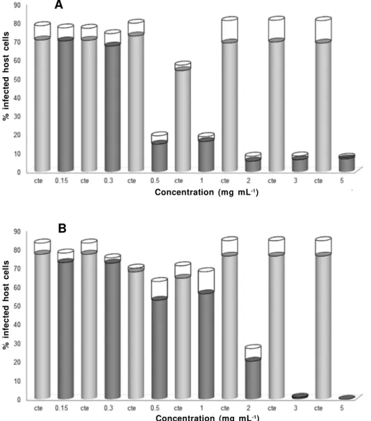
of aqueous extracts used in this study was similar to other data (Benencia et al., 2000) and no toxic effects were observed in the host cell. Neem progressively reduced the infection (Figure 1) by 2 mg mL-1 (60%),
3 mg mL-1 (75%) and 5 mg mL-1 (85%), respectively
(Figure 1a). This decrease in the infection is associated with the elimination of intracellular tachyzoites (Figure 1b). At lower concentrations (0.1 and 1 mg mL-1), no
significant effect was observed on the infection or intracellular parasite reduction (Figure 1a-b).
Similarly to neem, cinnamon also drastically reduced the infection, but anti-Toxoplasma effects
started at the lower concentration of 0.5 mg mL-1
(Figure 2a). This infection reduction (70%) was similar to that seen for the 1 mg mL-1 concentration. At higher
doses, a reduction of about 90% was observed (Figure 2a) and the number of intracellular parasites also
FIGURE 1. Percentage of infected cells (A) and mean number of intracellular tachyzoites (B) after treatment with
progressive concentration of neem extract for 24 h. Untreated infected cells (gray column); treated infected cells (dark column); standard error (open column). ***p<0.001; **p<0.01.
Concentration (mg mL-1)
Concentration (mg mL-1)
%
i
n
fe
c
te
d
h
o
s
t
c
e
ll
s
%
i
n
fe
c
te
d
h
o
s
t
c
e
ll
drastically reduced (Figure 2). Together, these results suggest that the antiparasitic effect of neem and cinnamon extracts is dependent on the concentration, leading to effective Toxoplasma elimination at
concentrations higher than 2 mg mL-1 and 0.5 mg
mL-1, respectively.
The reduction in the number of intracellular parasites is related to the accentuated and progressive morphologic disorganisation of tachyzoites within PV (Figure 3). Simultaneously to the increase in the concentration of extracts, there was a direct increase in the mean number of PV-containing disorganised parasites, which was more accentuated in treatment with cinnamon than in that with neem (Figure 3). These results suggest that in the presence of natural plant extracts PV-containing disorganised tachyzoites
B
FIGURE 2. Percentage of infected cells (A) and mean number of intracellular tachyzoites (B) after treatment with
progressive concentration of cinnamon extract for 24 h. Untreated infected cells (gray column); treated infected cells (dark column); standard error (open column). ***p<0.001; **p<0.01.
undergo progressive elimination.
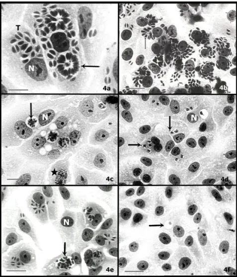
This morphological feature of parasite disorganisation was observed under light microscopy. In fact, the alteration in the morphology of intravacuolar tachyzoites promoted their gradual elimination (Figure 4). Untreated cultures showed tachyzoites (T) with a crescent shape (Figure 4a), multiplying inside the PV and forming a rosaceous structure (Figure 4a - arrow). Treatments of infected cells with lower concentrations of both extracts caused no alteration in the parasites, as exemplified by Melia treatment at 0.3 mg mL-1
(Figure 4b). The main features of disorganised parasites were the progressive disruption of the rosaceous structure, which became rounded (Figure 4c). Cinnamon treatment (1 mg mL-1) rapidly induced
morphologic disorganisation and elimination of the
Concentration (mg mL-1)
Concentration (mg mL-1)
M
e
a
n
n
u
m
b
e
r
o
f
in
tr
a
c
e
ll
u
la
r
ta
c
h
y
z
o
it
e
s
M
e
a
n
n
u
m
b
e
r
o
f
in
tr
a
c
e
ll
u
la
r
ta
c
h
y
z
o
it
e
FIGURE 3. Mean number of parasitophorous vacuole (PV) containing tachyzoite morphologically disorganized
during treatment with neem (gray line) and cinnamon (dark line).
FIGURE 4. Morphological aspects of intravacuolar parasites observed under light microscopy (a - f) during treatment
of extracts. Untreated cells (a); infected cells treated with 1 mg mL-1(c) and 2 mg mL-1 (e) of neem; cells treated for
24 h with 0.3 mg mL-1(b), 1 mg mL-1(d) and 3 mg mL-1(f) of cinnamon. Parasitophorous vacuole (arrow); tachyzoites
(T); N=nucleus; star= chromosomes; bar (a) =10
m; bar (b-f) = 40
m.Melia
intravacuolar tachyzoites (Figure 4d), while for neem treatment intravacuolar parasites with crescent shapes could be observed (Figure 4e). These antiparasitic effects of the extract in the infection were irreversible (data not shown). Despite the effective action of the extracts in arresting the parasite multiplication, host cells did not interrupt their cell cycle during treatment (Figure 4c -star). These results show that a progressive increase in the concentration of extracts induced no toxicity effects on host cells but led to an intense disorganisation and elimination of intravacuolar tachyzoites.
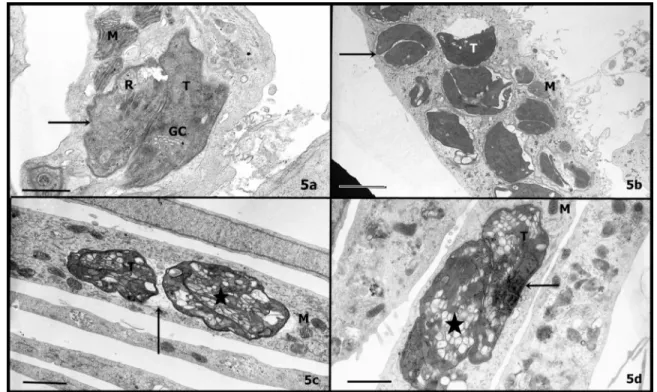
In addition, the morphologic observation of the effects caused by the extract treatments and the ultrastructural analyses of the infected cells and parasites were performed by transmission electronic microscopy. Host cells showed a homogeneous
cytoplasm, where organelles such as mitochondria and endoplasmic reticulum did not change their morphologic features (Figure 5), similarly to untreated cultures (Figure 5a). On the other hand, intravacuolar tachyzoites developed progressive vacuolisation. The incubation with a low concentration of neem (1 mg mL-1)
allowed the observation of a preliminary disorganisation in the intravacuolar parasites (Figure 5b), in spite of any alteration observed by optical microscopy at this same concentration, as shown before. In addition, at those concentrations where parasites were seen altered by optical microscopy, electron microscopy revealed high vacuolisation in the parasite cytoplasm (Figure 5c-d). The secretory systems of parasites, Golgi complex, rhoptries, micronemes and dense granule - were totally disrupted and a strong vesiculation could be noticed
FIGURE 5. Ultrastructural aspects of infected cells (a) treated with 1 mg mL-1(b) and 2 mg mL-1(d) of neem or 1 mg
mL-1 of cinnamon (c) for 24h. Host cell mitochondria (m); parasite Golgi complex (GC); vesiculation (star); Tachyzoite
(T); Parasitophorous vacuole (arrow). Bar (Figure a, c, d): 2
m. Bar (Figure b): 5
min the cytoplasm (Figure 5c-d). These vesicles have a membrane enclosing them and the parasite’s nucleus is highly condensed (Figure 5d).
These results suggest that, in the presence of aqueous extracts of neem or cinnamon, efficient interruption of parasite development and elimination of intracellular T. gondii occurred without induced toxic
alterations in the host cells.
DISCUSSION
Traditionally, neem extract exhibited insecticidal, spermicidal, antitumor, antiparasitic,
anthelmintic and anti-inflammatory properties. Cinnamon extract shows insecticidal, antiparasitic and antimicrobial properties. However, nothing is known about the action of these extracts on the intracellular development of Toxoplasma gondii.
Our results showed that the aqueous extracts of neem and cinnamon caused a progressive disorganisation in the intracellular tachyzoites, causing their elimination while the host cell suffered no toxic effects.
cinnamon are limonoids such as azadirachtins (AZ) (Huang et al., 1996; Nakatani et al., 1998; Kaushik,
2002; D’Ambrosio & Guerriero, 2002). The general biological action of these extracts is the induction of lipid peroxidation, generation of antiproliferative and antioxidant effects and detoxication of enzymes (Akudugu et al., 2001; Kumar et al., 2006). T. gondii
has a high lipid concentration in plasma and intraparasite membranes, including organelle content (Foussard et al., 1991). The integrity of the secretory system of T. gondii, as the Apicomplexan protozoan,
is vital to its invasion, survival and development. Our results showed that the secretory system of the parasite suffered drastic disorganisation and vesiculation, possibly because of the action of the chemical components of the extracts on their highly lipidic membranes. The anti-toxoplasma action of cinnamon was more efficient than that of neem, since its effect on the parasites was seen at a lower concentration. The disorganisation of the Golgi complex and any secretory organelles interrupts the survival of T. gondii in the cellular medium (Carvalho et al., 2009).
Other authors have also demonstrated that the interruption of secretory organelles has antiparasitic effects (Udenya, 2004; Totino et al., 2008; Sturm et al., 2009). We have demonstrated that antiproliferative drugs that arrest intracellular development lead to protozoan death and elimination (Melo et al., 2000; Melo & Beiral, 2003; Tenorio et al., 2005; De Aquino et al., 2008). Host cell strategies to eliminate intracellular parasites in the presence of antiproliferative drugs would be determinate, but in cases of hydroxyurea treatment, T. gondii was
eliminated via PV-containing parasites with lysosomes (Carvalho & Melo, 2006). However, previous studies also determined that under drug pressure the autophagic process has also been used as a microbicidal mechanism to eliminate virus, protozoa and bacteria and induce death by parasite cytoplasm vesiculation (Bera et al., 2003; Edinger & Thompson, 2004; Levine & Deretic, 2007; Totino et al., 2008). T. gondii vesiculations were observed during the treatment
with plant extracts, suggesting an autophagic process. In fact, such an autophagy has also been previously demonstrated as a microbicidal process in T. gondii
elimination by primed macrophages, where high intraparasite vesiculations were observed before their elimination (Ling et al., 2006; Andrade et al., 2006). However, new studies will be carried out to understand this disorganisation process and the relationship of the majority of the chemical components of the extracts, as well as the microbicidal mechanism involved in the elimination of intracellular T. gondii.
ACKNOWLEDGMENT
This work was supported by “Fundação Carlos
REFERENCE
AKUDUGU, J.; GÄDE, G.; BÖHM, L. Cytotoxicity of azadirachtin A in human glioblastoma cell lines. Life Sciences, v.68, p.1153-60, 2001.
ANDRADE, R.M. et al. CD40 induces macrophage anti-Toxoplasma gondii activity by triggering autophagy dependent fusion of pathogen-containing vacuoles and lysosomes. Journal of Clinical Investigation, v.116, n.9, p.2366-77, 2006.
ANTHONY, J.P.; FYFE, L.; SMITH, H. Plant active components - a resource for antiparasitic agents? Trends in Parasitology, v.21, n.10, p.462-8, 2005. BENENCIA, F.; COURREGES, M.C.; COULOMBIE, F.C. Anti-inflammatory activities of Thichilia glabra aqueous leaf. Jounal of Ethanopharmacology, v.71, p.293-300, 2000. BERA, A. et al. Induction of autophagic cell death in
Leishmania donovani by antimicrobial peptides.
Molecular & Biochemical Parasitology, v.127, p.23-35, 2003.
CARVALHO, C.S.; FIGUEIREDO, G.R.; MELO, E.J.T. Golgi-Disturbing Agents Lead to the Elimination Toxoplasma gondii. The Open Biology Journal, v.2, p.10-9, 2009. CARVALHO, C.S.; MELO, E.J.T. Acidification of the parasitophorous vacuole containing Toxoplasma gondii in the presence of hydroxyurea. Annals of the Brazilian Academy of Science, v.78, n.3, p.475-84, 2006. D’AMBROSIO, M.; GUERRIERO, A. Degraded limonoids from Melia azedarach and biogenetic Implications. Phytochemistry, v.60, n.4, p.419-24, 2002.
DE AQUINO, T.M. et al. Synthesis; anti-Toxoplasma gondii and antimicrobial activities of benzaldehyde 4-phenyl-3-thiosemicarbazones and 2-[(phenylmethylene) hydrazono]-4-oxo-3-phenyl-5-thiazolidineacetic acids. Bioorganic & Medicinal Chemistry Letters, v.16, p.446-56, 2008.
DHAR, R. el al. Inhibition of the growth and development of asexual and sexual stages of drug-sensitive and resistant strains of the human malaria parasite Plasmodium falciparum by Neem (Azadirachta indica) fractions. Journal of Ethnopharmacology, v.61, p.31-9, 1998.
EDINGER, A.L.; THOMPSON, C.B. Death by design: apoptosis; necrosis and autophagy. Current Opinion in Cell Biology, v.16, p.663-9, 2004.
FOUSSARD, F.; LERICHE, M.A.; DUBREMETZ, J.F. Characterization of the lipid content of Toxoplasma gondii rhoptries. Parasitology, v.102, p.367-70, 1991.
HUANG, R.C. et al. Limonoids from Melia azedarach. Phytochemistry, v.43, n.3, p.581-3, 1996.
KAUSHIK, N. Determination of azadirachtin and fatty acid methyl esters of Azadirachta indica seeds by HPLC and GLC. Analytical and Bioanalytical Chemistry, v.374, p.1199-204, 2002.
KHAN, M.R.; KIHARA, M.; OMOLOSO, A.D. Antimicrobial
Chagas Filho de Amparo à Pesquisa do Estado do Rio de Janeiro” (FAPERJ - Proc. No E-26/111.616/
activity of Horsfieldia helwigii and Melia azedarach; Fitoterapia, v.72, p.423-7, 2001.
KOLODZIEJ, H.; KIDERLEN, A.F. Antileishmanial activity and immune modulatory effects of tannins and related compounds on Leishmania parasitized RAW 264.7 cells; Phytochemistry, v.66, n.17, p.2056-71, 2005.
KUMAR, S. et al. Anticancer effects of ethanolic neem leaf extract on prostate cancer cell line (PC-3). Journal
of Ethnopharmacology, v.105, p.246-50, 2006.
LEVINE, B.; DERETIC, V. Unveiling the roles of autophagy in innate and adaptive immunity. Nature Reviews in Immunology, v.3, p.1-11, 2007.
LING, Y.M. et al. Vacuolar and plasma membrane stripping and autophagic elimination of Toxoplasma gondii in primed effector macrophages. Journal of Experimental Medicine, v.203, p.2063-7, 2006.
LIRUSSI, D. et al. Inhibition of Trypanosoma cruzi by plant extracts used in Chinese medicine. Fitoterapia, v.75, p.718-23, 2004.
McALLISTER, M.M. A decade of discoveries in veterinary protozoology changes our concept of ‘‘subclinical’’ toxoplasmosis. Veterinary Parasitology, v.132, p.241-7, 2005.
MELO, E.J.T.; BEIRAL, H.J. Effect of hydroxyurea on the intracellular multiplication of Toxoplasma gondii;
Leishmania amazonensis and Trypanosoma cruzi.
Brazilian Journal of Medical and Biological Research,
v.36, p.1-5, 2003.
MELO, E.J.T.; MAYERHOFFER, R.O.; DE SOUZA, W.
Hydroxyurea inhibits intracellular Toxoplasma gondii multiplication. FEMS Microbiology Letters, v.185, p.79-82, 2000.
NAKATANI, M. et al. Degraded limonoids from Melia azedarach. Phytochemistry, v.49, n.6, p.1773-6, 1998. NEWMAN, D.J.; CRAGG, G.M.; SNADER, K.M. Natural Products as Sources of New Drugs over the Period 1981-2002. Journal Natural Products, v.66, p.1022-37, 2003. SALEHZADEH, A. et al. The antimitotic effect of the neem terpenoid azadirachtin on cultured insect cells. Insect Biochemical and Molecular Biology, v.33, p.681-9, 2003. STURM, A. et al. Alteration of the parasite plasma membrane and the parasitophorous vacuole membrane during exo-erythrocytic development of malaria parasites. Protist, v.160, p.51-63, 2009.
SUKTHANA, Y. Toxoplasmosis: beyond animals to humans. Trends in Parasitology, v.22, n.3, p.137-42, 2006. TENÓRIO, R.P. et al. Synthesis of thiosemicarbazone and 4-thiazolidinone derivates and their in vitro anti-Toxoplasma gondii activity. Bioorganic & Medicinal Chemistry Letters, v.15, p.2575-8, 2005.
TOTINO, P.R.R. et al. Plasmodium falciparum: Erythrocytic stages die by autophagic-like cell death under drug pressure. Experimental Parasitology, v.118, p.478-86, 2008. UDENYA, I.J. et al. An antimalarial extract from neem leaves is antiretroviral. Royal Society of Tropical Medicine and Hygiene, v.98, p.435-7, 2004.