Dimensions and dynamics in
integrin function
1Craniofacial Developmental Biology and Regeneration Branch,
National Institute of Dental and Craniofacial Research, National Institutes of Health, Bethesda, MD, USA
2Basic Science Division, Fox Chase Cancer Center, Philadelphia, PA, USA
K.M. Yamada1,
R. Pankov1 and
E. Cukierman1,2
Abstract
Integrins play crucial roles in cell adhesion, migration, and signaling
by providing transmembrane links between the extracellular matrix
and the cytoskeleton. Integrins cluster in macromolecular complexes
to generate cell-matrix adhesions such as focal adhesions. In this
mini-review, we compare certain integrin-based biological responses and
signaling during cell interactions with standard 2D cell culture versus
3D matrices. Besides responding to the composition of the matrix,
cells sense and react to physical properties that include
three-dimen-sionality and rigidity. In routine cell culture, fibroblasts and
mesen-chymal cells appear to use focal adhesions as anchors. They then use
intracellular actomyosin contractility and dynamic, directional
inte-grin movements to stretch cell-surface fibronectin and to generate
characteristic long fibrils of fibronectin in “fibrillar adhesions”. Some
cells in culture proceed to produce dense, three-dimensional matrices
similar to
in vivo
matrix, as opposed to the flat, rigid, two-dimensional
surfaces habitually used for cell culture. Cells within such more
natural 3D matrices form a distinctive class of adhesion termed
“3D-matrix adhesions”. These 3D adhesions show distinctive morphology
and molecular composition. Their formation is heavily dependent on
interactions between integrin
α
5ß
1and fibronectin. Cells adhere much
more rapidly to 3D matrices. They also show more rapid
morphologi-cal changes, migration, and proliferation compared to most 2D
matri-ces or 3D collagen gels. Particularly notable are low levels of tyrosine
phosphorylation of focal adhesion kinase and moderate increases in
activated mitogen-activated protein kinase. These findings underscore
the importance of the dimensionality and dynamics of matrix
sub-strates in cellular responses to the extracellular matrix.
Correspondence K.M. Yamada CDBRB, NIDCR, NIH Building 30, Room 421 30 Convent Drive MSC 4370 Bethesda, MD 20892-4370 USA
Fax: +1-301-402-0897 E-mail: [email protected]
Presented at SIMEC 2002 (International Symposium on Extracellular Matrix), Angra dos Reis, RJ, Brazil, October 7-10, 2002.
Received February 27, 2003 Accepted April 15, 2003
Key words
•Extracellular matrix •Cell adhesion •Integrin •Fibronectin •Signal transduction •Migration
Introduction
Integrins are the major vertebrate
recep-tors for extracellular matrix molecules.
Be-sides mediating cell adhesion and migration,
they help to regulate critical cellular
signal-ing processes that range from cell growth
will focus on roles of integrins in the
forma-tion of a fibrillar extracellular matrix and
then on the cellular responses to
extracellu-lar matrix from our personal perspective. We
will review studies showing that, besides its
biochemical composition, important features
of the extracellular matrix that govern the
cellular responses it elicits include its
three-dimensionality and its deformability or
ri-gidity. In-depth reviews of other aspects of
integrin-based cell-matrix interactions with
many more citations than possible in this
mini-review have been published previously
(1-10,14-19).
Formation of extracellular matrices
-the importance of dynamics
Although the capacity of purified
col-lagen to self-assemble into fibrils in the
ab-sence of cells has been a central conceptual
paradigm of matrix biochemistry, the way in
which collagen molecules produced by
liv-ing cells actually become organized into
ex-tracellular fibrillar matrices
in vivo
appears
to be under cellular control. In fact, both
older and very recent studies provide strong
evidence that the fibrillar organization of
collagens type I and III, at least in the early
stages of organization, depends on the
pre-ceding organization of another matrix
pro-tein - fibronectin (20-22). Since other
extra-cellular molecules also often co-localize with
fibronectin fibrils, it is obviously important
to understand how cells form fibrils of
fibro-nectin. Much is now known about the
bio-chemistry of fibronectin matrix assembly
(7,23). Crucial elements identified to date
include the binding of soluble fibronectin
molecules by integrins and then exposure of
cryptic fibronectin self-assembly sites to
pro-mote self-association; the mechanism for
exposure of specific cryptic sites is thought
to involve tension (24,25), i.e., stretching of
fibronectin by intracellular contractility (7,23,
26-29).
Integrin and fibronectin dynamics on the
cell surface can be tracked by using
non-perturbing antibody tracers and directly
la-beled fibronectin (30). Different patterns of
integrin behavior were identified for two
types of integrin receptors. The multi-ligand
receptor
α
Vß
3remains within focal
adhe-sions. In contrast, the fibronectin receptor
α
5ß
1translocates out of focal contacts at 6.5
µm/h along fibrillar adhesions (previously
termed “extracellular matrix contacts”) in
parallel to actin microfilament bundles (30).
Fibronectin is translocated in a very similar
pattern (28,30). Tensin is a primary
cyto-skeletal component of fibrillar adhesions,
and it also moves in a parallel fashion (31).
In fact, a dominant-negative inhibitor of
tensin blocks integrin translocation, fibrillar
adhesion formation, and fibronectin
fibrillo-genesis without affecting focal adhesions
(30). As shown diagrammatically in Figure
1, we proposed that translocating
α
5ß
1inte-grins induce initial fibronectin
fibrillogen-esis by transmitting cytoskeleton-generated
tension to extracellular fibronectin molecules.
In fact, blocking this integrin translocation
by a variety of treatments prevents both the
formation of fibrillar adhesions and
fibro-nectin fibrillogenesis. Thus, fibrofibro-nectin
fibrils appear to be generated by a process of
localized, directional, integrin translocation,
which exposes cryptic domains on the
fibro-nectin molecule, allowing polymerization
into fibers and resulting in matrix assembly.
Cellular responses to extracellular
matrices - the importance of
dimensions
in collagen gels under different degrees of
tension behave quite differently (40-43), and
epithelial cells in three-dimensional
envi-ronments show major differences in
biosyn-thesis of differentiated products (44) or in
responses to apoptotic stimuli (45,46).
Although a variety of systems have been
developed to mimic physiological
three-di-mensional environments (36,37), a recent
approach involves using tissue- or cell
cul-ture-derived three-dimensional matrices for
analyzing the behavior of migratory and
mes-enchymal cells (47,48). The logic behind
this approach is that cells do not normally
encounter a pure collagen matrix, nor a
multi-millimeter-thick basement membrane.
In-stead, they exist in complex environments
comprised of multiple molecules and a
sub-stantial degree of fibrillar organization with
complexity beyond that provided by
col-lagen gels. Recent findings indicate that cells
respond differently to such complex 3D
ma-trices compared to collagen gels or flat tissue
culture substrates.
Cryostat sections of tissues of embryos
from which cellular contents have been
extracted with alkaline detergent solution
provide a well-organized fibrillar
extracellu-lar matrix that can be used as a system for
culturing fibroblastic cells in three
dimen-sions. It can be mimicked effectively by
tracting morphologically similar dense
ex-tracellular matrices accumulated by certain
cultured fibroblasts after multiple days of
cell culture. In both cases, a key component
of these matrices is fibronectin, along with
collagen I and heparan sulfate proteoglycan
(Cukierman E, unpublished results). Cells
plated into such matrices show greater than
6-fold rates of cell attachment compared to
purified collagen gels, as well as compared
to substrates of purified fibronectin,
lami-nin, or collagen I (47). As initially
estab-lished for fibroblasts and other cells plated
in collagen gels (32,33,49), they acquire an
in vivo
-like fibroblastic shape that is slender
and elongated, rather than flattened and
tri-angular (Figure 2). However, the rate at
which human fibroblasts acquire such
mor-phology is much faster in the natural 3D
matrices compared to collagen gels; the final
morphology is also slightly different.
Inter-estingly, even rates of cell proliferation
ap-pear to be augmented more than 2-fold above
proliferation rates on the excellent cell
cul-ture substrate fibronectin. Finally, cell
mi-gration is augmented on such matrices
com-pared to fibronectin, collagen, or 3D
col-lagen gels (47).
Are the effects of complex matrices due to
the individual molecular components or to the
be a combination: flattening the 3D matrices
by mechanical force (using a
Teflon-cov-ered lead weight) or by coating culture
sub-strates with all of the same molecules using a
mixture of 3D matrix components
solubi-lized by guanidine (followed by dialysis) (48)
could not mimic any of the enhancing effects
of 3D matrix on attachment, spreading, or
proliferation. Conversely, not only does
pro-viding a collagenous three-dimensional
ma-trix not suffice, but even a fibrillar mama-trix
formed from purified fibronectin does not,
implying that some molecular component in
the 3D matrix besides fibronectin is needed
(47).
The specific type of integrin used by cells
for interactions with an extracellular matrix
seems to be matrix-specific. In collagen gels,
the predominant functional integrin is
α
2ß
1(50). In contrast, in each of the dramatic
effects observed by 3D matrices on cell
at-tachment, morphology, migration, and
pro-liferation, the dominant integrin involved is
the fibronectin receptor
α
5ß
1integrin (47).
Another fibronectin receptor, the
α
Vß
3inte-grin, is also important for cell interactions
with purified fibronectin itself (requiring
antibody inhibition by both anti-
α
Vand
α
5integrins), yet 65-88% of the cellular
re-sponses of human fibroblasts to 3D matrices
can be suppressed by antibody against
α
5while only 27-36% inhibition is observed by
the same antibody for fibroblasts on 2D
fi-bronectin. This intriguing finding identifies
a striking dominance or preference for this
integrin when cells interact with 3D matrix
-it suggests that there may be e-ither a
hyperactivation of function of the
α
5ß
1inte-grin, a suppression of functions of other
integrins, or some combination of these novel
actions in an intriguing system of integrin
crosstalk.
The adhesions formed by fibroblastic,
mesenchymal, and neural crest cells to 3D
matrices are distinct from both the focal
adhesions and the fibrillar adhesions
tradi-tionally described and studied in cell culture
(7,16,47,51) (Figure 2). These novel
“3D-matrix adhesions” contain many of the same
plaque proteins as are found in focal
adhe-sions, such as paxillin, vinculin, and focal
adhesion kinase (FAK). However, they lack
the
α
Vß
3integrin, and they are instead based
on the
α
5ß
1integrin (Table 1).
Most intriguingly, unlike the very
well-studied tyrosine-phosphorylated forms of
FAK in focal adhesions, the FAK in
3D-matrix adhesions is poorly phosphorylated
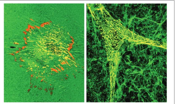
Figure 2. Comparison of fibro-blast cell shapes and localization of components in 2-dimensional and 3-dimensional environ-ments. Cells on 2D substrates tend to spread onto the sub-strate in a flattened morphology (left panel), whereas cells at-taching to 3D matrices rapidly assume an elongated morpholo-gy (right panel) that tends to mimic shapes of fibroblastic and mesenchymal cells in vivo. The α5 integrin (red) localizes to
at its major tyrosine 397 phosphorylation
site. This discrepancy in signaling in 2D
versus 3D contexts is specific in that paxillin
is equally phosphorylated at tyrosine 31 in
both 3D-matrix adhesions and focal
adhe-sions. Moreover, the extent of
mitogen-acti-vated protein kinase (extracellular
signal-regulated kinase, ERK) phosphorylation is
moderately elevated (47), which may help
contribute to the enhanced rate of
prolifera-tion in 3D matrix. Differences in signaling
processes have also been reported in cells
suspended in three-dimensional collagen gels
when compared to 2D cultures (52-54), or
under different loading conditions for
col-lagen gel contraction measurements (40-42).
The important conclusion from these reports
is that studies of signaling in regular cell
culture may provide misleading results: cells
may have different signaling responses when
they exist in three-dimensional settings that
mimic
in vivo
conditions more closely.
Fu-ture studies of cell interactions with
extra-cellular matrix and signal transduction should
therefore ideally be designed with these
prin-ciples in mind.
Cellular responses to extracellular
matrices - the importance of matrix
rigidity
A previous analysis of the role of matrix
pliability/rigidity revealed that the ability of
cells to physically move and rearrange
fibro-nectin was important in determining the
na-ture of the cell adhesions that form on a 2D
substrate. If fibronectin is cross-linked to a
substrate, rather than being adsorbed and
able to be remodeled, it results in
exagger-ated focal adhesions and trapping of the
α
5integrin in these adhesions rather than
translo-cating away to form fibrillar adhesions (55).
Not surprisingly, fibronectin matrix assembly
was blocked in the absence of these latter
adhesions involved in generating fibronectin
fibrils (30,55). This response to immobilized
fibronectin may be due to the development
of increased tension within cells against the
fixed fibronectin substrate. Effects of local
tension can be mimicked by experimentally
applying external physical tension near focal
adhesions, which causes enlargement of these
structures; there is a dynamic relationship
between the forces on adhesions and their
size and function (7,56,57).
If 3D matrices are stiffened and
immobi-lized by chemical cross-linking, a similar
enhancement of focal adhesion-like
struc-tures occurs. Normal 3D-matrix adhesions
are lost, and
α
5integrins remain confined to
focal adhesions (47). Thus, one can view the
pliability or capacity for rearrangement of
matrix components as being another
impor-tant element of cellular responses to the
extracellular matrix, along with its
biochemi-cal composition and three-dimensionality
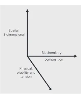
(Figure 3).
Molecule Focal
adhesion (mature)
Fibrillar
adhesion 3D-matrixadhesion
α5 integrin
ß1 integrin
ß3 integrin
paxillin
tensin
talin
vinculin phospho-tyrosine FAK
phospho-FAK [Y397]
Table 1. Molecular compositions of three types of cell adhesions.
Spatial: 3-dimensional
Biochemistry:
composition
Physical: pliability and
tension Figure 3. Cells respond to distinct physical and biochemical characteristics of the
extracellular matrix. Although it is well known that cells respond in differing fashion to distinct matrix molecules (i.e., composition of the matrix), they also respond to differences in pliability of the matrix, to tension forces, and to the spatial nature of the matrix (3D versus 2D). These responses result in a variety of adhesion structures formed in response to diverse environmental cues.
Figure 4. Summary of cellular re-sponses to the extracellular ma-trix. The responses of cells to extracellular matrix differ de-pending on its organization and the type of cell adhesion. Focal adhesions are localized sites of firm anchorage to a relatively rigid substrate molecule such as vitronectin, in which integrins such as αVß3 organize intracellu-lar complexes of adhesion com-plex components that include fo-cal adhesion kinase (FAK), paxillin, and tensin, as well as classical signaling molecules such as Src and ERK. In addition, novel components such as kinectin (58) are being discov-ered; the number of components is already greater than thirty, and more are expected. Fibrillar ad-hesions extend towards the cell center from the edges of focal adhesions. They consist of the α5ß1 fibronectin receptor in
Concluding remarks
Integrin-mediated cell interactions with
extracellular matrix now appear to be
con-siderably more complex and sophisticated
than originally envisioned. The existence of
different types of adhesions in the same cell,
as well as differences in the molecular
com-position of adhesions, signaling, and
bio-logical functions in two-dimensional versus
three-dimensional environments (Figure 4)
provides both enlightenment and a concern.
It seems obvious in retrospect that different
functions of cell-matrix interactions should
require different types of adhesions (i.e.,
focal adhesions for the strongest level of
adhesion and fibrillar adhesions for
permit-ting dynamic assembly of matrix fibrils). On
the other hand, focal adhesions can
them-selves translocate under certain conditions
(59), and the existence of multiple types of
adhesions forces us to re-think concepts about
the actual sites from which
integrin-medi-ated signaling may originate. A concern,
which is probably healthy, is that studies of
different cell types and different model
ma-trices, whether 2D or 3D, are likely to give
different answers to experimental questions
as the cells utilize different types of adhesion
structures. However, the comforting aspect
of this complexity is that it is now clear that
cells have many ways in which to
accom-plish the subtle variations and major changes
in function that they require for maintaining
normal physiological function in the intact
organism and in responding to injury and
other challenges.
References
1. Giancotti FG & Ruoslahti E (1999). Integrin signaling.Science, 285: 1028-1032.
2. Humphries MJ (2000). Integrin structure.Biochemical Society Trans-actions, 28: 311-339.
3. Hynes RO (2002). Integrins: Bidirectional, allosteric signaling ma-chines.Cell, 110: 673-687.
4. Miranti CK & Brugge JS (2002). Sensing the environment: A histori-cal perspective on integrin signal transduction.Nature Cell Biology, 4: E83-E90.
5. Yamada KM & Danen EHJ (2000). Integrin signaling. In: Gutkind JS (Editor), Signaling Networks and Cell Cycle Control. Humana Press, Totowa, NJ, USA, 1-25.
6. Schwartz MA & Ginsberg MH (2002). Networks and crosstalk: Integrin signalling spreads.Nature Cell Biology, 4: E65-E68. 7. Geiger B, Bershadsky A, Pankov R & Yamada KM (2001).
Trans-membrane cross-talk between the extracellular matrix and the cyto-skeleton.Nature Reviews. Molecular Cell Biology, 2: 793-805. 8. Danen EH & Yamada KM (2001). Fibronectin, integrins, and growth
control. Journal of Cellular Physiology, 189: 1-13.
9. Damsky CH & Ilic D (2002). Integrin signaling: It’s where the action is.Current Opinion in Cell Biology, 14: 594-602.
10. Aplin AE, Howe AK & Juliano RL (1999). Cell adhesion molecules, signal transduction and cell growth.Current Opinion in Cell Biology, 11: 737-744.
11. Stupack DG & Cheresh DA (2002). Get a ligand, get a life: Integrins, signaling and cell survival.Journal of Cell Science, 115: 3729-3738. 12. Springer TA (2002). Predicted and experimental structures of inte-grins and beta-propellers.Current Opinion in Structural Biology, 12: 802-813.
13. Arnaout M, Goodman S & Xiong J (2002). Coming to grips with integrin binding to ligands.Current Opinion in Cell Biology, 14:
641-651.
14. Brown E (2002). Integrin-associated proteins. Current Opinion in Cell Biology, 14: 603-607.
15. Howe AK, Aplin AE & Juliano RL (2002). Anchorage-dependent ERK signaling - mechanisms and consequences.Current Opinion in Ge-netics and Development, 12: 30-35.
16. Sastry SK & Burridge K (2000). Focal adhesions: A nexus for intra-cellular signaling and cytoskeletal dynamics.Experimental Cell Re-search, 261: 25-36.
17. Shimaoka M, Takagi J & Springer TA (2002). Conformational regula-tion of integrin structure and funcregula-tion.Annual Review of Biophysics and Biomolecular Structure, 31: 485-516.
18. Yamada KM & Even-Ram S (2002). Integrin regulation of growth factor receptors.Nature Cell Biology, 4: E75-E76.
19. Takagi J & Springer TA (2002). Integrin activation and structural rearrangement.Immunological Reviews, 186: 141-163.
20. McDonald JA, Kelley DG & Broekelmann TJ (1982). Role of fibro-nectin in collagen deposition: Fab’ to the gelatin-binding domain of fibronectin inhibits both fibronectin and collagen organization in fibroblast extracellular matrix.Journal of Cell Biology, 92: 485-492. 21. Velling T, Risteli J, Wennerberg K, Mosher DF & Johansson S
(2002). Polymerization of type I and III collagens is dependent on fibronectin and enhanced by integrins alpha 11 beta 1 and alpha 2 beta 1.Journal of Biological Chemistry, 277: 37377-37381. 22. Sottile J & Hocking DC (2002). Fibronectin polymerization regulates
the composition and stability of extracellular matrix fibrils and cell-matrix adhesions.Molecular Biology of the Cell, 13: 3546-3559. 23. Schwarzbauer JE & Sechler JL (1999). Fibronectin fibrillogenesis: A
paradigm for extracellular matrix assembly.Current Opinion in Cell Biology, 11: 622-627.
Structural insights into the mechanical regulation of molecular rec-ognition sites.Trends in Biotechnology, 19: 416-423.
25. Hocking DC, Sottile J & McKeown-Longo PJ (1998). Activation of distinct alpha5beta1-mediated signaling pathways by fibronectin’s cell adhesion and matrix assembly domains.Journal of Cell Biology, 141: 241-253.
26. Zhong C, Chrzanowska-Wodnicka M, Brown J, Shaub A, Belkin AM & Burridge K (1998). Rho-mediated contractility exposes a cryptic site in fibronectin and induces fibronectin matrix assembly.Journal of Cell Biology, 141: 539-551.
27. Pankov R & Yamada KM (2002). Fibronectin at a glance.Journal of Cell Science, 115: 3861-3863.
28. Ohashi T, Kiehart DP & Erickson HP (2002). Dual labeling of the fibronectin matrix and actin cytoskeleton with green fluorescent protein variants.Journal of Cell Science, 115: 1221-1229. 29. Ohashi T, Kiehart DP & Erickson HP (1999). Dynamics and elasticity
of the fibronectin matrix in living cell culture visualized by fibronec-tin-green fluorescent protein.Proceedings of the National Academy of Sciences, USA, 96: 2153-2158.
30. Pankov R, Cukierman E, Katz BZ, Matsumoto K, Lin DC, Lin S, Hahn C & Yamada KM (2000). Integrin dynamics and matrix assembly: Tensin-dependent translocation of α5ß1 integrins promotes early fibronectin fibrillogenesis.Journal of Cell Biology, 148: 1075-1090. 31. Zamir E, Katz M, Posen Y et al. (2000). Dynamics and segregation of
cell-matrix adhesions in cultured fibroblasts.Nature Cell Biology, 2: 191-196.
32. Hay ED (1991). Cell Biology of Extracellular Matrix. 2nd edn. Vol. 1. Plenum Press, New York.
33. Friedl P & Brocker EB (2000). The biology of cell locomotion within three-dimensional extracellular matrix. Cellular and Molecular Life Sciences, 57: 41-64.
34. Friedl P & Brocker EB (2000). T cell migration in three-dimensional extracellular matrix: Guidance by polarity and sensations. Develop-mental Immunology, 7: 249-266.
35. Bissell MJ & Radisky D (2001). Putting tumours in context.Nature Reviews. Cancer, 1: 46-54.
36. Cukierman E, Pankov R & Yamada KM (2002). Cell interactions with three-dimensional matrices. Current Opinion in Cell Biology, 14: 633-639.
37. Walpita D & Hay E (2002). Studying actin-dependent processes in tissue culture.Nature Reviews. Molecular Cell Biology, 3: 137-141. 38. Bissell MJ, Radisky DC, Rizki A, Weaver VM & Petersen OW (2002). The organizing principle: Microenvironmental influences in the nor-mal and nor-malignant breast.Differentiation, 70: 537-546.
39. Hagios C, Lochter A & Bissell MJ (1998). Tissue architecture: The ultimate regulator of epithelial function? Philosophical Transactions of the Royal Society of London. Series B: Biological Sciences, 353: 857-870.
40. Halliday NL & Tomasek JJ (1995). Mechanical properties of the extracellular matrix influence fibronectin fibril assembly in vitro. Experimental Cell Research, 217: 109-117.
41. Grinnell F (2000). Fibroblast-collagen-matrix contraction: Growth-factor signalling and mechanical loading.Trends in Cell Biology, 10: 362-365.
42. Grinnell F & Ho CH (2002). Transforming growth factor beta stimu-lates fibroblast-collagen matrix contraction by different mechan-isms in mechanically loaded and unloaded matrices.Experimental Cell Research, 273: 248-255.
43. Brown RA, Sethi KK, Gwanmesia I, Raemdonck D, Eastwood M & Mudera V (2002). Enhanced fibroblast contraction of 3D collagen lattices and integrin expression by TGF-beta1 and -beta3:
Mechanoregula-tory growth factors? Experimental Cell Research, 274: 310-322. 44. Aggeler J, Park CS & Bissell MJ (1988). Regulation of milk protein
and basement membrane gene expression: The influence of the extracellular matrix.Journal of Dairy Science, 71: 2830-2842. 45. Weaver VM, Lelievre S, Lakins JN, Chrenek MA, Jones JC, Giancotti
F, Werb Z & Bissell MJ (2002). Beta4 integrin-dependent formation of polarized three-dimensional architecture confers resistance to apoptosis in normal and malignant mammary epithelium. Cancer Cell, 2: 205-216.
46. Debnath J, Mills KR, Collins NL, Reginato MJ, Muthuswamy SK & Brugge JS (2002). The role of apoptosis in creating and maintaining luminal space within normal and oncogene-expressing mammary acini.Cell, 111: 29-40.
47. Cukierman E, Pankov R, Stevens DR & Yamada KM (2001). Taking cell-matrix adhesions to the third dimension.Science, 294: 1708-1712.
48. Cukierman E (2002). Preparation of extracellular matrices produced by cultured fibroblasts. In: Bonifacino JS, Dasso M, Lippincott-Schwartz J, Harford JB & Yamada KM (Editors), Current Protocols in Cell Biology. John Wiley & Sons, New York, 10.19.11-10.19.14. 49. Elsdale T & Bard J (1972). Collagen substrata for studies on cell
behavior.Journal of Cell Biology, 54: 626-637.
50. Maaser K, Wolf K, Klein CE, Niggemann B, Zanker KS, Brocker EB & Friedl P (1999). Functional hierarchy of simultaneously expressed adhesion receptors: Integrin alpha2beta1 but not CD44 mediates MV3 melanoma cell migration and matrix reorganization within three-dimensional hyaluronan-containing collagen matrices. Molec-ular Biology of the Cell, 10: 3067-3079.
51. Geiger B & Bershadsky A (2002). Exploring the neighborhood: Ad-hesion-coupled cell mechanosensors.Cell, 110: 139-142. 52. Ishii I, Tomizawa A, Kawachi H et al. (2001). Histological and
func-tional analysis of vascular smooth muscle cells in a novel culture system with honeycomb-like structure. Atherosclerosis, 158: 377-384.
53. Ravanti L, Heino J, Lopez-Otin C & Kahari V-M (1999). Induction of collagenase-3 (MMP-13) expression in human skin fibroblasts by three-dimensional collagen is mediated by p38 mitogen-activated protein kinase.Journal of Biological Chemistry, 274: 2446-2455. 54. Vaalamo M, Mattila L, Johansson N, Kariniemi AL,
Karjalainen-Lindsberg ML, Kahari VM & Saarialho-Kere U (1997). Distinct popu-lations of stromal cells express collagenase-3 (MMP-13) and colla-genase-1 (MMP-1) in chronic ulcers but not in normally healing wounds.Journal of Investigative Dermatology, 109: 96-101. 55. Katz BZ, Zamir E, Bershadsky A, Kam Z, Yamada KM & Geiger B
(2000). Physical state of the extracellular matrix regulates the struc-ture and molecular composition of cell-matrix adhesions.Molecular Biology of the Cell, 11: 1047-1060.
56. Riveline D, Zamir E, Balaban NQ, Schwarz US, Ishizaki T, Narumiya S, Kam Z, Geiger B & Bershadsky AD (2001). Focal contacts as mechanosensors: Externally applied local mechanical force induces growth of focal contacts by an mDia1-dependent and ROCK-inde-pendent mechanism.Journal of Cell Biology, 153: 1175-1186. 57. Balaban NQ, Schwarz US, Riveline D et al. (2001). Force and focal
adhesion assembly: A close relationship studied using elastic micropatterned substrates.Nature Cell Biology, 3: 466-472. 58. Tran H, Pankov R, Tran SD, Hampton B, Burgess WH & Yamada KM
(2002). Integrin clustering induces kinectin accumulation.Journal of Cell Science, 115: 2031-2040.