www.bjournal.com.br
Volume 42 (9) 776-869 September 2009
Institutional Sponsors
The Brazilian Journal of Medical and Biological Research is partially financed by
Braz J Med Biol Res, September 2009, Volume 42(9) 796-803
Immune strategies using single-component LipL32 and
multi-component recombinant LipL32-41-OmpL1 vaccines against
leptospira
Immune strategies using single-component
LipL32 and multi-component recombinant
LipL32-41-OmpL1 vaccines against leptospira
C.Y. Feng
1,2,
Q.T. Li
1,
X.Y. Zhang
1,
K. Dong
1, B.Y. Hu
1and X.K. Guo
11Department of Microbiology and Parasitology, Institutes of Medical Sciences,
Shanghai Jiao Tong University School of Medicine, Shanghai, China
2Department of Basic Medicine, Zhejiang Medical College, Zhejiang Hangzhou, China
Abstract
Genes encoding lipoproteins LipL32, LipL41 and the outer-membrane protein OmpL1 of leptospira were recombined and cloned into a pVAX1 plasmid. BALB/c mice were immunized with LipL32 and recombined LipL32-41-OmpL1 using DNA, DNA-protein and DNA-protein-DNA-protein strategies, respectively. Prime immunization was on day 1, boost immunizations were on day 11 and day 21. Sera were collected from each mouse on day 35 for antibody, cytokine detection and microscopic agglutination test while spleen cells were collected for splenocyte proliferation assay. All experimental groups (N = 10 mice per group) showed
statistically significant increases in antigen-specific antibodies, in cytokines IL-4 and IL-10, as well as in the microscopic agglu -tination test and splenocyte proliferation compared with the pVAX1 control group. The groups receiving the recombined LipL32-41-OmpL1 vaccine induced anti-LipL41 and anti-OmpL1 antibodies and yielded better splenocyte proliferation values than the groups receiving LipL32. DNA prime and protein boost immune strategies stimulated more antibodies than a DNA-DNA immune strategy and yielded greater cytokine and splenocyte proliferation than a protein-protein immune strategy. It is clear from these results that recombination of protective antigen genes lipL32, lipL41, and ompL1 and a DNA-protein immune strategy resulted in better immune responses against leptospira than single-component, LipL32, or single DNA or protein immunization.
Key words: Immune strategy; Recombinant vaccine; Leptospira; Lipoproteins LipL32 and LipL41
Introduction
Correspondence: Q.T. Li, Department of Microbiology and Parasitology, Institutes of Medical Sciences, Shanghai Jiao Tong University School of Medicine, Shanghai 200025, China. E-mail: qingtianli@gmail.com
Research supported in part by the National Natural Science Foundation of China (#30830002, #30670102, and #30770820), the National Key Program for Infectious Diseases of China (#2008ZX10004-002, #2008ZX10004-009, #2009ZX10004-712), Program of Shanghai Subject Chief Scientist (#09XD1402700) and the National High Technology Research and Development Program of China.
Received November 26, 2008. Accepted May 27, 2009.
Leptospirosis is a severe spirochetal zoonosis with global impact. Reservoir hosts with chronic renal tubular infection transmit pathogenic leptospira species to new hosts through urinary shedding. Recently, leptospirosis has become prevalent in cities with sanitation problems and in large populations of urban rodent reservoirs (1). This acute
febrile disease can cause flu-like episodes with severe renal
and hepatic damage, such as hemorrhage and jaundice. In more severe cases, massive pulmonary hemorrhages or even fatal sudden hemoptysis can occur.
Whole-cell leptospirosis vaccines have been used to protect against several serovars of leptospira, including
icterohaemorrhagiae, grippotyphosa and pomona. Un-fortunately, currently available whole-cell vaccines fail to induce long-term protection against leptospirosis and cannot provide cross-protection against infection with the more than 250 known leptospira serovars (2). The success of subunit vaccines has been hampered by weak or short-term immunity and unavailability of nontoxic, potent adjuvants (3). Thus, new vaccine strategies are needed to prevent leptospirosis. The complete genomic DNA sequence of pathogenic Leptospira interrogans serovar Lai strain Lai (4)
and Copenhageni (5) represents a new, unexploited field for
Strategies for leptospira vaccine 797
of immunoreactivity of outer-membrane lipoproteins in the host environment.
Many molecular and cellular studies have been carried out on leptospires, including potential virulence factors and features of lipopolysaccharide and outer membrane proteins (OMPs). Studies of leptospiral OMPs (6) have
identified them as providing an important approach to finding vaccine candidates. Given their exposed location
at the interface between leptospires and the mammalian host, they are potentially relevant in pathogenesis. Three
classes of leptospiral OMPs have been identified thus
far: 1) lipoproteins, the most abundant class, comprising LipL32, LipL41, LipL48, LipL36 and LipL21 (2,7-10) and the temperature-regulated Qlp42 (11); 2) transmembrane protein OmpL1 (12); 3) peripheral membrane proteins such as LipL45 (13). LipL32 is the most abundant constituent of the L. interrogans serovar Lai outer-membrane proteome and is shared by pathogenic Leptospira genomospecies but is not present in saprophytic genomospecies. Previous
research (14) has shown that LipL32 induces significant
protection against leptospiral challenge in a hamster model. Results of surface immunoprecipitation studies suggest that OmpL1 and LipL41 are exposed on the surface (6,7). Both are expressed during infection of the mammalian host and are conserved among pathogenic leptospira species but not in saprophytic leptospira species. Studies have demonstrated that when they are expressed as membrane
proteins OmpL1 and LipL41 together provide a significant
level of protection against homologous challenge in a hamster model of leptospirosis (15).
DNA vaccines take advantage of the fact that plasmid DNA can directly transfect animal cells and produce pro-teins in vivo (16). This makes it possible to induce immune responses by direct injection of DNA plasmids encoding antigenic proteins into animal cells. DNA vaccines provide
prolonged antigen expression, leading to amplification of
the immune response, and they appear to offer several advantages such as easy construction, low mass production cost, high temperature stability, and the ability to elicit both humoral and cell-mediated immune responses (17,18). This method has been used to elicit protective antibodies and cell-mediated immune responses in many diseases.
In the present study, we used a linking primer PCR method to construct a lipL32-41-ompL1 fusion gene and studied the immunogenicity of different vaccination strate-gies with LipL32 and LipL32-41-OmpL1.
Material and Methods
Animals, bacterial strains and plasmids
Balb/c mice (4 weeks of age) were purchased from
Shanghai Laboratory Animal Co. Ltd. (China). Pathogenic
L. interrogans serovar Lai strain Lai was provided by the Institute for Infectious Disease Control and Prevention (China). Plasmid pVAX1 and plasmid pET-28b(+) were from the Department of Medical Microbiology and Parasi-tology, Shanghai Jiao Tong University School of Medicine (China).
Construction and preparation of the plasmids Genomic DNA of L. interrogans serovar Lai strain
Lai was used as a template for PCR amplification of the
target sequences. Primers and PCR were used to amplify and recombine the lipL32, lipL41 and ompL1 genes for construction of eukaryotic and prokaryotic expression vectors. First, lipL32, lipL41 and ompL1 were amplified with primers P1 (CCGGAATTCTATGAAAAAACTTTCGATT) and P2 (TCGACTGTAGCCTTAGTCGCGTCAG), P3 (TTCTGACGCGACTAAGGCTACAGTC) and P4 (ATA TGTTTTCTTTGCGTTGCTTTCA), P5 (GAAAGCAACGCA AAGAAAACATATG) and P6 (CCGCTCGAGTTAGAGTTC GTGTTTATA), respectively. The three PCR products were
then purified, mixed and used as templates in the next PCR
assay, using P1 and P6 as primers. There were overlaps between P2 and P3, and P4 and P5; thus, a full-length recombinant LipL32-41-OmpL1 protein could be produced.
Purified PCR products and plasmids pVAX1 and pET-28b(+)
were digested with restriction enzymes (Takara Bio Inc., Japan), ligated (T4 ligase, Takara Bio Inc.) and sequenced with an ABI DNA sequencer (Perkin-Elmer, USA). Among these plasmids, 28b(+)/LipL32-41-OmpL1, pET-28b(+)/LipL32, pET-28b(+)/LipL41 and pET-28b(+)/OmpL1 were prepared for protein injection after protein expression
and purification. For in vitro transfection and DNA injection, pVAX1/LipL32 and pVAX1/LipL32-41-OmpL1 were prepared with an endotoxin-free Qiagen Plasmid Mega Kit (Qiagen, Germany). Plasmid stocks were stored at -20°C.
Expression of pVAX1/LipL32 and pVAX1/LipL32-41-OmpL1 in HEK293 cells
HEK293 cells were transfected with plasmids pVAX1/ LipL32 and pVAX1/LipL32-41-OmpL1 in a 6-well plate us-ing Lipofectamine™ 2000 (Invitrogen, USA) accordus-ing to manufacturer protocol. First, 1.5 µL Lipofectamine™ 2000 and 36 µL serum-free medium were mixed and incubated for 5 min at room temperature and 500 ng DNA was added to the Lipofectamine™ 2000 mixture and incubated 20 min. Then, another 500 µL serum-free medium was added, and
the final mixture was added to a 6-well plate and incubated
glycerol, 100 μg/mL PMSF, 10 μg/mL leupeptin, and 0.1%
bromophenol blue) for 10 min on ice. Cell lysates were boiled and centrifuged at 12,000 g at 4°C for 10 min. The supernatant solutions were collected and analyzed by Western blot.
Expression and purification of proteins
Plasmid pET-28b(+) containing lipL32 or lipL32-41-ompL1 genes was transformed into E. coli BL21. Expression of the 6*His fusion proteins was induced using isopropyl-
β-D-thiogalactopyranoside (IPTG). An overnight culture
was diluted 50-fold in fresh medium at 37°C in Luria-Bertani
medium containing 50 μg/mL kanamycin. IPTG was added
when the culture reached an absorbanceof 0.6 to 0.8 at 600 nm. Cells were harvested, aliquots (1 mL) were cen-trifuged, and pellets were resuspended in 1X SDS lysis buffer. Samples were then electrophoresed on 12 and 10%
SDS-polyacrylamide gels. In order to confirm the correct
expression of the fusion proteins in BL21 cells, Western blot analysis was done with a monoclonal antibody against
the 6*His tag. His6 fusion proteins were purified by affinity
chromatography according to the protocol of Qiaexpres-sionist (Qiagen). Ni-NTA slurry and the lysate containing the recombinant proteins were mixed gently at 4°C for 60 min and loaded onto the column. After the two wash steps, elution buffer was added to elute the protein. The wash fractions and eluate were analyzed by SDS-PAGE.
Animal immunization
Four-week-old BALB/c mice were randomly divided into eight groups of 10 mice each. The animal immunization procedure, prime on day 1, boost on day 11 and day 21, is shown in Table 1. Fifty micrograms of plasmid was adminis-tered intramuscularly (im) into the thigh quadriceps muscle.
For protein immunization, 10 μg of recombined protein with
complete Freund adjuvant was injected subcutaneously(sc)
for prime immunization, and 10 μg of recombined protein
with incomplete Freund adjuvant was injected sc for boost immunization. On day 35, serum samples were collected from each mouse for microscopic agglutination test and antibody and cytokine detection; spleen cells were collected for splenocyte proliferation assay.
Microscopic agglutination test
Serum samples were tested for the presence of anti-leptospiral antibodies using the microscopic aggluti-nation test (MAT). The antigens used were 5- to 7-day-old auto-agglutination-free cultures grown in Ellinghausen-McCullough-Johnson-Harris medium (BD, USA) at 28°C with approximately 1 to 2 x 10⁸ organisms/mL. MAT was
carried out by doubling dilutions starting from 1 in 50 and reaching 1 in 6400. One “+” indicates that 25% of
lep-tospirae were precipitated or dissolved. The highest final
serum dilution titer when 50% of antigens appeared to be agglutinated (++) with antibodies was considered to be the agglutination titer. Normal saline was used as a negative control, and leptospira-immunized rabbit serum was used as a positive control.
ELISA for antigen-specific antibody production The wells of microtiter plates were coated with 50 µL
(10 μg/mL) of purified LipL32, LipL41, or OmpL1 per well at
37°C overnight. The plates were then washed three times with PBS-T (PBS containing 0.05% Tween-20 and 1% nonfat dried milk) and blocked with blocking buffer at 37°C for 1 h. After the plates were washed three times with PBS-T, 50 µL of serum was added to each well in order to bind LipL32, LipL41 or OmpL1. Following incubation at 37°C for 2 h, antimurine IgG conjugated with alkaline phosphatase was added (Sigma-Aldrich, USA) at 37°C for 1 h. Substrate solu-tion in 20 mM carbonate buffer, pH 9.8, containing 2.5 mM p-nitrophenylphosphate, disodium salt and para-nitrophenyl phosphate (Sigma-Aldrich) was added. The reaction was stopped with 2 M H2SO4. The absorbance value of each
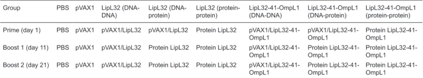
Table 1. Immunization procedure.
Group PBS pVAX1 LipL32 (DNA-DNA)
LipL32 (DNA-protein)
LipL32 (protein-protein)
LipL32-41-OmpL1 (DNA-DNA)
LipL32-41-OmpL1 (DNA-protein)
LipL32-41-OmpL1 (protein-protein)
Prime (day 1) PBS pVAX1 pVAX1/LipL32 pVAX1/LipL32 Protein LipL32 pVAX1/LipL32-41-OmpL1
pVAX1/LipL32-41-OmpL1
Protein LipL32-41-OmpL1
Boost 1 (day 11) PBS pVAX1 pVAX1/LipL32 Protein LipL32 Protein LipL32 pVAX1/LipL32-41-OmpL1
Protein LipL32-41-OmpL1
Protein LipL32-41-OmpL1
Boost 2 (day 21) PBS pVAX1 pVAX1/LipL32 Protein LipL32 Protein LipL32 pVAX1/LipL32-41-OmpL1
Protein LipL32-41-OmpL1
Protein LipL32-41-OmpL1
Strategies for leptospira vaccine 799
well was read at 450 nm with a microplate reader (Tecan Spectra III, Australia). Wells without coating antigens were used as a blank control.
ELISA for cytokine induction
The collected sera were used to detect cytokines IL-2,
IL-4, IL-10, and INF-γ with homologous ELISA kits (Boster,
China). First, 100 µL of serum was added to each well and incubated for 90 min at 37°C. Serum was then discarded from each well, and 100 µL of biotin-antibody reagent was added. Plates were placed in an incubator at 37°C for another 60 min. After the plates were washed three times with PBS, 100 µL of avidin-biotin-complex reagent was added to each well, followed by 30 min of incubation.
Plates were then washed five times with PBS. The plates
were incubated for 30 min and protected from light after TMB reagent was added to each well. The reaction was stopped with the addition of 100 µL TMB stop reagent to each well and absorbance was measured at 450 nm with an ELISA reader.
Splenocyte proliferation assay
Spleen cells isolated from mice in each group were
resuspended at 2 x 10⁴ cells/mL. A 100-µL aliquot contain-. A 100-µL aliquot contain- A 100-µL aliquot
contain-ing 2 x 10³ cells was immediately added to each well of
a 96-well flat-bottomed microtiter plate in triplicate. rIL-2
(R&D, USA) and homologous LipL32 or LipL32-41-OmpL1
proteins were added to the wells at a final concentration of 0.2 ng/mL (rIL-2) and 10 μg/mL (proteins), respectively.
rIL-2 was added to three more wells as a control. After 48 h of incubation at 37°C in 5% CO2, splenocyte
prolifera-tion was assayed with a CCK-8 kit (Beyotime, China). The
stimulation index is defined as: absorbance for the test/
absorbance value for the control where absorbance was measured at 450 nm.
Statistical analysis
Statistical analysis was performed by the Student-Newman-Keuls t-test. Values were compared between immunization groups. P values <0.05 were considered to
be statistically significant.
Results
PCR amplification and nucleotide sequencing of the
fusion gene
The recombinant fusion gene lipL32-41-ompL1 was con-structed by PCR with linking primers and cloned into pVAX1 and pET-28b(+) plasmids. A BLAST search of the GenBank database revealed an identical nucleotide sequence when compared with the available complete genome sequence database of L. interrogans serovar Lai strain Lai.
In vitro expression of protein LipL32 and
LipL32-41-OmpL1 in mammalian cells
Expression of LipL32 and LipL32-41-OmpL1 proteins was demonstrated by transient transfection of HEK293 cells followed by Western blot analysis. The constructed
lipL32 and lipL32-41-ompL1 genes yielded high expression levels of the corresponding proteins with apparent molecu-lar masses of 32 and 101 kDa, respectively. The molecumolecu-lar mass of 101 kDa was equivalent to the combined molecular masses of LipL32, LipL41 and OmpL1 (Figure 1).
Humoral immunity induced by DNA immunization The IgG humoral immune responses of the mice were analyzed by ELISA against recombinant LipL32, LipL41 and OmpL1 proteins (Figure 2). Because there were no differ-ences in any parameters between the PBS group and the pVAX1 group, data from the PBS group are not shown. All
experimental groups showed significant increases in anti -bodies compared with the pVAX1 group. Groups receiving protein prime or boost showed stronger antibody responses, including LipL32 (DNA-protein or protein-protein) and
LipL32-41-OmpL1 (DNA-protein or protein-protein). There
was no significant difference in anti-LipL32 levels between
the LipL32 group and the LipL32-41-OmpL1 group, but anti-LipL41 and anti-OmpL1 levels were much higher in mice immunized with LipL32-41-OmpL1 than in those im-munized with only LipL32.
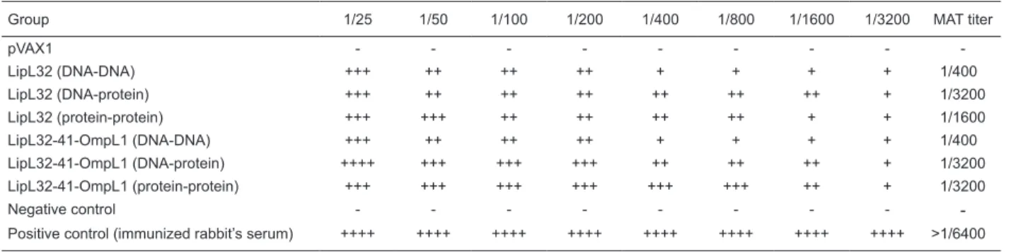
The antibody titers of serum samples from another MAT test are shown in Table 2. The agglutination titers were ob-served up to 1 in 1600 and 1 in 3200 in the protein-boosted groups, whereas among the DNA-boosted groups they were seen at 1 in 400 but absent in the PBS and pVAX1 control groups. The MAT results agreed with the ELISA
results for antigen-specific antibody production, which
demonstrated that mice boosted with protein showed an increased humoral immune response compared with those boosted with DNA plasmids.
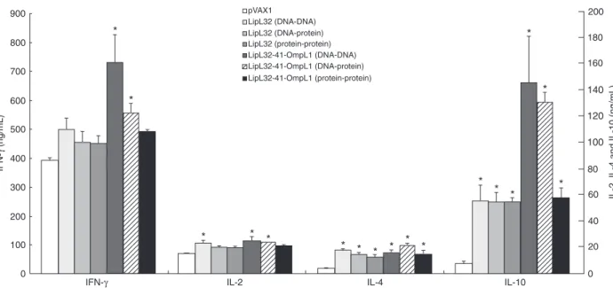
To evaluate T helper 2 cell (Th2) immune responses, ELISA assays were used to describe production of cytok-ines IL-4 and IL-10 (Figure 3). All vaccine groups showed
significant increases in IL-4 and IL-10 levels compared
with the PBS and pVAX1 control groups. Furthermore, the mice vaccinated with LipL32-41-OmpL1 had higher IL-10 levels than those vaccinated with LipL32. There were no
significant differences in IL-4 production between the vac -cinated groups. Better humoral immune responses were elicited when mice were immunized with the fusion DNA plasmid or protein.
Cellular immunity induced by DNA immunization IL-2 and INF-γ were used as markers to detect Th1
immune responses (Figure 3). In the LipL32-41-OmpL1
group (DNA-DNA or DNA-protein), IL-2 and INF-γ were significantly induced compared with the pVAX1 group. There
were no differences between the protein-protein groups (LipL32 or LipL32-41-OmpL1) and the pVAX1 group. Mice immunized with LipL32-41-OmpL1 and LipL32 (DNA-DNA)
showed higher IL-2 levels than those immunized with ho-mologous proteins. Mice vaccinated with LipL32-41-OmpL1 (DNA-protein) also had higher IL-2 levels than those im-munized with homologous proteins. The LipL32-41-OmpL1 (DNA-DNA) group presented the best immune responses
in terms of INF-γ production.
A CCK-8 kit was used to detect splenocyte proliferation. The stimulation indices analyzed at 450 nm with an ELISA
reader are shown in Figure. 4. All test groups showed signifi -cant differences compared with the pVAX1 control group (P < 0.01). Mice boosted with DNA plasmids showed a greater cell immune response than those boosted with protein, and the splenocyte proliferation of the fusion LipL32-41-OmpL1 groups was stronger than that of the LipL32 groups.
Figure 2. ELISA for anti-LipL32, anti-LipL41 and anti-OmpL1 antibody production. Groups of 10 mice each were immunized with DNA (50 µg/mouse) or protein (10 µg/mouse) on day 1. Mice were boosted with equimolar amounts of DNA or protein on days 11 and 21 and bled on day 35. Anti-LipL32, anti-LipL41, and anti-OmpL1 were then detected. Data are reported as geometric means ± SD. *P < 0.05 compared to the pVAX1 group (Student-Newman-Keuls t-test).
Table 2. Microscopic agglutination test titers in each group.
Group 1/25 1/50 1/100 1/200 1/400 1/800 1/1600 1/3200 MAT titer
pVAX1 - - -
-LipL32 (DNA-DNA) +++ ++ ++ ++ + + + + 1/400
LipL32 (DNA-protein) +++ ++ ++ ++ ++ ++ ++ + 1/3200
LipL32 (protein-protein) +++ +++ ++ ++ ++ ++ + + 1/1600
LipL32-41-OmpL1 (DNA-DNA) +++ ++ ++ ++ + + + + 1/400
LipL32-41-OmpL1 (DNA-protein) ++++ +++ +++ +++ ++ ++ ++ + 1/3200
LipL32-41-OmpL1 (protein-protein) +++ +++ +++ +++ +++ +++ ++ + 1/3200
Negative control - - -
-Positive control (immunized rabbit’s serum) ++++ ++++ ++++ ++++ ++++ ++++ ++++ ++++ >1/6400
Strategies for leptospira vaccine 801
Discussion
Leptospiral outer membrane proteins have a number of advantages over current leptospiral vaccines, and highly
conserved outer membrane proteins have special signifi -cance in vaccine development for leptospirosis. Studies have demonstrated that LipL32, LipL41 and OmpL1 are highly conserved among various pathogenic leptospira species but not in saprophytic leptospira species (2,7). This suggests that these antigens play an important role in virulence and pathogenesis. Since most potential cross-protective immunogens should have conserved sequences, we chose these three OMPs of L. interrogans serovar Lai to construct the recombinant fusion gene, lipL32-41-ompL1.
DNA vaccines have been described against tumors (19) and several intracellular and extracellular pathogens, including HIV (20), SARS (21), pertussis (22), and lep-tospires (23). The presence of pathogenic leplep-tospires in the intracellular compartment has already been demonstrated
in vitro (23). Monocyte/macrophage-like and Vero cells have been shown to be permissive host cells for virulent leptospire invasion (24). We therefore decided to use the eukaryotic vector pVAX1 to construct a DNA vaccine and to evaluate the cellular and humoral immune responses induced by different vaccination strategies.
With regard to the humoral immune response of the immunized mice described here, all the antibodies elicited in the protein-boosted groups showed levels that were
significantly higher than those in the groups that were not
protein-boosted. This indicates that a protein-boost strat-egy could improve the immunogenicity of DNA vaccines against leptospirosis and could thus be a valuable strategy
Figure 4. Proliferation values of the splenocyte-proliferation as-say.Spleen cells (2 x 10³)were added to each well of a 96-well flat-bottomed microtiter plate in triplicate. rIL-2 and homologous proteins were added to the wells to a final concentration of 0.2 ng/mL (rIL-2) and 10 μg/mL (proteins). Another three wells con -tained rIL-2 as a control. After 48 h of incubation at 37°C in 5% CO2, splenocyte proliferation was assayed with a CCK-8 kit. The
stimulation index is defined as: absorbance value for the test cul -ture/control absorbance value at 450 nm. Data are reported as means ± SD for 10 animals per group. *P < 0.05 compared to the pVAX1 group (Student-Newman-Keuls t-test).
Figure 3. ELISA for cytokine production. Groups of 10 mice each were immunized with DNA (50 µg/mouse) or protein (10 µg/mouse)
on day 1. Mice were boosted with equimolar amounts of DNA or protein on days 11 and 21 and bled on day 35. INF-γ, 2, 4 and
for inducing better immune responses to DNA vaccines. In the present study, the LipL32-41-OmpL1 immunization groups also showed abundant anti-LipL41 and anti-OmpL1 antibodies in addition to the anti-LipL32 antibodies seen in the LipL32 groups. These results indicate that the LipL32-41-OmpL1 vaccination groups showed better responses than the single-gene groups (LipL32, LipL41 or OmpL1). This
is especially significant because other studies have shown
that the protective effects of immunization with OmpL1 and LipL41 are synergistic (15).
The MAT test is the standard experiment for diagnosis and immune analysis of leptospirosis. In this study, the highest MAT titers were observed in the LipL32 group (DNA-protein), the LipL32-41-OmpL1 group (DNA-protein) and the LipL32-41-OmpL1 group (protein-protein). This also indicates the advantages of recombinant DNA vaccine and DNA-protein strategies in leptospirosis immunization.
It is known that subsets of Th cells can be distinguished by the pattern of cytokines that they produce. Th1 cells
produce IL-2 and INF-γ and play a critical role in directing
cell-mediated immune responses, which are important for clearance of intracellular pathogens. Th2 cells produce IL-4 and IL-10, which are important for humoral responses (25). In our cytokine assays for IL-4 and IL-10, all vaccination
groups showed significantly increased levels compared with the control groups. With respect to IL-2 and INF-γ, the
LipL32-41-OmpL1 (DNA-DNA and DNA-protein) groups elicited greater cytokine production than the control groups. Our results indicated that the LipL32-41-OmpL1 vaccination groups could elicit stronger cellular and humoral responses than the corresponding LipL32 groups. In other acellular vaccine studies (26,27), the immune responses induced
by recombinant or multiple-component vaccines have been better than those induced by a single-component vaccine. A recombination strategy might be better than a combination strategy for multiple-component vaccines because the former strategy has lower costs and higher
efficacy than the latter.
In the present study, we used a splenocyte-proliferation assay to monitor cellular immune responses. The results
showed that all test groups were significantly different from
the pVAX1 control group. The splenocyte-proliferation re-actions in the LipL32-41-OmpL1 vaccination groups were stronger than those of LipL32 groups, indicating that an enhanced cellular immune response was elicited in the LipL32-41-OmpL1 vaccination groups. The mechanism of this phenomenon remains unknown.
Our study has some limitations. First, BALB/c mice are used to evaluate the immune response of leptospira vaccine candidates. To determine the precise protective effects of the vaccine candidates and the immune strategies, hamster or guinea pig models should be used (28). Second, some new candidates might be included as recombinant DNA and protein vaccines, such as leptospira
immunoglobulin-like protein A (29,30). However, there is still no definite
information regarding vaccine composition and immune strategies for leptospirosis.
In conclusion, recombinant LipL32-41-OmpL1 could be a useful vaccine candidate against leptospirosis and might be better than a single-component LipL32 vaccine. The DNA prime-protein boost strategy induced both humoral and cellular immune responses against leptospira and should be studied further.
References
1. Bharti AR, Nally JE, Ricaldi JN, Matthias MA, Diaz MM, Lovett MA, et al. Leptospirosis: a zoonotic disease of global importance. Lancet Infect Dis 2003; 3: 757-771.
2. Haake DA, Chao G, Zuerner RL, Barnett JK, Barnett D, Ma-zel M, et al. The leptospiral major outer membrane protein LipL32 is a lipoprotein expressed during mammalian infec-tion. Infect Immun 2000; 68: 2276-2285.
3. Faisal SM, Yan W, McDonough SP, Chang YF. Leptospira
immunoglobullike protein A variable region (LigAvar) in-corporated in liposomes and PLGA microspheres produces a robust immune response correlating to protective immu-nity. Vaccine 2009; 27: 378-387.
4. Ren SX, Fu G, Jiang XG, Zeng R, Miao YG, Xu H, et al. Unique physiological and pathogenic features of Leptospira interrogans revealed by whole-genome sequencing. Nature
2003; 422: 888-893.
5. Nascimento AL, Verjovski-Almeida S, Van Sluys MA, Montei-ro-Vitorello CB, Camargo LE, Digiampietri LA, et al. Genome
features of Leptospira interrogans serovar Copenhageni.
Braz J Med Biol Res 2004; 37: 459-477.
6. Haake DA, Walker EM, Blanco DR, Bolin CA, Miller MN, Lovett MA. Changes in the surface of Leptospira interrogans
serovar grippotyphosa during in vitro cultivation. Infect Im-mun 1991; 59: 1131-1140.
7. Shang ES, Summers TA, Haake DA. Molecular cloning and sequence analysis of the gene encoding LipL41, a surface-exposed lipoprotein of pathogenic Leptospira species. Infect Immun 1996; 64: 2322-2330.
8. Haake DA, Matsunaga J. Characterization of the leptospiral outer membrane and description of three novel leptospiral membrane proteins. Infect Immun 2002; 70: 4936-4945. 9. Haake DA, Martinich C, Summers TA, Shang ES, Pruetz
Strategies for leptospira vaccine 803
10. Cullen PA, Haake DA, Bulach DM, Zuerner RL, Adler B. LipL21 is a novel surface-exposed lipoprotein of pathogenic
Leptospira species. Infect Immun 2003; 71: 2414-2421. 11. Nally JE, Artiushin S, Timoney JF. Molecular characterization
of thermoinduced immunogenic proteins Q1p42 and Hsp15 of Leptospira interrogans. Infect Immun 2001; 69: 7616-7624.
12. Dong H, Hu Y, Xue F, Sun D, Ojcius DM, Mao Y, et al. Char-acterization of the ompL1 gene of pathogenic Leptospira
species in China and cross-immunogenicity of the OmpL1 protein. BMC Microbiol 2008; 8: 223.
13. Matsunaga J, Young TA, Barnett JK, Barnett D, Bolin CA, Haake DA. Novel 45-kilodalton leptospiral protein that is pro-cessed to a 31-kilodalton growth-phase-regulated peripheral membrane protein. Infect Immun 2002; 70: 323-334. 14. Seixas FK, da Silva EF, Hartwig DD, Cerqueira GM, Amaral
M, Fagundes MQ, et al. Recombinant Mycobacterium bovis
BCG expressing the LipL32 antigen of Leptospira interro-gans protects hamsters from challenge. Vaccine 2007; 26: 88-95.
15. Haake DA, Mazel MK, McCoy AM, Milward F, Chao G, Matsunaga J, et al. Leptospiral outer membrane proteins OmpL1 and LipL41 exhibit synergistic immunoprotection.
Infect Immun 1999; 67: 6572-6582.
16. Wolff JA, Malone RW, Williams P, Chong W, Acsadi G, Jani A, et al. Direct gene transfer into mouse muscle in vivo. Sci-ence 1990; 247: 1465-1468.
17. Mor G, Eliza M. Plasmid DNA vaccines. Immunology, toler-ance, and autoimmunity. Mol Biotechnol 2001; 19: 245-250.
18. Alpar HO, Bramwell VW. Current status of DNA vaccines and their route of administration. Crit Rev Ther Drug Carrier Syst
2002; 19: 307-383.
19. Stevenson FK, Ottensmeier CH, Johnson P, Zhu D, Buchan SL, McCann KJ, et al. DNA vaccines to attack cancer. Proc Natl Acad Sci U S A 2004; 101 (Suppl 2): 14646-14652. 20. Barouch DH, Santra S, Steenbeke TD, Zheng XX, Perry HC,
Davies ME, et al. Augmentation and suppression of immune responses to an HIV-1 DNA vaccine by plasmid cytokine/Ig administration. J Immunol 1998; 161: 1875-1882.
21. Zhao P, Cao J, Zhao LJ, Qin ZL, Ke JS, Pan W, et al. Immune responses against SARS-coronavirus nucleocapsid protein induced by DNA vaccine. Virology 2005; 331: 128-135. 22. Li QT, Zhu YZ, Chu JY, Dong K, He P, Feng CY, et al.
Granulocyte-macrophage colony-stimulating factor DNA
prime-protein boost strategy to enhance efficacy of a recom -binant pertussis DNA vaccine. Acta Pharmacol Sin 2006; 27: 1487-1494.
23. Branger C, Chatrenet B, Gauvrit A, Aviat F, Aubert A, Bach JM, et al. Protection against Leptospira interroganssensu lato challenge by DNA immunization with the gene encod-ing hemolysin-associated protein 1. Infect Immun 2005; 73: 4062-4069.
24. Merien F, Baranton G, Perolat P. Invasion of Vero cells and induction of apoptosis in macrophages by pathogenic
Leptospira interrogans are correlated with virulence. Infect Immun 1997; 65: 729-738.
25. Gor DO, Rose NR, Greenspan NS. TH1-TH2: a procrustean paradigm. Nat Immunol 2003; 4: 503-505.
26. Doria-Rose NA, Haigwood NL. DNA vaccine strategies: can-didates for immune modulation and immunization regimens.
Methods 2003; 31: 207-216.
27. Williamson ED, Bennett AM, Perkins SD, Beedham RJ, Miller J, Baillie LW. Co-immunisation with a plasmid DNA cocktail primes mice against anthrax and plague. Vaccine
2002; 20: 2933-2941.
28. Silva EF, Santos CS, Athanazio DA, Seyffert N, Seixas FK, Cerqueira GM, et al. Characterization of virulence of
Leptospira isolates in a hamster model. Vaccine 2008; 26: 3892-3896.
29. Faisal SM, Yan W, Chen CS, Palaniappan RU, McDonough SP, Chang YF. Evaluation of protective immunity of Lep-tospira immunoglobulin like protein A (LigA) DNA vaccine against challenge in hamsters. Vaccine 2008; 26: 277-287. 30. Choy HA, Kelley MM, Chen TL, Moller AK, Matsunaga J,
Haake DA. Physiological osmotic induction of Leptospira interrogans adhesion: LigA and LigB bind extracellular