The Brazilian Journal of
INFECTIOUS DISEASES
w w w . e l s e v i e r . c o m / l o c a t e / b j i d
Review article
Neurologic complications of HIV in the HAART era:
where are we?
Monica M. Gomes da Silva
∗Universidade Federal do Paraná, Curitiba, PR, Brazil
a r t i c l e
i n f o
Keywords:
HIV
Neurological complication HAND
Cognitive impairment
a b s t r a c t
Human immunodeficiency virus (HIV)-associated neurological complications continue to occur despite the development in antiretroviral treatment. New forms of old opportunistic infections and increased prevalence of neurocognitive disorders are the challenges that infectious diseases specialists face in daily clinic. How to screen and treat these disorders are subject of debate and new studies are underway to answer these questions. This review focuses on a brief discussion about opportunistic infections still present in late diagnosed HIV-infected patients and describes new forms of HIV-related neurological complications.
© 2012 Elsevier Editora Ltda. All rights reserved.
Introduction
The central nervous system (CNS) is a common site of involvement in HIV patients. The spectrum of neurological complications of HIV is very broad, including complications caused by HIV itself as well as complications due to oppor-tunistic pathogens and neoplasms.1
Following the introduction of highly active antiretroviral therapy (HAART), the prevalence of opportunistic infections (OIs) decreased in many areas of the world. However, in devel-oped countries patients continue to present with OIs as the initial manifestation of HIV infection because of late diagnosis of HIV, non-adherence to antiretroviral or prophylactic treat-ments or unavailability of therapeutic and preventive options. Moreover, syndromes such as HIV-associated neurocogni-tive disorder (HAND) have become more prevalent, according to some studies.2 New forms were described, and more
often infectious diseases specialists face the challenge to
∗ Corresponding author at:Universidade Federal do Paraná, Rua Desembargador Vieira Cavalcanti, 1089, Curitiba, Paraná, 80810050, Brazil.
E-mail address:monica.mgomes@gmail.com
investigate patients with few, if any, neurological complaints, even in the presence of suppressed viral load.
This review focuses on recent developments in the field of neurological complications of HIV in the HAART era, and will discuss old OIs and the new forms of HIV-related neurological complications.
The “old OIs”
The most common intra-cranial lesions in acquired immun-odeficiency syndrome (AIDS) patients are toxoplasma encephalitis (TE), cryptococcal meningitis with mucinous pseudocysts formation (CM), primary central nervous system lymphoma (PCNSL), and progressive multifocal leukoen-cephalopathy (PML).3In developing countries such as Brazil,
neurotuberculosis is an important differential diagnosis for TE, being the second most common HIV intracranial complication.4 Table 1shows the main MRI patterns of the
1413-8670/$ – see front matter © 2012 Elsevier Editora Ltda. All rights reserved.
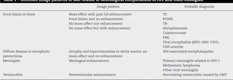
Table 1 – Common image patterns in MRI related to neurological complications of HIV and main etiologies.
Image pattern Probable diagnosis Focal lesion in brain Mass effect with post Gd enhancement
Focal lesion and no enhancement No mass affect nor enhancement No mass effect but with enhancement
TE PCNSL TB
Histoplasmosis Criptococcosis PML
Viral encephalitis (HSV, CMV, VZV) CNS arteritis
Diffuse disease in encephalic parenchima
Atrophy and hiperintensities in white matter, no mass effect and no enhancement
HIV-associated encephalopathy Meningitis Meningeal enhancement Primary meningitis related to HIV-1
Methastatic lymphoma Other viral meningitis
Ventriculitis Periventricular enhancement Necrotizing ventriculitis caused by CMV TE, toxoplasma encephalitis; PCNSL, primary central nervous system lymphoma; TB, tuberculosis; PML, progressive multifocal leukoen-cephalopathy; HSV, herpes simplex virus; CMV, cytomegalovirus; VZV, varicella-zoster virus; CNS, central nervous system.
most common etiologies of CNS involvement in HIV-infected patients.
TE is the most common cause of CNS mass lesions in patients infected with HIV. The seroprevalence of toxoplasma varies from 10% to 40% in the United States to 70% to 90% in Europe.Toxoplasma gondii (TG) is a protozoan which can also cause disease in immunocompetent patients, but in this setting, the disease is usually benign and does not affect the CNS.5,6
Usually, the CD4 cell count of patients with HIV presenting with TE is less than 100, but some patients can develop the disease with CD4 cell counts between 100 and 200 cells/mm3.7
The prevalence of TE has decreased with the use of trimetho-prim sulfamethoxazole for prophylaxis againstPneumocystis jirovecii.8Table 2shows the diagnosis and treatment
charac-teristics of CNS OIs.
Clinical characteristics of TE are nonspecific and depend on where the lesion is located in the brain.6Most commonly,
lesions from TE are located in the basal ganglia or subcortical white matter, although any part of the CNS can be involved. Concomitant diffuse atrophy disproportionate to the patient’s age is very common, present in more than 1/3 of patients. TE lesions are usually abscesses of more than 3 cm in diameter that most commonly show enhancement after contrast injec-tion. On brain CT, lesions are hypodense and surrounded by edema and enhance with contrast, classically presenting “ring enhancement” or “asymmetric target sign”. MRI can often reveal multiple lesions in a patient presenting with a single lesion on CT.5
The diagnosis of TE is based on clinical and laboratory find-ings. A high degree of suspicion is warranted for a patient with HIV infection, CD4 less than 100 cells/mm3, positive serum
anti-toxoplasma IgG (which demonstrates previous contact with the protozoan), and focal signs on neurological exam. Brain CT or MRI should be performed with pre- and post-contrast images, but in many resource-limited settings these may not be available. If the diagnosis is suspected, an empiric treatment with sulphadiazine, pyrimetamine, and folinic acid should be instituted.6
Usually, TE shows a rapid response to therapy as shown by image resolution or clinical improvement. Treatment is
considered a failure if no clinical and radiographic improve-ments are observed after two weeks of therapy. In such cases, a brain biopsy is indicated to search for an alternative diagnosis.6
The principal differential diagnosis of TE in some countries is PCNSL.9This is a neoplasm linked to infection with
Epstein-Barr virus (EBV) and occurs in advanced HIV disease (CD4 less than 100 cells/mm3). It causes mass lesions with multiple
patterns of contrast enhancement on radiographic imaging, very similar to TE. Diagnosis can be made by CSF analysis with presence of neoplastic cells on flow cytometry, associ-ated with a positive EBV DNA by polymerase chain reaction (PCR). Chemotherapy and radiotherapy are the treatment.3
Leptomeningitis is very common in the clinical picture of CNS TB. Also, CNS TB can cause infarction, abscess for-mation, and granuloma.10 Usually, the lesions present with
mass-effect and IV contrast enhancement on CT and MRI.11
CSF tests can help to make the diagnosis, with a mononu-clear pleocytosis, low glucose, and increased protein levels. CSF must be sent forM. tuberculosisculture, but this can take weeks to produce a result.M. tuberculosisDNA by PCR in CSF may lead to a faster diagnosis, but the sensitivity is variable. Additionally, a search for extra-neural sites of tuberculosis can be helpful. Around 20% of patients will present with extra-neural signs of active tuberculosis, such as lung lesions, that are more easily accessible for collection of material to confirm the diagnosis.10
Cryptococcus neoformans, besides being a very common cause of meningitis, can also cause gelatinous pseudocyst formation, with the deposit of a thick material in the perivas-cular spaces of periventriperivas-cular white-matter (Virchow-Robin spaces). Usually, these do not enhance after IV contrast injection, and they cause no mass effect. Although not pathognomonic, their appearance is highly suggestive of cryptococcosis.12
Table 2 – Intra-cranial diseases, their etiologic agents, useful tests for diagnosis and treatment options.
Disease Agent Diagnostic tests Primary treatment Supressive treatment Toxoplasmosis Toxoplasma gondii MRI showing a mass-effect
lesion located in basal ganglia or cortex, with ring or asymmetrical-target enhancement, Positive anti-toxoplasman serum and CSF IgG, CSFToxoplasma gondii
DNA by PCR
Sulfadiazine 1-1.5 g q6h + pyrimethamine 200 mg first day followed by pyrimethamine
50-75 mg/day + folinic acid 10-20 mg/day
Sulphadiazine 500-1000 mg q6h + pyrimethamine 25-50 mg/day + folinic acid 10-20 mg/day
Central nervous system lymphoma
Related to EBV MRI showing a mass-effect lesion in periventricular white-matter, with ring or nodular contrast
enhancement, positive CSF EBV DNA by PCR
Chemotherapy / radiotherapy (role of gancyclovir?)
N/A
Tuberculoma or tuberculous abscess
Mycobacterium tuberculosis
MRI showing mass-effect lesion with multiple patterns of contrast enhancement, positive CSFM. tuberculosis
culture, positive CSFM. tuberculosisDNA by PCR
Treat according to local guidelines, local data on resistance and patientˇıs history of pre-exposure to anti-TB treatment
RFP 600 mg/day + INH 5 mg/Kg/day + PZD 25-30 mg/Kg/day (+- etambutol 15-25 mg/Kg/day) + B6 vitamin*
N/A
Cryptococcal
meningoencephalitis
Cryptococcussp. MRI showing gelatinous pseudocysts located in basal ganglia, thamalus or periventricular areas, non-enhancing and no significant mass-effect
IV fluconazol 400-800 mg/day or IV amphotericin B 0.7 mg/Kg/day
Oral Fluconazol 200 mg/day
PML JC virus MRI demonstrating white
matter changes, no mass effect and no contrast enhancement (in case of classical PML), positive CSF DNA JC virus
HAART N/A
Cysticercosis Cysticercus(larval
cysts of pork
tapewormTaenia
solium)
MRI showing cystic lesion (concomitant calcified lesions can suggest diagnosis), serological tests: serum and CSF (immunoblot)
Depending on location of lesions and stage,
dexamethasone + - albendazol N/A
MRI, magnetic resonance imaging; CSF, cerebrospinal fluid; PCR, polymerase chain reaction; EBV, Epstein-Barr virus; N/A, not available; TB, tuber-culosis; RFP, rifampin; INH, isoniazid; PZD, pyrazinamide; PML, progressive multifocal leukoencephalopathy; HAART, highly active antiretroviral therapy.
∗ B6 vitamin is used to antagonized neurotoxic effects of INH.
The prevalence of neurocysticercosis is not higher among HIV-infected individuals.14However, some atypical cases have
been described, mostly in resource-limited settings. Treat-ment should be started with corticosteroids and/or cysticidal therapy (praziquantel or albendazol), according to the stage and topography of the disease in the CNS.
What is new with the old CNS OIs?
Progressive multifocal leukoencephalopathy (PML) is a disease caused by the polyomavirus JC virus (JCV), and is associated with severe immunosupression (CD4 less than 100 cells/mm3),
although some cases with a higher CD4 count have been described. The main feature is a progressive lesion of white matter causing variable neurologic signs and symptoms, no fever, and a progressive disease. Diagnosis is suggested by the
typical appearance in MRI complemented by a positive CSF JCV DNA by nested PCR. There is no specific treatment, but improvement or at least stability has been reported in some patients due to HAART.15
Koralnik et al. described an HIV-infected patient with JCV infection restricted to granule cell neurons of the cerebellum and with compatible symptoms. That was a description of a new syndrome called JCV granule cell neuronopathy. Patients with this new variant have no typical white matter changes on MRI but present with cerebellar atrophy.16
Another variant of the “classical” PML is the “inflamma-tory” PML. Patients with previous diagnosis of PML who start HAART develop worsening of previous known PML.17In some
cases, patients have undiscovered subclinical PML during HAART.18In both cases, image tests are different from
ANI
MCMD
HAD
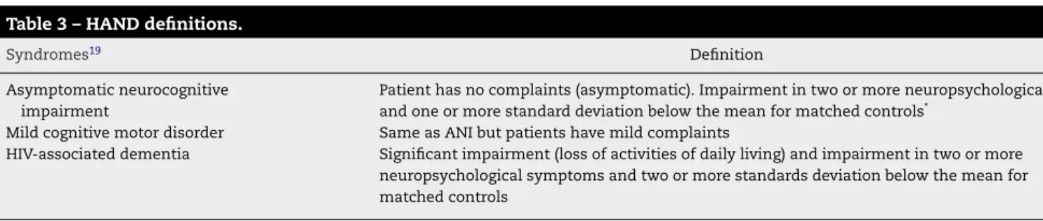
Fig. 1 – Clinical stages of cognitive impairment in HIV-infected patients. ANI, asymptomatic neurocognitive impairment; MCMD, minor cognitive motor disorder; HAD, HIV-associated dementia.
associated with fatal cases of PML. Steroids may help to reduce the exacerbated inflammatory response and contribute to bet-ter outcomes.
HIV-associated neurocognitive disorder
HIV-associated neurocognitive disorder (HAND) is the new definition for what used to be called AIDS-dementia complex – the phenotypic form of HIV encephalitis. In the 1993 Centers for Disease Control and Prevention (CDC) classification, HAND was defined as an AIDS-defining illness.
HAND is the most common cause of dementia in young people, and one of the few treatable dementias. It usually has an insidious onset and a chronic, progressive course, bring-ing disability to many HIV patients. Also, this syndrome may cause poor adherence to HIV care, job loss, decline of driv-ing ability, and lesser adherence with HIV medications, with increased morbidity and mortality.
In 2007, new criteria were published defining how to diagnose HAND, including a wide spectrum of HIV-related cog-nitive impairments19(Table 3,Fig. 1) as follows:
• Asymptomatic neurocognitive impairment (ANI) • Mild cognitive motor disorder (MCMD)
• HIV-associated dementia (HAD).
In the HAART era, the incidence of HAND has decreased, but the prevalence has paradoxically increased, with mild forms of the disease observed more commonly than before. In developing countries, patients often died before the diag-nosis of HAND could be made. However, with the broad use of HAART, this scenario is changing.
Although HAND is observed more frequently in patients with severe immunosupression (usually a CD4 count less than 100 cells/mm3), it can occur in patients with higher CD4 cell
counts, especially in its subtle forms. CD4 nadir less than 50 cels/mm3 is a risk factor for development of neurocognitive
disorder in HIV patients.20
HAND is characterized by the triad of cognitive impair-ment, behavioral abnormality, and motor dysfunction. Clas-sically, HIV dementia is considered a subcortical dementia, characterized by more motor symptoms and less memory and language impairment than is commonly seen in cortical dementias such as Alzheimer’s disease.21 Recently,
asymp-tomatic and mild forms of neurocognitive impairment were shown to actually increase the risk for future symptomatic decline. This may be a reason why infectious diseases special-ists should actively screen HIV patients with no neurological or cognitive complains in daily practice.22
The clinical diagnosis of HAND is ideally based on the patient’s performance on neurocognitive tests, as described. Unfortunately these tests need to be applied by specially trained professionals (such as psychologists or neurologists), and in many resource-limited countries they are not widely available. Another way to screen patients for neurocognitive impairment is through the use of scales, such as the HIV Dementia Scale or the International HIV Dementia Scale. The latter is a bedside test that has been developed and applied in populations from the United States and Uganda, show-ing high specificity and sensitivity. This scale was also useful when applied to HIV-infected patients from India. This test can be administered by non-neurologists, and is a useful tool for detection of cognitive impairment. If a patient screens positive by this scale, he/she can be referred for further neurocognitive evaluation with additional neuropsychological testing.23
CSF findings in HAND patients are non-specific, usu-ally showing normal or mild pleocytosis, normal or mildy increased protein and normal glucose. It is important to exclude opportunistic infections. CSF HIV-1 viral load is not reliable in the diagnosis of cognitive disorder caused by HIV infection itself but, when positive, it may suggest a CSF HIV escape and may guide therapy to a regimen with improved CSF penetration, according to penetration score.24
Cranial CT and MRI may show cortical atrophy, but the degree of atrophy has not been correlated with the grade of dementia. MRI may show diffuse white-matter hyperintensi-ties in the deep white matter with atrophy of bilateral caudate nucleus, but these results are non-specific. Care should be taken with the use of imaging studies, because patients with
Table 3 – HAND definitions.
Syndromes19 Definition
Asymptomatic neurocognitive impairment
Patient has no complaints (asymptomatic). Impairment in two or more neuropsychological and one or more standard deviation below the mean for matched controls*
Mild cognitive motor disorder Same as ANI but patients have mild complaints
HIV-associated dementia Significant impairment (loss of activities of daily living) and impairment in two or more
neuropsychological symptoms and two or more standards deviation below the mean for matched controls
ANI, asymptomatic neurocognitive impairment.
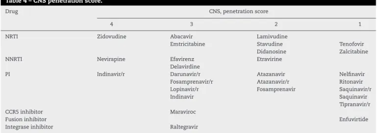
Table 4 – CNS penetration score.
Drug CNS, penetration score
4 3 2 1
NRTI Zidovudine Abacavir Lamivudine
Emtricitabine Stavudine Tenofovir Didanosine Zalcitabine NNRTI Nevirapine Efavirenz Etravirine
Delavirdine
PI Indinavir/r Darunavir/r Atazanavir Nelfinavir Fosamprenavir/r Atazanavir/r Ritonavir Lopinavir/r Fosamprenavir Saquinavir/r Indinavir Saquinavir
Tipranavir/r CCR5 inhibitor Maraviroc
Fusion inhibitor Enfuvirtide Integrase inhibitor Raltegravir
CNS, central nervous system; NRTI, nucleoside reverse transcriptase inhibitor; NNRTI, non-nucleoside reverse transcriptase inhibitor.
HAND may have a normal MRI and patients with bilateral white matter hyperintensities may have an alternative diag-nosis. Image studies must rule out alternative diagdiag-nosis.
There is no specific treatment for HAND. Antiretroviral drugs that penetrate the blood brain barrier (BBB) are cur-rently considered the best choices for treatment of these patients.25,26
Penetration of ARV through the BBB: the CPE
score
Many drugs were investigated in the treatment of HAND with no good results so far. At this moment, the best treatment for HAND is ARV with good CNS penetration. In order to evalu-ate general characteristics of available ARV to penetrevalu-ate the CNS, Letendre et al. developed a CNS penetration score (CPE) (Table 4), which showed that higher scores were related to lower CSF viral load.25,26
Other studies presented discordant results when ana-lyzing neuro-HAART (HAART constituted by drugs with improved BBB penetration) ability to improve neurocognitive impairment, with one study showing worsening of cognitive performance in patients with higher CPE scores.27This result
questioned neural toxicity but, overall, more randomized clin-ical trials are needed to better answer the question of whether neuro-HAART can really improve cognitive performance in HAND patients.
At this moment, starting neuro-HAART in patients with no cognitive impairment is not indicated, but some guide-lines already suggest changing the ARV regimen in patients diagnosed with HAND in order to improve their cognitive performance.28
In conclusion, neurological involvement in HIV-infected patients is still important, even in the post-HAART era, with not only new forms or atypical presentations of old OIs, but also milder forms of neurocognitive impairment. New clini-cal trials investigating easier diagnostic tools for patients with cognitive impairment, the utility of neuro-HAART, and clini-cal evaluation of those patients are ongoing to answer many doubts the scientific community still has.
Conflict of interest
The author declares to have no conflict of interest.
r e f e r e n c e s
1. Sacktor N. The epidemiology of human immunodeficiency virus-associated neurological disease in the era of highly active antiretroviral therapy. J Neurovirol. 2002;8 Suppl. 2:115–21.
2. Heaton RK, Franklin DR, Ellis RJ, et al., CHARTER Group; HNRC Group. HIV-associated neurocognitive disorders before and during the era of combination antiretroviral therapy: differences in rates, nature, and predictors. J Neurovirol. 2011;17:3–16.
3. Gomes da Silva MM, Cunha CA. Neurological complications of HIV infection. In: Zelalem T, editor. Fundamentals of Global HIV Medicine. USA: IHL Press; 2009.
4. Oliveira JF, Greco DB, Oliveira GC, Christo PP, Guimarães MD, Oliveira RC. Neurological disease in HIV-infected patients in the era of highly active antiretroviral treatment: a Brazilian experience. Rev Soc Bras Med Trop. 2006;39:146–51. 5. Ramsey RG, Gean AD. CNS Toxoplasmosis. In: Donovan Post
MJ, editor. Neuroimaging Clinics of North America. Philadelphia: WB Saunders Company; 1997.
6. Luft BJ, Sivadas R. Toxoplasmosis. In: Scheld WM, Whitley RJ, Marra CM, editors. Infections of the central nervous system. Philadelphia: Lippincott Willians and Wilkins; 2004. 7. Nascimento LV, Stollar F, Tavares LB, et al. Risk factors for
toxoplasmic encephalitis in HIV-infected patients: a case-control study in Brazil. Ann Trop Med Parasitol. 2001;95:587–93.
8. Antinori A, Larussa D, Cingolani A, et al. Prevalence, associated factors, and prognostic determinants of
AIDS-related toxoplasmic encephalitis in the era of advanced highly active antiretroviral therapy. Clin Infect Dis.
2004;39:1681–91.
9. Collazos J. Opportunistic infections of CNS in HIV-infected individuals. In: Roos KL, editor. Principles of neurologic infectious diseases. USA: The MacGraw-Hill Companies; 2005. 10. Rodrigues MG, da Rocha AJ, Masruha MR, Minett TS.
11. Whiteman MLH. Neuroimaging of CNS tuberculosis in HIV-infected patients. In: Donovan Post MJ, editor. Neuroimaging clinics of North America. Philadelphia: WB Saunders Company; 1997.
12. Jarvis JN, Harrison TS. HIV-associated cryptococcal meningitis. AIDS. 2007;21:2119–29.
13. Serpa JA, Moran A, Goodman JC, Giordano TP, White Jr AC. Neurocysticercosis in the HIV era: a case report and review of the literature. Am J Trop Med Hyg. 2007;77:113–7.
14. Delobel P, Signate A, El Guedj M, et al. Unusual form of neurocysticercosis associated with HIV infection. R Eur J Neurol. 2004;11:55–8.
15. Skiest DJ. Focal neurological disease in patients with acquired immunodeficiency syndrome. Clin Infect Dis. 2002;34:103–15. 16. Koralnik IJ, Wüthrich C, Dang X, et al. JC virus granule cell
neuronopathy: a novel clinical syndrome distinct from progressive multifocal leukoencephalopathy. Ann Neurol. 2005;57:576–80.
17. Tan K, Roda R, Ostrow L, McArthur J, Nath A. PML-IRIS in patients with HIV infection: clinical manifestations and treatment with steroids. Neurology. 2009;72:1458–64. 18. Sidhu N, McCutchan JA. Unmasking of PML by HAART:
unusual clinical features and the role of IRIS. J Neuroimmunol. 2010;219:100–4.
19. Antinori A, Arendt G, Becker JT, et al. Updated research nosology for HIV associated neurocognitive disorders. Neurology. 2007;69:1789–99.
20. Ellis RJ, Badiee J, Vaida F, et al., CHARTER Group. CD4 nadir is a predictor of HIV neurocognitive impairment in the era of combination antiretroviral therapy. AIDS. 2011;25:1747–51.
21. Gendelman HE. Biomarkers, laboratory, and animal models for the design and development of adjunctive therapies for HIV-1 dementia and other neuroinflammatory disorders. J Neuroimmune Pharmacol. 2007;2:8–13.
22. Heaton R, Franklin D, Woods S, et al. Asymptomatic HIV associated neurocognitive disorder increases risk for future symptomatic decline: a CHARTER longitudinal study. In: 19th
Conference on Retroviruses and Opportunistic Infections. 2012.
23. Sacktor NC, Wong M, Nakasujja N, et al. The international HIV dementia scale: a new rapid screening test for HIV dementia. AIDS. 2005;19:1367–74.
24. Cysique LAJ, Maruff P, Brew BJ. Antiretroviral therapy in HIV infection: are neurologically active drugs important? Arch Neurol. 2004;61:1699–704.
25. Letendre S, Marquie-Beck J, Capparelli E, et al. Validation of the CNS penetration-effectiveness rank for quantifying antiretroviral penetration into the central nervous system. Arch Neurol. 2008;65:65–70.
26. Letendre S, Ellis RJ, Best B, et al. Penetration and effectiveness of antiretroviral therapy in the central nervous system. Antiinflamm Antiallergy Agents Med Chem. 2009;8:169–83. 27. European Guidelines for treatment of HIV infected adults in
Europe 2011. Available from:
http://www.europeanaidsclinicalsociety.org/images/stories/ EACS-Pdf/eacsguidelines-v6 english.pdf