DOI:
10.1590/0004-282X20150071
ARTICLE
VIEW AND REVIEW
Cerebrospinal fluid analysis in the HIV
infection and compartmentalization of HIV in
the central nervous system
Análise de LCR na infeção pelo HIV e compartimentalização do HIV no sistema
nervoso central
Sérgio Monteiro de Almeida
1,2Almost forty years after it began, the HIV epidemic
re-mains a challenging public health problem. he incidence
of infection is increasing in some vulnerable groups, mainly
young men who have sex with men (MSM) and older
peo-ple
1,2. he nervous and the immune systems are HIV tar
-get organs
3,4,5. he introduction of highly active antiretrovi
-ral therapy (HAART) has changed the clinical situation for
patients with AIDS, decreased the incidence of
opportu-nistic infections, and thus lowered mortality. However, the
incidence of neurological complications directly related to
HIV has not decreased proportionally
6, probably because
of the low penetration of antiretroviral (ARV) drugs into
the central nervous system (CNS), the neuronal toxicity of
ARVs, or the persistence of neuronal lesions caused by HIV
before treatment
7.
The purpose of this review is to discuss the
indica-tions for cerebrospinal fluid (CSF) analysis in HIV
infec-tion in clinical practice. CSF analysis during HIV infecinfec-tion
is indicated for the diagnosis of opportunistic infections
and co-infections; the diagnosis of meningitis, acute or
chronic, caused by HIV; the quantification of HIV viral
load in CSF; and the analysis of HIV
compartmentaliza-tion in the CNS
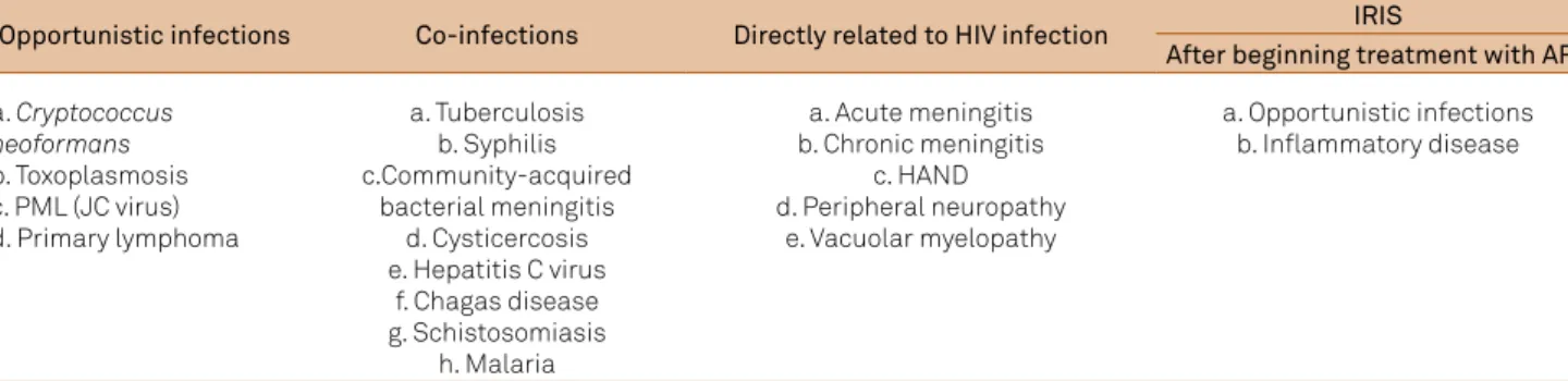
5. Table 1 summarizes CNS complications
in HIV infection.
1Universidade Federal do Paraná, Departamento de Patologia Médica; Hospital de Clínicas, Laboratório de Clínica Patológica, Seção de Virologia, Curitiba PR, Brazil; 2Instituto de Pesquisa Pelé Pequeno Príncipe, Curitiba PR, Brazil.
Correspondence: Sérgio Monteiro de Almeida; Hospital de Clínicas, Seção de Virologia, Setor Análises Clínicas, Universidade Federal do Paraná; Rua Padre Camargo, 280; 80060-240 Curitiba PR, Brasil; E-mail: [email protected]
Conflict of interest: There is no conlict of interest to declare.
Received 19 November 2014; Received in inal form 16 March 2015; Accepted 06 April 2015.
ABSTRACT
The nervous system plays an important role in HIV infection. The purpose of this review is to discuss the indications for cerebrospinal luid
(CSF) analysis in HIV infection in clinical practice. CSF analysis in HIV infection is indicated for the diagnosis of opportunistic infections and
co-infections, diagnosis of meningitis caused by HIV, quantiication of HIV viral load, and analysis of CNS HIV compartmentalization. Although
several CSF biomarkers have been investigated, none are clinically applicable. The capacity of HIV to generate genetic diversity, in association
with the constitutional characteristics of the CNS, facilitates the generation of HIV quasispecies in the CNS that are distinct from HIV in the
systemic circulation. CSF analysis has a well-deined and valuable role in the diagnosis of CNS infections in HIV/AIDS patients. Further research
is necessary to establish a clinically applicable biomarker for the diagnosis of HIV-associated neurocognitive disorders.
Keywords:
HIV, AIDS, central nervous system, cognitive impairment, cerebrospinal luid, viral load, biomarkers.
RESUMO
O sistema nervoso representa um papel importante na infecção pelo HIV. O objetivo desta revisão é discutir as indicações para análise do
líquido cefalorraquidiano (LCR) na infecção pelo HIV na prática clínica. A análise do LCR na infecção pelo HIV é indicada para o diagnóstico
de infecções oportunistas e co-infecções, meningite pelo HIV, quantiicação da carga viral de HIV e compartimentalização do HIV no SNC.
Uma série de biomarcadores no LCR foi investigada, na literatura, porém não apresentam aplicabilidade clínica. A grande capacidade do
HIV de gerar diversidade genética, associado a características constitucionais do SNC propicia o desenvolvimento quasiespécies distintas
no SNC das circulantes sistemicamente. A análise do LCR na infecção pelo HIV é bem estabelecida no diagnóstico de infecções no CNS,
contudo mais pesquisas é necessária para estabelecer a aplicabilidade clínica dos biomarcadores no diagnóstico de desordens cognitivas
associadas ao HIV.
OPPORTUNISTIC INFECTIONS AND CO-INFECTIONS
CSF analysis is important for the diagnosis of
opportunis-tic infections and co-infections in the CNS. Several diagnosopportunis-tic
methods can be used. he operational characteristics of each
method difer according to the etiological agents under in
-vestigation
8,9,10. In AIDS, more than one opportunistic
infec-tion can co-exist because of immunosuppression.
Co-infections are prevalent in the population. hey can
occur in association with HIV infection, independent of
im-munosuppression
11. Table 1 shows the most common
eti-ologies of CNS co-infections that occur with HIV in Brazil.
CNS COMPLICATIONS DIRECTLY RELATED TO HIV
INFECTION
Acute meningitis caused by HIV
Acute meningitis is present in 5%–10% of HIV-infected
patients, usually before seroconversion, as well as during or
after the observation of mononucleosis-like signs and
symp-toms. Its clinical evolution is self-limited. Acute meningitis
can be asymptomatic or accompanied by focal neurological
signs such as peripheral facial palsy.
he CSF has a biochemical and cellular pattern compatible
with that of acute viral meningitis
6. Anti-HIV enzyme-linked
immunosorbent assays (ELISA) often yield negative results
with blood and CSF samples. Because HIV/AIDS is a systemic
infection, diagnostic tests (triage and conirmatory) must be
performed using peripheral blood samples, not CSF samples
1.
Patients with a risk of HIV infection are advised to have a
second serological diagnosis test for HIV after 4–6 weeks
1. No
speciic biomarker in the CSF or blood helps diagnose acute
meningitis caused by HIV. he p24 antigen test for early di
-agnosis of HIV using blood samples is neither sensitive nor
standardized for CSF analysis.
Chronic meningitis caused by HIV
Chronic meningitis is present in 40% of infected
indi-viduals at any infection stage, while 59% are asymptomatic.
Because it is an exclusion diagnosis, other causes of chronic
meningitis must be ruled out. he CSF presents with pleo
-cytosis (5–50 cells/mm³) and increased total protein level
(50–100 mg/dL); glucose, the CSF/blood glucose ratio, and
lactic acid are in the normal range
8,12,13.
Other neurologic complications associated with HIV
he use of CSF analysis for the speciic diagnosis of
HIV-associated neurocognitive disorders (HAND), HIV
my-elopathy, and immunological recovery inlammatory syn
-drome (IRIS-HIV) is of limited clinical importance. Speciic
biomarkers for the diagnosis of these diseases have not been
identiied, although several have been studied.
HAND is the main neurologic complication directly
asso-ciated with HIV infection
14; it is a clinical diagnosis
6,7,15. CSF
analysis is important for ruling out opportunistic infections
and co-infections. Several biomarkers in CSF with the
poten-tial to diagnose HAND have been studied. However, they are
not clinically applicable
16,17,18,19, 20and are only used in research.
With the aging of the HIV population, other biomarkers have
become important and have been investigated
21,22,23,24. he
same can be applied to polyneuropathies and myelopathy
as-sociated with HIV. HIV vacuolar myelopathy occurs in 10%
of patients with AIDS; it is an exclusion diagnosis. Other
frequent causes of myelopathy, such as neurosyphilis, HTLV,
CMV, and speciic endemic etiologies, must be ruled out.
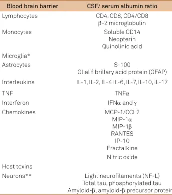
Neurons do not have a CD4 receptor, the main receptor
involved in HIV cell invasion; consequently, HIV does not
in-fect neurons. Neuronal injury in HIV inin-fection results from
an indirect mechanism due to the complex interaction of
anions, inlammatory and neuronal injury proteins and also
constitutional HIV proteins
4. hese proteins are investigated
as biomarkers in CSF or serum for CNS HIV infection and
are summarized in Table 2. It is possible that pyroptosis plays
an important role in neuronal lesion and death. Pyroptosis
markers in the CSF of patients infected with HIV have not yet
been studied. he mechanism of cell death, namely, apopto
-sis, which here can be described as the death of cells with a
productive infection caused by HIV, does not depend on the
inlammatory process; it is associated with the activation
Table 1.
Central nervous system complications in HIV infection.
Opportunistic infections
Co-infections
Directly related to HIV infection
IRIS
After beginning treatment with ARV
a.
Cryptococcus
neoformans
b. Toxoplasmosis
c. PML (JC virus)
d. Primary lymphoma
a. Tuberculosis
b. Syphilis
c.Community-acquired
bacterial meningitis
d. Cysticercosis
e. Hepatitis C virus
f. Chagas disease
g. Schistosomiasis
h. Malaria
a. Acute meningitis
b. Chronic meningitis
c. HAND
d. Peripheral neuropathy
e. Vacuolar myelopathy
a. Opportunistic infections
b. Inlammatory disease
of caspase-3. Recently, a mechanism of cell death in
HIV-infected cells, called pyroptosis, has been described. he
term pyroptosis refers to proinlammatory programmed cell
death, a form of cells death intermediate between apoptosis
and necrosis. Pyroptosis is dependent on caspase-1, which
in turn depends on inlammatory processes. Caspase-1 is
not involved in apoptotic cell death, and caspase-1-deicient
cells respond normally to most apoptotic signals. he
HIV-1-induced death of CD4 T lymphocytes is mediated by
pyroptosis. he two mechanisms of programmed cell death,
apoptosis and pyroptosis, are not exclusive
25,26,27.
he CD4 and CD8 lymphocyte counts in the peripheral
blood and the CD4/CD8 ratio are valuable biomarkers for
immunological status in HIV/AIDS. In the CSF, they are of
limited value because their changes relect the changes in the
peripheral blood
28. For CSF analysis, some authors have
sug-gested combining biomarkers for diferent conditions, such
as neopterin to assess immunoactivation and light neuroila
-ments to evaluate neurodegeneration
29.
IRIS-HIV is characterized by the occurrence of
oppor-tunistic infections or non-infectious inlammatory disease
within weeks of initiating or changing ARV treatment,
de-spite improvements in immunological status. About 15%–
25% of patients that receive ARV treatment develop
IRIS-HIV within the irst few months of therapy. he nervous
system is a frequent target. Biomarkers in the CSF that
diag-nose IRIS have not yet been identiied; the diagnosis is based
on clinical criteria
30,31.
HIV VIRAL LOAD IN THE CSF
HIV particles present in the CSF can have diferent ori
-gins: they can drain from perivascular spaces or infected cells
of the meninges, or they can originate in the plasma and pass
through the choroid plexus during CSF production,
particu-larly in the case of damage to or inlammation of the choroid
plexus
12. In clinical practice, determining the HIV viral load in
the CSF is important for monitoring the therapeutic efects
of ARV treatment, for identifying patients with CNS escape
(compartmentalization), and for determining the diferential
diagnosis in psychiatric disorders
5.
he correlation between HAND and the HIV viral load in
the CSF is dependent on the degree of immunosuppression.
In cases with a CD4 count of > 200 cells/mm³, the HIV viral
load in the CSF correlates positively with that in the peripheral
blood, but not with HAND. After the initiation of ARV therapy,
the HIV viral load in the CSF and blood decreases. In patients
with a CD4 count of < 200 cells/mm³, the HIV viral load in
the CSF correlates positively with cognitive symptoms, but
not with the peripheral blood viral load. he initiation of ARV
treatment leads to a decrease in the CSF viral load, although
the decrease is slower than the decrease in the blood
32,33,34.
Opportunistic infections and co-infections increase the
HIV viral load in the peripheral blood. Some opportunistic
infections or co-infections in the CNS can increase the viral
load in the CSF, as noted in patients with neurosyphilis
35.
Automated methods, commercially available, for
quanti-fying HIV RNA in blood samples have not been standardized
for CSF analysis.
COMPARTMENTALIZATION OF HIV IN THE CNS
Speciic CNS immunological characteristics, the
blood-brain barrier (BBB), rapid mutation and
recombina-tion of HIV, and poor ARV penetrarecombina-tion in CNS contribute to
the compartmentalization of HIV in the CNS.
Some organs, such as the CNS, genital tract, and
gas-trointestinal lymphoid tissue, are viral compartments and
reservoirs that allow HIV to persist despite ARV therapy that
eliminates the virus from the peripheral blood. Compartments
are deined as anatomical regions that restrict the genetic
low of HIV, thereby enabling viral evolution and divergence
from the virus circulating in the peripheral blood. On the
other hand, reservoirs are cells or anatomical sites where
HIV or HIV-infected cells survive because the viral kinetics
is slower than that in the peripheral blood. Compartments
and reservoirs protect HIV from speciic immune responses,
Table 2.
Biomarkers of HIV and central nervous system (CNS)
infection
15, 17, 18, 19, 59.
Blood brain barrier
CSF/ serum albumin ratio
Lymphocytes
CD4, CD8, CD4/CD8
β
-2 microglobulin
Monocytes
Soluble CD14
Neopterin
Quinolinic acid
Microglia*
Astrocytes
S-100
Glial ibrillary acid protein (GFAP)
Interleukins
IL-1, IL-2, IL-4 IL-6, IL-7, IL-10, IL-17
TNF
TNF
α
Interferon
IFN
α
and
γ
Chemokines
MCP-1/CCL2
MIP-1
α
MIP-1
β
RANTES
IP-10
Fractalkine
Nitric oxide
Host toxins
Neurons**
Light neuroilaments (NF-L)
Total tau, phosphorylated tau
Amyloid-
β
, amyloid-
β
precursor proteins
GFAP: Glial ibrillary acid protein; IL: interleukin; TNF: tumor
necrosis factor; IFN: interferon; MCP-1/CCL2: monocyte
chemotactic protein-1/ chemokine (C-C motif) ligand 2;
MIP-1: macrophage inlammatory protein 1; RANTES: regulated on
activation normal T cell expressed and secreted; IP-10: Interferon
gamma-induced protein 10; NF-L: Light neuroilaments.
ARV therapy, and biochemical changes, thereby providing an
environment for pathogen-host interactions
36,37.
he CNS serves as an important reservoir for HIV
36,38,39.
Several constitutional characteristics speciic to the CNS
support the view that the CNS is an immunologically
privileged site. he BBB is composed of endothelial cells that
selectively restrict the passage of cell components and
mac-romolecules from the systemic circulation to the CNS. CNS
cells rarely express proteins with immunological
proper-ties, such as MHC class I and II. In addition, the CNS lacks a
lymphatic system
40,41,42,43.
HIV can infect two types of cells in the CNS: cells
de-rived from monocytes (microglia and macrophages) and
astrocytes
3. he neurological symptoms of HIV, which are the
same as systemic symptoms, change dramatically after the
introduction of ARV treatment.
In treatments with a combination of ARV drugs, the
main objective is to suppress HIV replication in all cells and
tissues
7. Efective treatment of HAND probably entails com
-plete suppression of HIV replication in the CNS. Incom-plete
suppression of the virus in the CNS, caused by factors such as
lack of ARV penetration in the CNS, can promote viral
muta-tions and resistance to ARV drugs, both of which allow the
virus to redistribute to non-CNS tissues. In this context, the
CNS is considered a possible reservoir or sanctuary for HIV
44.
HIV exhibits high genetic and antigenic variability.
Mutation and recombination are the main mechanisms that
underlie the genetic diversity of HIV and the evolution of the
HIV-1 pandemic
45,46. he high genetic diversity of HIV can
be attributed to the lack of a control mechanism for reverse
transcriptase activity and the consequently high error rate
(0.2–2 mutations per genome per cycle), in association with a
high replication rate accompanied by rapid viral turnover
47,48.
Reverse transcriptase does not have 3
′
-exonuclease
regula-tory activity and therefore cannot ensure that the DNA
tran-scribed is an accurate copy of the RNA
49.
he
env
and
gag
genes in the HIV-1 subtypes difer by ap
-proximately 20% and 15%, respectively
50; fewer diferences,
greater than 1%, characterize quasispecies. he nucleotide
sequences of diferent groups (M, N, O, and P) difer by 30%,
and the nucleotide sequences of diferent types (HIV-1 and
HIV-2) difer by approximately 50%
51.
he failure of some ARV drugs to penetrate the CNS con
-tributes to persistent neurocognitive deicits and allows for
slow replication in the CNS. he improvement of neurocogni
-tive performance after 12 weeks of HAART is greater in those
who receive ARV drugs with better CNS penetration
52.
Despite the limited CNS penetration of most ARV drugs,
HAART is partially efective in suppressing HIV replication
in the CNS. Although some HAART-based treatments are
better than others with regard to CNS penetration
34, ARV
drugs must be selected to prevent viral activity in the CNS,
limit neuron dysfunction, and prevent or treat HIV-infected
patients with HAND. In addition, these strategies may help
prevent the development of ARV resistance
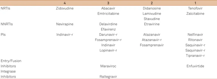
53. Table 3 shows
the ARV CNS penetration according to the index of ARV CNS
penetration efectiveness (CPE)
53.
Despite the efective suppression of viremia with ARV
therapy, HIV can still replicate in the CNS, with the
develop-ment of resistant strains in the CNS in patients with acute and
sub-acute neurological manifestations
38,39,54. Disagreement
between the HIV viral loads in the plasma and CSF is deined
by detectable levels of HIV RNA in the CSF, indicative of a
vi-ral load of > 200 copies/mL, when the vivi-ral load in the plasma
is < 50 copies/mL or by an HIV RNA viral load in the CSF that
is ≥ 1 log higher than that in the plasma
55.
Diferent concentrations of ARV drugs have been found
in the CSF. herefore, the use of ARV drugs that penetrate the
BBB is considered necessary to control infection in the CNS
in patients at an advanced stage of the disease,
particular-ly those with neurological problems. Compartmentalization
of HIV-1 infection in the CNS can afect the response to
Table 3.
Index of ARV central nervous system penetration effectiveness (CPE)
53.
4
3
2
1
NRTIs
Zidovudine
Abacavir
Didanosine
Tenofovir
Emtricitabine
Lamivudine
Zalcitabine
Stavudine
NNRTIs
Nevirapine
Delavirdine
Etravirine
Efavirenz
PIs
Indinavir-r
Darunavir-r
Atazanavir
Nelinavir
Fosamprenavir-r
Atazanavir-r
Ritonavir
Indinavir
Fosamprenavir
Saquinavir-r
Lopinavir-r
Saquinavir-r
Tipranavir-r
Entry/Fusion
Inhibitors
Maraviroc
Enfuvirtide
Integrase
Inhibitors
Raltegravir
treatment, which can lead to the development of varying
degrees of resistance to ARV drugs in both compartments.
Although tests for HIV-1 resistance in the CSF are not
recom-mended during routine treatment of individuals with ARV
failure, the choice of treatment in patients with neurological
problems requires knowledge of the resistance proile of the
virus in the CSF
56,57.
Because lumbar puncture is an invasive procedure,
re-searchers have attempted to develop a predictive model
with which to estimate the risk of detectable RNA in the CSF
(threshold of > 50 copies/mL) and help identify HIV-positive
patients who would beneit most from CSF monitoring. he
variables included in the predictive model are race, major
de-pressive disorder (MDD), adherence to ARV, AVR CNS
pen-etration efectiveness (CPE), plasma HIV RNA quantiica
-tion, and duration of current antiretroviral therapy (ART).
However, the CSF-HIV risk score for assessing HIV activity in
the CNS requires further validation
58.
Final remarks, CSF analysis has a well-deined and valu
-able role in the diagnosis of CNS opportunistic infections
and co-infections in HIV/AIDS patients. Although further
research is necessary to establish a clinically applicable
bio-marker for HAND diagnosis, promising CSF biobio-markers
in-clude neopterin, MCP-1, IP-10, and light neuroilaments.
Because of its constitutional and immunological
char-acteristics, the CNS can act as a reservoir for HIV. HIV can
replicate in the CNS, independent of HIV in the
periph-eral blood, and compartmentalization can occur, with the
development of quasispecies. Determination of the HIV
viral load in the CSF is important for assessing the
com-partmentalization of HIV in the CNS and for monitoring
therapeutic effects.
References
1. Ministério da Saúde (BR). Programa Nacional de DST/AIDS. Diagnostico da infecção pelo HIV. Manual técnico para o diagnóstico da infecção pelo HIV Brasília, DF: Ministério da Saúde; 2013 [cited 2013 10 01]. Available from: http://www.aids.gov.br/assistencia/manualdst/item12.htm
2. AVERTing HIV and AIDS. HIV & AIDS in USA. West Sussex: AVERT; 2014 [cited 2014 Mar]. Available from: http://www.avert.org/hiv-aids-usa.htm#footnote27_sdg3l0s
3. Price RW. The two faces of HIV infection of
cerebrospinal luid. Trends Microbiol. 2000;8(9):387-91. http://dx.doi.org/10.1016/S0966-842X(00)01821-7
4. Brew BJ, Wesselingh SL, Gonzales M, Heyes MP, Price RW. Managing HIV. How HIV leads to neurological disease. Med J Aust. 1996;164(4):233-4.
5. Tyler KL, McArthur JC. Through a glass, darkly: cerebrospinal luid viral load measurements and the pathogenesis of human immunodeiciency virus infection of the central nervous system. Arch Neurol.
2002;59(6):909-12. http://dx.doi.org/10.1001/archneur.59.6.909
6. Heaton RK, Clifford DB, Franklin DR Jr, Woods SP, Ake C, Vaida F et al. HIV-associated neurocognitive disorders persist in the era of potent antiretroviral therapy: CHARTER Study. Neurology 2010;75(23):2087-96. http://dx.doi.org/10.1212/WNL.0b013e318200d727
7. Dore GJ, Correll PK, Li Y, Kaldor JM, Cooper DA, Brew BJ. Changes to AIDS dementia complex in the era of highly active antiretroviral therapy. AIDS 1999;13(10):1249-53. http://dx.doi.org/10.1097/00002030-199907090-00015
8. Fishman RA. Cerebrospinal luid in diseases of the nervous system. 2nd ed. Philadelphia: WB Saunders; 1992.
9. Almeida SM, Ribeiro CE, Pessa LFC, Moreira SR, Vidal LR, Nogueira MB et al. Incidence of neurological manifestations as AIDS deining clinical conditions in Brazil. BMC Proceedings. 2008;2(Supppl 1):45. http://dx.doi.org/10.1186/1753-6561-2-s1-p45
10. Steiner I, Schmutzhard E, Sellner J, Chaudhuri A, Kennedy PGE. EFNS-ENS guidelines for the use of PCR technology for the diagnosis of infections of the nervous system. Eur J Neurol. 2012;19(10):1278-91. http://dx.doi.org/10.1111/j.1468-1331.2012.03808.x
11. Almeida SM, Zavala JA, Gabardo BM, Ribeiro CE, Rossoni AM, Araújo JM. Acute bacterial meningitis in HIV, patients in southern Brazil: Curitiba, Paraná, Brazil. Arq Neuropsiquiatr. 2007;65(2A):273-8. http://dx.doi.org/10.1590/S0004-282X2007000200016
12. Zink MC, Clements JE. The two faces of HIV infection of cerebrospinal luid: response. Trends Microbiol. 2000;8(9):390-1. http://dx.doi.org/10.1016/S0966-842X(00)01822-9
13. Almeida SM, Boritza K, Cogo LL, Pessa L, França J, Rota I et al.
Quantiication of cerebrospinal luid lactic acid in the differential diagnosis between HIV chronic meningitis and opportunistic meningitis. Clin Chem Lab Med. 2011;49(5):891-6. http://dx.doi.org/10.1515/CCLM.2011.131
14. Almeida SM, Ribeiro CE, Pereira AP, Badiee J, Cherner M, Smith D et al. Neurocognitive impairment in HIV-1 clade C- versus B-infected individuals in Southern Brazil. J Neurovirol. 2013;19(6):550-6. http://dx.doi.org/10.1007/s13365-013-0215-5
15. Antinori A, Arendt G, Becker JT, Brew BJ, Byrd DA, Cherner M et al. Updated research nosology for HIV-associated neurocognitive disorders. Neurology 2007;69(18):1789-99. http://dx.doi.org/10.1212/01.WNL.0000287431.88658.8b
16. Brew BJ, Letendre SL. Biomarkers of HIV related central nervous system disease. Int Rev Psychiatry. 2008;20(1):73-88. http://dx.doi.org/10.1080/09540260701878082
17. Almeida SM, Letendre S, Zimmerman J, Lazzaretto D, McCutchan A, Ellis R. Dynamics of monocyte chemoattractant protein type one (MCP-1) and HIV viral load in human cerebrospinal luid and plasma. J Neuroimmunol. 2005;169(1-2):144-52. http://dx.doi.org/10.1016/j.jneuroim.2005.07.012
18. Cinque P, Brew BJ, Gisslen M, Hagberg L, Price RW. Cerebrospinal luid markers in central nervous system HIV infection and AIDS dementia complex. Handb Clin Neurol. 2007;85:261-300. http://dx.doi.org/10.1016/S0072-9752(07)85017-2
19. Gisslen M, Hagberg L, Brew BJ, Cinque P, Price RW, Rosengren L. Elevated cerebrospinal luid neuroilament light protein concentrations predict the development of AIDS dementia complex. J Infect Dis. 2007;195(12):1774-8. http://dx.doi.org/10.1086/518043
20. Hagberg L, Cinque P, Gisslen M, Brew BJ, Spudich S, Bestetti A et al. Cerebrospinal luid neopterin: an informative biomarker of central nervous system immune activation in HIV-1 infection. AIDS Res Ther. 2010;7(1):15. http://dx.doi.org/10.1186/1742-6405-7-15
21. Brew BJ, Pemberton L, Blennow K, Wallin A, Hagberg L. CSF amyloid beta42 and tau levels correlate with AIDS dementia complex. Neurology. 2005;65(9):1490-2. http://dx.doi.org/10.1212/01.wnl.0000183293.95787.b7
22. Gisslén M, Krut J, Andreasson U, Blennow K, Cinque P, Brew BJ et al. Amyloid and tau cerebrospinal luid biomarkers in HIV infection. BMC Neurol. 2009;9(1):63. http://dx.doi.org/10.1186/1471-2377-9-63
24. Krut JJ, Gisslen M, Hagberg L, Zetterberg H, Price RW, Nilsson S et al. Hyperphosphorylated Tau in cerebrospinal luid: a biomarker for neurological aging in HIV? In: 21st Conference of Retrovirus and Opportunistic Infections; 2014 Mar 3-6; Boston. New York: National AIDS Treatment Advocacy Project; 2014. Poster 453.
25. Doitsh G, Galloway NLK, Geng X, Yang Z, Monroe KM, Zepeda O et al. Cell death by pyroptosis drives CD4 T-cell depletion in HIV-1 infection. Nature. 2014;505(7484):509-14. http://dx.doi.org/10.1038/nature12940
26. Doitsh G, Galloway NLK, Geng X, Yang Z, Monroe KM, Zepeda O et al. Pyroptosis drives CD4 T-cell death and chronic inlammation in HIV-1 infected lymphoid tissues. In: 21st Conference of Retrovirus and Opportunistic Infections; 2014 Mar 3-6; Boston. New York: National AIDS Treatment Advocacy Project; 2014. Oral presentation 76.
27. Monroe KM, Yang Z, Johnson JR, Geng X, Doitsh G, Krogan NJ et al. IFI16 DNA sensor is required for death of lymphoid CD4 T cells abortively infected with HIV. Science. 2014;343(6169):428-32. http://dx.doi.org/10.1126/science.1243640
28. Neuenburg JK, Cho TA, Nilsson A, Bredt BM, Hebert SJ, Grant RM et al. T-cell activation and memory phenotypes in cerebrospinal luid during HIV infection. J Acquir Immune Deic Syndr. 2005;39(1):16-22. http://dx.doi.org/10.1097/01.qai.0000155036.03004.a0
29. Gisslen M, Hagberg L, Rosengren L, Brew BJ, Cinque P, Spudish S et al. Deining and evaluating HIV-related neurodegenerative disease and its treatment targets: a combinatorial approach to use of cerebrospinal luid molecular biomarkers. J Neuroimmune Pharmacol. 2007;2(1):112-9. http://dx.doi.org/10.1007/s11481-006-9035-1
30. Lipman M, Breen R. Immune reconstitution inlammatory syndrome in HIV. Curr Opin Infect Dis. 2006;19(1):20-5. http://dx.doi.org/10.1097/01.qco.0000200543.80712.01
31. Venkataramana A, Pardo CA, McArthur JC, Kerr DA, Irani DN, Grifin JW et al. Immune reconstitution inlammatory syndrome in the CNS of HIV-infected patients. Neurology. 2006;67(3):383-8. http://dx.doi.org/10.1212/01.wnl.0000227922.22293.93
32. Ellis RJ, Hsia K, Spector SA, Nelson JA, Heaton RK, Wallace MR et al. Cerebrospinal luid human immunodeiciency virus type 1 RNA levels are elevated in neurocognitively impaired individuals with acquired immunodeiciency syndromeAnn Neurol. 1997;42(5):679-88. http://dx.doi.org/10.1002/ana.410420503
33. Ellis RJ, Moore DJ, Childers ME, Letendre S, McCutchan JA, Wolfson T et al. Progression to neuropsychological impairment in human immunodeiciency virus infection predicted by elevated cerebrospinal luid levels of human immunodeiciency virus RNA. Arch Neurol. 2002;59(6):923-8. http://dx.doi.org/10.1001/archneur.59.6.923
34. Langford D, Marquie-Beck J, Almeida S, Lazzaretto D, Letendre S, Grant I et al. Relationship of antiretroviral treatment to postmortem brain tissue viral load in human immunodeiciency virus-infected patients. J Neurovirol. 2006;12(2):100-7. http://dx.doi.org/10.1080/13550280600713932
35. Almeida SM, Ellis R, Letendre S, McCutchan JA, Grant I. Cerebrospinal luid human immunodeiciency virus viral loaad in patients with neurosyphilis. J Neurovirol. 2010;16(1):6-12. http://dx.doi.org/10.3109/13550280903514776
36. Karris MA, Smith DM. Tissue-speciic HIV-1 infection: why it matters. Future Virol. 2011;6(7):869-82. http://dx.doi.org/10.2217/fvl.11.48
37. North TW, Higgins J, Deere JD, Hayes TL, Villalobos A, Adamson L et al. Viral sanctuaries during highly active antiretroviral therapy in a nonhuman primate model for AIDS. J Virol. 2010;84(6):2913-22. http://dx.doi.org/10.1128/JVI.02356-09
38. Zárate S, Pond SLK, Shapshak P, Frost SDW. Comparative study of methods for detecting sequence compartmentalization in human immunodeiciency virus type 1. J Virol. 2007;81(12):6643-51. http://dx.doi.org/10.1128/JVI.02268-06
39. Haggerty S, Stevenson M. Predominance of distinct viral genotypes in brain and lymph node compartments of HIV-1-infected individuals. Viral Immunol. 1991;4(2):123-31. http://dx.doi.org/10.1089/vim.1991.4.123
40. Clyde F, Barker CF, Billingham RE. Immunologically privileged sites. Adv Immunol. 1978;25:1-54. http://dx.doi.org/10.1016/S0065-2776(08)60930-X
41. Kreutzberg GW. Microglia: a sensor for pathological events in the CNS. Trends Neurosci. 1996;19(8):312-8. http://dx.doi.org/10.1016/0166-2236(96)10049-7
42. Sedgwick JD, Ford AL, Foulcher E, Airriess R. Central nervous system microglial cell activation and proliferation follows direct interaction with tissue-iniltrating T cell blasts. J Immunol. 1998;160(11):5320-30.
43. Shrikant P, Benveniste EN. The central nervous system as an immunocompetent organ: role of glial cells in antigen presentation. J Immunol. 1996;157(5):1819-22.
44. Abbott NJ. Glia and the blood: brain barrier. Nature. 1987;325(6101):195. http://dx.doi.org/10.1038/325195a0
45. Thomson MM, Nájera R. Molecular epidemiology of HIV-1 variants in the global AIDS pandemic: an update. AIDS Rev. 2005;7(4):210-24.
46. Nájera R, Delgado E, Pérez-Alvarez L, Thomson MM. Genetic recombination and its role in the development of the HIV-1 pandemic. AIDS. 2002;16 Suppl 4:S3-16.
47. Korber B, Gaschen B, Yusim K, Thakallapally R, Kesmir C, Detours V. Evolutionary and immunological implications of contemporary HIV-1 variation. Br Med Bull. 2001;58(1):19-42. http://dx.doi.org/10.1093/bmb/58.1.19
48. Domingo E. Quasispecies and the implications for virus persistence and escape. Clin Diagn Virol. 1998;10(2-3):97-101. http://dx.doi.org/10.1016/S0928-0197(98)00032-4
49. Perelson AS, Neumann AU, Markowitz M, Leonard JM, Ho DD. HIV-1 dynamics in vivo: virion clearance rate, infected cell life-span, and viral generation time. Science. 1996;271(5955):1582-6. http://dx.doi.org/10.1126/science.271.5255.1582
50. Brew BJ. HIV neurology. Oxford, UK: Oxford University Press; 2001.
51. Ariën KK, Vanham G, Arts EJ. Is HIV-1 evolving to a less virulent form in humans? Nat Rev Microbiol. 2007;5(2):141-51. http://dx.doi.org/10.1038/nrmicro1594
52. Cysique LA, Vaida F, Letendre S, Gibson S, Cherner M, Woods SP et al. Dynamics of cognitive change in impaired HIV-positive patients initiating antiretroviral therapy. Neurology. 2009;73(5):342-8. http://dx.doi.org/10.1212/WNL.0b013e3181ab2b3b
53. Letendre S, Ellis R, Deuthsch R, et al (Autor, favor citar mais três autores e informar se são somente os 6. Devem ser citados seis autores e et al. para mais). In: 17th Conference on Retrovirus and Opportunistic Infections; 2010 Feb 16-19; San Francisco. New York: National AIDS Treatment Advocacy Project; 2010. Poster 430.
54. Harrington PR, Schnell G, Letendre SL, Ritola K, Robertson K, Hall C et al. Cross-sectional characterization of HIV-1 env compartmentalization in cerebrospinal luid over the full disease course. AIDS. 2009;23(8):907-15. http://dx.doi.org/10.1097/QAD.0b013e3283299129
55. Canestri A, Lescure FX, Jaureguiberry S, Moulignier A, Amiel C, Marcelin AG et al. Discordance between cerebral spinal luid and plasma HIV replication in patients with neurological symptoms who are receiving suppressive antiretroviral therapy. Clin Infect Dis. 2010;50(5):773-8. http://dx.doi.org/10.1086/650538
56. Antinori A, Cingolani A, Giancola ML, Forbici F, De Luca A, Perno CF. Clinical implications of HIV-1 drug resistance in the neurological compartment. Scand J Infect Dis Suppl. 2003;35(s106):41-4. http://dx.doi.org/10.1080/03008870310009650
57. Rotta I, Raboni SM, Ribeiro CEL, Riedel M, Winhescki MG, Smith DM et al. Cerebrospinal luid can be used for HIV genotyping when it fails in blood. Arq Neuropsiquiatr. 2014;72(7):506-9. http://dx.doi.org/10.1590/0004-282X20140093
58. Hammond ER, Crum RM, Treisman GJ, Mehta SH, Marra CM, Clifford DB et al. The cerebrospinal luid HIV risk score for assessing central nervous system activity in persons with HIV. Am J Epidemiol. 2014;180(3):297-307. http://dx.doi.org/10.1093/aje/kwu098