Authors
Ester Miranda Pereira 1 Adalberto Socorro da Silva 1
Anatália Labilloy 1 José Tiburcio do Monte Neto 1
Semiramis Jamil Hadad do Monte 1
1 Universidade Federal do Piauí.
Submitted on: 06/24/2015. Approved on: 10/27/2015.
Correspondence to: Ester Miranda Pereira. Laboratório de Imunogenética e Biologia Molecular da Universidade Federal do Piauí. Avenida Universitária - lado ímpar, Teresina, PI, Brazil. CEP: 64049-550.
E-mail: [email protected]
Podocyturia in Fabry disease
Podocitúria na doença de Fabry
Introdução: A doença de Fabry (DF) é uma desordem lisossômica ligada ao cromossomo X ocasionada por mutações no gene que codifica a enzima lisossômica α-galactosidase A (α-GAL). A redução ou ausência da atividade dessa enzima leva ao acúmulo progressivo de gb3. A doença renal é uma importante consequência clínica da acumulação de Gb3. Podócito é o tipo celular mais afetado na doença renal, que mostra apenas uma resposta parcial à Terapia de Reposição Enzimática. Além disso, a disfunção podocitária é a principal contribuinte para a perda progressiva da função renal e pode ser encontrada alterada mesmo antes do início da microalbuminúria. Assim, a podocitúria na DF pode ser uma ferramenta importante para prever a doença renal. Objetivo: O objetivo deste estudo foi quantificar a excreção urinária de podócitos em pacientes com DF (V269M, n = 14) e controles saudáveis (n = 40), e relacioná-las com as variáveis sexo, idade, tempo de terapia e a razão albumina: creatinina (AUC). Métodos: Podócitos urinários foram identificados utilizando imunofluorescência para podocalixina e DAPI. O número de células podocalixina positivo foi contado e o número médio foi utilizado (faixa normal 0-0.6 podócitos/
mL). Resultados: O número médio de
podócitos na urina de pacientes com DF foi significativamente maior do que os controles saudáveis (p < 0.0001). Observou-se uma correlação positiva entre podocitúria e AUC (p = 0.004; r2 =
0.6417). Conclusão: A podocitúria pode ser uma ferramenta adicional para avaliar a progressão da doença renal em pacientes que se espera que tenha um fenótipo mais agressivo.
R
ESUMOPalavras-chave: doença de fabry, podóci-tos, técnica indireta de fluorescência para anticorpo.
Introduction: Fabry disease is a lysosomal storage disorder due to abnormalities in the GLA gene (Xq22). Such changes result in the reduction/absence of activity of the lysosome enzyme α-GAL, whose function is to metabolize globotriaosylceramide (Gb3). Renal disease is a major clinical outcome of the accumulation of Gb3. Podocyte injury is thought to be a major contributor to the progressive loss of the renal function and may be found altered even before the onset of microalbuminuria.
Objective: The aim of this study was to quantify the urinary excretion of podocytes in Fabry disease patients (V269M, n = 14) and healthy controls (n = 40), and to correlate podocyturia with the variables gender, age, time of therapy and albumin: creatinine ratio (ACR).
Methods: Urinary podocytes were stained using immunofluorescence to podocalyxin and DAPi. The number of podocalyxin-positive cells was quantified and the average number was taken (normal range 0-0.6 podocytes/mL). Results: The average number of podocytes in the urine of Fabry disease patients was significantly higher than in healthy controls (p < 0.0001). We observed a positive correlation between podocyturia and ACR (p = 0.004; (r2 = 0.6417). We found no correlation between podocyturia and gender, age or duration of therapy. Conclusion: Podocyturia is an important parameter in the assessment of renal disease in general, and it may serve as an additional early tool for monitoring Fabry disease nephropathy even before changes in ACR are seen. This may prove to be a useful tool to assess disease progression in patients expected to have a more aggressive phenotype.
A
BSTRACTKeywords: fabry disease; fluorescent anti-body technique, indirect; podocytes.
I
NTRODUCTIONFabry disease is a lysosomal storage disorder caused by abnormalities in the GLA gene,1,2 which is mapped to the X chromosome in humans (Xq22).3,4 Such changes result in either a reduction or an absence of the activity of the hydrolase α-galactosidase A (α-gal A) or the inability of α-gal A to enter the lysosome.5 This enzyme metabolizes neutral glycosphingolipids with D-galactosyl residues, mainly globotriaosylceramide (Gb3), which is the major lipid that accumulates in Fabry disease.6
Progressive glycosphingolipid accumulation within cells results in multi-organ dysfunction. Fabry nephropathy is one of the most severe clinical consequences of this accumulation.7,8 Podocytes are the most affected kidney cell type and show a suboptimal response to enzyme replacement therapy (ERT).9 Whereas the natural history and histopathological findings of Fabry nephropathy have been largely described over the past decades, strikingly, the precise mechanism linking the initial biochemical insult and kidney failure remains poorly understood.
Podocytes are highly specialized epithelial cells that serve as key components of the glomerular filtration barrier, and podocyte injury leads to loss of the integrity of the barrier and progression to chronic kidney disease.10 Foot process effacement has been shown to precede microalbuminuria in Fabry disease, and this phenomenon seems to be reversible by early clearance of the Gb3 deposits in podocytes.11 Podocytes can detach from the glomeruli, move into the urinary space, traverse the tubules and enter the urine, a phenomenon called podocyturia. In fact, podocyte loss to levels below a certain threshold is known to promote glomerulosclerosis. Ichickwa and colleagues have speculated that injuries can be fully reversed by correcting the initial insult only when damage occurs in no more than 25% of the podocyte population.12 Thus, podocytopenia predicts the prognosis of kidney glomerular diseases in both experimental and human clinical scenarios.10
Podocytes can be retrieved from the urine in a minimal amount in a healthy state but can be found in greater numbers as an early finding of glomerular diseases.13 The assessment of podocyturia by cytological or molecular methods allows for a valuable, noninvasive and inexpensive approach to assess glomerular disease status and progression.
Determining the status and extent of podocyturia in Fabry disease can be a relevant issue for predicting renal disease during phases in which histological and functional reversibility might still be achieved. In addition, podocyte loss via the urine in FD can be useful in monitoring disease progression and renal failure to respond to standard therapies. The aim of this study was to quantify the urinary excretion of podocytes in FD patients (V269M, n = 14) and healthy controls (n = 40) and to correlate podocyturia with the variables gender, age, time of therapy and the albumin: creatinine ratio (ACR).
M
ATERIALSANDM
ETHODSThis study was approved by the Universidade Federal do Piauí Institutional Board Review (0404.0.045.000-10), and all patients have provided written informed consent prior to enrollment. Demographics and clinical data were obtained from reviewing the patients’ medical records.
SUBJECTS
The subjects of the study were 14 patients diagnosed with Fabry disease receiving ERT (0.2 mg/Kg biweekly), all of them from a single family in Piauí, Brazil. The patients were traced back from a pedigree composed of 610 members, seventy nine of which have a confirmed molecular diagnosis of FD (V269M). Each member of the pedigree was traced based using the index case identified in 2005 in Teresina - Piauí, Brazil.
PODOCYTURIA
Next, the samples were incubated with the primary antibody Mouse anti-Podocalyxin (Invitrogen) for 1 h and with the secondary antibody Alexa Fluor® 647 Goat Anti-Mouse IgG (Invitrogen) for 45 min. All steps were performed at room temperature. DAPI was used to stain nucleic acids. Slides were viewed with a fluorescent microscope. Two independent healthcare providers evaluated each sample, and the average value of the two measurements was taken. Podocyturia was defined as the number of podocalyxin positive cells per slide per mL of urine.
ALBUMIN/CREATININE RATIO (ACR)
The albumin and creatinine concentrations in the urine samples were determined using the LabTestAlbumin kit and the LabTestCreatinine Kit (Minas Gerais, Brazil), respectively, according to the manufacturer’s protocol. The urine ACR was calculated from the concentrations of urine albumin and urine creatinine, and the values are expressed in mg/g.
STATISTICAL ANALYSIS
Statistical analysis was performed using IBM SPSS Statistics software v.21. A t-test was performed to assess differences in the median, considering a significance level of p < 0.05, and a Pearson correlation coefficient was calculated to explore the linear correlation between variables.
R
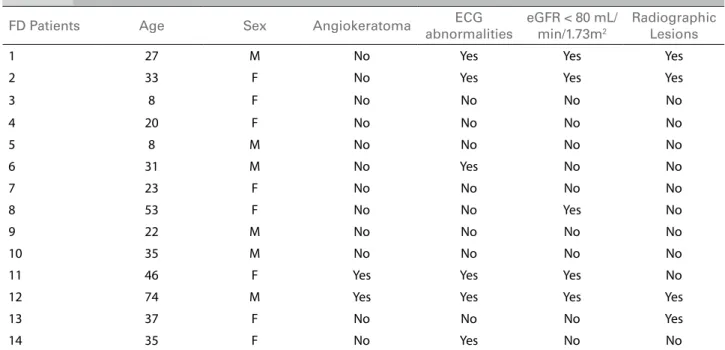
ESULTSThe patients’ demographics can be found in Table 1. Fourteen patients (seven males and seven females) with the V269M mutation in exon 6 of the GLA gene were included in the study. Their ages ranged from 8-74 years, and the duration of therapy with agalsidase alfa ranged from 1-24 months. We also obtained urine from 40 healthy individuals with no known history of kidney disease, who served as controls.
Podocytes that detach into the urinary space and are retrieved in the urine can be visualized and quantified using cytological methods combined with immunofluorescence staining for podocyte surface proteins.14 As observed in other glomerulopathies,15-18 we observed a significant loss of podocytes via the urine (p = 0.0001) of the FD patients (Median ± SEM: 0.8193 ± 0.1090), which was greater than what we observed in healthy controls (Median ± SEM: 0.4450 ± 0.03754) (Figure 1).
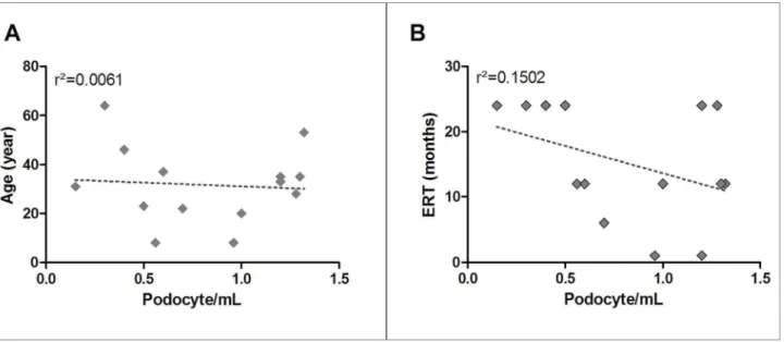
Furthermore, the number of podocytes in the urine positively correlated (r2 = 0.6417) with the urine ACR (p = 0.0006, 95% CI = 0.4703 to 0.9345) (Figure 2). However, no correlation was found with variables age and time of therapy (Figure 3).
D
ISCUSIONNephrologists treating Fabry disease are faced with two important questions: the first is concerned with the optimum time to start ERT. In fact, there is no medical consensus regarding this issue because some physicians start the treatment soon after the diagnosis and others wait for the appearance of the first symptoms.19-22 Because the current gold standard for investigating nephropathy is proteinuria and proteinuria is a consequence of podocytopenia, we realized that testing for podocyturia could yield an earlier diagnosis.
In this study, we investigated the degree of podocyturia in FD patients with the classic phenotype. Our results suggest that FD patients of both genders presented significant urinary podocyte excretion compared to healthy controls. Determining the status and extent of podocyturia in Fabry disease can be relevant in predicting renal disease during phases in which histological and functional reversibility might still be achieved.
This pilot study demonstrated the feasibility and clinical relevance of assessing podocyturia in patients with Fabry disease. However, this study was cross-sectional and involved a limited number of patients, all with the same disease genotype. Further longitudinal follow-up studies are needed, especially with the inclusion of a genotypically diverse population of patients with Fabry disease before the development of clinically overt proteinuria. These studies could be used to better assess the predictive value of podocyturia in relation to disease progression and histopathological changes, as well as these events’ temporal relationship with known markers of glomerular disease, in the context of Fabry disease.
C
ONCLUSIONFigure 1. Fabry disease patients lose more podocytes in the urine than health controls. A fresh sample of the irst urine (50 - 100 mL) void was used. Urinary podocytes were stained using immunoluorescence to podocalyxin and DAPI. The number of podocalyxin-positive cells was counted by two healthcare professionals, and the average number was taken.
Figure 2. Podocyturia correlates with the albumin-creatinine ratio in Fabry disease patients. Podocytes were identiied through immunoluorescence staining against podocalyxin in fresh urine samples of FD patients, and the number of positive cells was quantiied (podocytes/mL). The dotted line represents the linear regression.
TABLE 1 DEMOGRAPHICANDCLINICALOFFABRYPATIENTS
FD Patients Age Sex Angiokeratoma ECG
abnormalities
eGFR < 80 mL/ min/1.73m2
Radiographic Lesions
1 27 M No Yes Yes Yes
2 33 F No Yes Yes Yes
3 8 F No No No No
4 20 F No No No No
5 8 M No No No No
6 31 M No Yes No No
7 23 F No No No No
8 53 F No No Yes No
9 22 M No No No No
10 35 M No No No No
11 46 F Yes Yes Yes No
12 74 M Yes Yes Yes Yes
13 37 F No No No Yes
14 35 F No Yes No No
FD: Fabry disease; M: Male; F: Female; ECG: Echocardiogram; eGFR: Estimated glomerular iltration ratio (calculated using the CDK-EPI formula).
phenotype, podocyte loss via the urine was significantly greater than for healthy controls and was strongly correlated with the patients’ urine ACR. Determining podocyturia in patients with Fabry disease with a classic phenotype may serve as a relevant tool to assess and predict renal disease in these patients, even before changes in the urine ACR are observed. However, future prospective
studies should be conducted to better examine the temporal relationship with known markers of glomerular disease and to validate our findings in a larger population of patients with early-stage Fabry nephropathy.
A
CKNOWLEDGMENTSFigure 3. There is no correlation between podocyturia and the variables age and time of treatment in Fabry disease patients. Podocytes were identiied through immunoluorescence staining against podocalyxin in fresh urine samples of FD patients, and the number of positive cells was quantiied (podocytes/mL) and correlated with A) age and B) enzyme repleacement therapy (ERT). The dotted line represents the linear regression.
R
EFERENCES1. Anderson W. A case of angeio-keratoma. Br J Dermatol 1898;10:113-7.
2. Fabry J. Ein Beitrag zur Kenntnis der Purpura haemorragica nodularis (Purpura papulosa hemorrhagica Hebrae). Arch Dermatol Syphilol 1898;43:187-200. DOI: http://dx.doi. org/10.1007/BF01986897
3. Bishop DF, Kornreich R, Desnick RJ. Structural organization of the human alpha-galactosidase A gene: further evidence for the absence of a 3' untranslated region. Proc Natl Acad Sci U S A 1988;85:3903-7. PMID: 2836863 DOI: http://dx.doi. org/10.1073/pnas.85.11.3903
4. Opitz JM, Stiles FC, Wise D, Race RR, Sanger R, Von Gemmingen GR, et al. The Genetics of Angiokeratoma Corporis Diffusum (Fabry's Disease) and Its Linkage Relations with the Xg Locus. Am J Hum Genet 1965;17:325-42. PMID: 17948499 5. Duve C. Exploring cells with a centrifuge. Science 1975;189:186-94.
PMID: 1138375 DOI: http://dx.doi.org/10.1126/science.1138375 6. Sweeley C, Klionsky B. Fabry’s disease: classification as
a sphingolipidosis and partial characterization of a novel glycolipid. J Biol Chem 1963;238:3148-50. PMID: 14081947
7. Fervenza FC, Torra R, Lager DJ. Fabry disease: an
underrecognized cause of proteinuria. Kidney Int 2008;73:1193-9. DOI: http://dx.doi.org/10.1038/sj.ki.5002677
8. Froissart M, Benistan K, Germain DP. Functional renal investigation in Fabry disease. Presse Med 2007;36:1S36-42. 9. Fervenza FC, Torra R, Warnock DG. Safety and efficacy of
enzyme replacement therapy in the nephropathy of Fabry disease. Biologics. 2008;2:823-43.
10. Pavenstädt H, Kriz W, Kretzler M. Cell biology of the glomerular podocyte. 2003;83:253-307. PMID: 12506131
11. Sanchez-Niño MD, Sanz AB, Carrasco S, Saleem MA,
Mathieson PW, Valdivielso JM, et al. Globotriaosylsphingosine actions on human glomerular podocytes: implications for Fabry nephropathy. Nephrol Dial Transplant 2011;26:1797-802. DOI: http://dx.doi.org/10.1093/ndt/gfq306
12. Ichikawa I, Ma J, Motojima M, Matsusaka T. Podocyte damage damages podocytes: autonomous vicious cycle that drives local spread of glomerular sclerosis. Curr Opin Nephrol Hypertens 2005;14:205-10. DOI: http://dx.doi.org/10.1097/01. mnh.0000165884.85803.e1
13. Vogelmann SU, Nelson WJ, Myers BD, Lemley KV. Urinary excretion of viable podocytes in health and renal disease. Am J Physiol Renal Physiol 2003;285:F40-8. PMID: 12631553 DOI: http://dx.doi.org/10.1152/ajprenal.00404.2002
14. Hara M, Yanagihara T, Kihara I, Higashi K, Fujimoto K, Kajita T. Apical cell membranes are shed into urine from injured podocytes: a novel phenomenon of podocyte injury. J Am Soc Nephrol 2005;16:408-16. DOI: http://dx.doi.org/10.1681/ ASN.2004070564
15. Hara M, Yanagihara T, Kihara I. Urinary podocytes in primary focal segmental glomerulosclerosis. Nephron 2001;89:342-7. PMID: 11598401 DOI: http://dx.doi. org/10.1159/000046097
16. Hara M, Yanagihara T, Kihara I. Cumulative excretion of urinary podocytes reflects disease progression in IgA nephropathy and Schönlein-Henoch purpura nephritis. Clin J Am Soc Nephrol 2007;2:231-8. DOI: http://dx.doi. org/10.2215/CJN.01470506
17. Kanno K, Kawachi H, Uchida Y, Hara M, Shimizu F, Uchiyama M. Urinary sediment podocalyxin in children with glomerular diseases. Nephron Clin Pract 2003;95:c91-9. PMID: 14646369 DOI: http://dx.doi.org/10.1159/000074322
18. Nakamura T, Ushiyama C, Suzuki S, Hara M, Shimada N, Ebihara I, et al. Urinary excretion of podocytes in patients with diabetic nephropathy. Nephrol Dial Transplant 2000;15:1379-83. DOI: http://dx.doi.org/10.1093/ndt/15.9.1379
19. Politei J, Schenone AB, Cabrera G, Heguilen R, Szlago M. Fabry disease and enzyme replacement therapy in classic patients with same mutation: different formulations - different outcome? Clin Genet 2015 Mar 26. [Epub ahead of print]
20. Sawai S. Fabry Disease: Pathogenesis, Clinical Symptoms, and Treatment with Enzyme Replacement Therapy. Brain Nerve 2015;67:1099-8.
21. Sanchez-Niño MD, Carpio D, Sanz AB, Ruiz-Ortega M,
Mezzano S, Ortiz A. Lyso-Gb3 activates Notch1 in human podocytes. Hum Mol Genet 2015;24:5720-32. DOI: http:// dx.doi.org/10.1093/hmg/ddv291