ORI GI N AL ARTI CLE
I sola t ion a nd susce pt ibilit y pr ofile of ba ct e r ia in dia be t ic foot a nd ve nous
st a sis ulce r of pa t ie nt s a dm it t e d t o t he e m e r ge ncy r oom of t he m a in
unive r sit y hospit a l in t he st a t e of Goiá s, Br a zil
Ly de Fr e it a s Fe r n a n de sI; Fa bia n a Cr ist ina Pim e nt aI I; Fe r na ndo de Fr e it a s Fe r na nde sI I I IPhysician. Specialist in General Surgery, Universidade Federal de Goiás ( UFG) , Goiânia, GO, Brazil. Specialist in Peripheral Vascular Surgery and Angiology, Universidade Federal de Uberlândia,
Uberlândia, MG, Brazil. MSc. student, Graduate Program in Tropical Medicine, I nstituto de Patologia Tropical e Saúde Pública ( I PTSP) , UFG, Goiânia, GO, Brazil. Vascular surgeon, Hospital das Clínicas, UFG, Goiânia, GO, Brazil.
I ICo- advisor. Associate professor, Departm ent of Microbiology, I m m unology, Parasitology and Pathology ( DMI PP) , Setor de Microbiologia, I PTSP, UFG, Goiânia, GO, Brazil.
I I IAdvisor. Associate professor, DMI PP, Setor de Parasitologia-Entom ologia, Laboratório de Artropodologia Médica e Veterinária, I PTSP, UFG, Goiânia, GO, Brazil.
Correspondence
J Vasc Bras. 2007; 6( 3) : 211-7.
ABSTRACT
Ba ck gr ound: I nfected lower lim b inj uries ( diabetic ulcers and venous stasis ulcers) cause great suffering and functional disability with social and econom ic im pact and increase in risk of severe com plications.
Obj e ct ive :To characterize the m icrobiota and determ ine the antim icrobial susceptibility profile of isolated bacteria in lower lim b inj uries secondary to the venous stasis ulcer and diabetic foot. M e t hods: Patients with lower lim b lesions were included in the study, both diabetics and patients with venous stasis ulcer, receiving care at the em ergency service of a university hospital in Goiânia ( Brazil) from February 2005 to August 2006. Sam ples were collected with cotton swab to perform culture and antim icrobial sensitivity test applying standardized techniques.
Re sult s: Presence of bacteria was detected in 88.46% of the sam ples. Gram-positive cocci were characterized as St aphylococcus aureus and St aphylococcus epiderm idis. Am ong Gram - negative rods, Pseudom onas aeruginosa, Escherichia coli, Prot eus m irabilisand Ent erobact er sp. were detected.
Ke yw or ds: Microbiota, venous stasis ulcer, diabetic foot, infection.
RESUM O
Cont e x t o: Lesões infectadas de m em bros inferiores ( úlceras diabéticas e úlceras de estase venosa) são causa de grande sofrim ento e incapacitação funcional com im pacto social, econôm ico e aum ento do risco de com plicações severas.
Obj e t ivo: Caracterizar a m icrobiota e determ inar o perfil de suscetibilidade antim icrobiana das bactérias isoladas de lesões de m em bros inferiores secundárias a úlcera de estase venosa e pé diabético.
M é t odos: Foram incluídos no estudo pacientes portadores de lesões de m em bros inferiores, sendo diabéticos, e pacientes com úlcera de estase venosa, atendidos em um serviço de urgência de um hospital universitário de Goiânia ( GO) , no período de fevereiro de 2005 a agosto de 2006. A coleta de m aterial foi realizada com swab de algodão para realização de cultura e teste de sensibilidade antim icrobiana, em pregando- se técnicas preconizadas.
Re sult a dos:Das am ostras analisadas, foi detectada a presença de bactérias em 88,46% . Os cocos gram - positivos foram caracterizados com o St aphylococcus aureuse St aphylococcus epiderm idis. Dentre os bastonetes gram -negativos, detectou-se Pseudom onas aeruginosa, Escherichia coli, Prot eus m irabilise Ent erobact er sp.
Conclusõe s: Os m icrorganism os isolados das lesões de m em bros inferiores ( pé diabético e úlcera de estase venosa) incluíram bactérias gram- positivas e negativas, sendo St aphylococcus aureus, Pseudom onas aeruginosa e Escherichia colias m ais freqüentes, com elevada resistência a diversos antim icrobianos.
Pa la vr a s- cha ve : Microbiota, úlcera de estase venosa, pé diabético, infecção.
I nt r oduct ion
Am ong the m ost com m on lower lim b lesions are diabetic and venous stasis ulcers.1 Plantar lesions
known as diabetic foot, a chronic and frequent com plication of diabetes m ellitus,1 result especially
from neuropathy and degenerative m icroangiopathy characterized by alteration in capillary structure and protective endothelial function.2 I ncreased plantar pressure, skin changes such as dryness,
fissures, m ycosis, osteoarticular deform ities, m uscle atrophy and bone prom inences, form ation of callus3 and repetition traum as can result in skin and subcutaneous tissue infection, abscesses and
deep layer phlegm s,4 significantly increasing risk of am putation,5 which is also associated with early
arteriosclerosis.6
Venous stasis ulcers are also frequent lesions7 and are related to physiopathological m echanism s of
chronic venous insufficiency.7 They generate social and econom ic im pact, work disability and
expenses associated with treatm ent.7, 8
Microorganism s associated with lower lim b lesions m entioned above are part of skin m icrobiota, and associations of anaerobic and facultative aerobic bacteria are com m on, resulting in m ixed
St aphylococcus aureus and St rept ococcus sp are present in moderate lower limb infections without
systemic toxicity, in superficial lesions with cellulitis, moderate ulceration and mild ischemia.10In
severe infections with extensive cellulitis, ulcer, lymphangitis and ischemia, gram-positive cocci are
present (St aphylococcus sp, St rept ococcus spand Ent erococcus sp), anaerobic bacteria, such as
bacteroids and facultative gram-negative (Escherichia coli,Ent erobact er sp, etc.), and
nonfermenting gram-negative rods (Pseudom onas and Acinet obact er) .10Our aim was to isolate and
characterize the microorganisms of lower limb lesions (diabetic foot and venous stasis ulcer), as well as to determine susceptibility profile of isolated bacteria.
M e t hod
The study population was comprised of patients with lower limb lesions (diabetic foot and venous stasis ulcer), who were admitted to a university hospital in Goi€nia, Brazil. The study was carried out after approval by the Ethics Committee and signing of a consent form by the patient or
responsible. Collection was performed in deep layers using cotton swab after skin disinfection with
physiologic solution and Povidine•, local anesthesia with 2% lidocaine without vasoconstrictor and
surgical debridement of devitalized tissues. The samples were conditioned in Stuart medium and sent to the laboratory for culture and antimicrobial sensitivity test (antibiogram).
The samples were sowed in sheep blood agar (5%) and incubated at 37 ‚C for 24-48 hours. Colonies were initially identified by gram staining, based on their development in selective and
nonselective culture mediums, biochemical/enzymatic tests11and techniques automated by the
MicroScan• system (Dade Behring – West Sacramento, California, USA). Susceptibility of isolated
bacteria was determined by the automated system, and the results were interpreted according to
recommendations by the Clinical and Laboratory Standards Institute.12
Re sult s
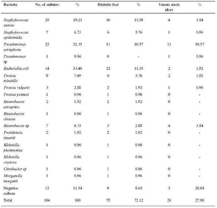
In this study, 79 cases of lower limb lesions were assessed: 50 diabetic foot and 29 stasis ulcers. A total of 104 cultures were performed, 92 (88.46%) of them being positive. In 65 cultures, gram-negative bacteria were isolated; of these, 42 (45.66%) were enterobacteria, 23 (25%) were nonfermenting rods and 27 (29.34%) were staphylococcus.
The 12 (11.54%) negative cultures corresponded to samples of the first collection from nine
Prevalent bacteria in lesions ( Figure 3) were: St aphylococcus aureus , St aphylococcus epiderm idis and gram -negative rods: Pseudom onas aeruginosa, Escherichia coli, Prot eus m irabilis and
Results of the susceptibility profile in the m ost frequent four bacteria are presented in Figures 4 to 7.S. aureus and P. aeruginosa were prevalent both in diabetic foot lesions and in venous stasis ulcers, whereas the third m ost isolated bacteria was E. coli in diabetic foot and Ent erobact er sp in venous stasis ulcer. All St aphylococcus aureuswere sensitive to vancom ycin, tobram ycin, Synercid ( quinupristin-dalfopristin) and linezolid. Sensitivity of S. aureusto gatifloxacin, am picillin/ sulbactam and cefazolin was 80% , whereas it was 77% for rifam picin ( Figure 4), with resistance to am picillin, penicillin, am ikacin, cephalothin, am oxicillin/ clavulanate and oxacillin. P. aeruginosa was sensitive to m eropenem , im ipenem e polym yxin B ( Figure 5) . E. coli was sensitive to im ipenem and
D iscussion
A high frequency of S. aureus, P. aeruginosa and enterobacteria was detected in assessed lesions, sim ilar to that reported by Assis et al.1 3 and Jorge et al.1 4 Regarding gram -positive cocci, thee was
prevalence of S. aureusand S. epiderm idis, in agreem ent with the reports by Goldstein et al.1 5,
Routh et al.1 6 and Slovenkai et al.1 7
Selection and dissem ination of m ultiresistant m icroorganism s have been occurring both in hospitals and in the com m unity and represent a great challenge in therapy.1 5 , 1 8 - 2 0 I n this study, m ethicillin-resistant S. aureus ( MRSA) had high prevalence ( 69% ) , different from the results found by Goldstein et al.1 5 and Carvalho et al.1 8, who found rates lower than 20% . I solation rate of
gram-negative bacteria in this study was sim ilar to that found by Carvalho et al.,1 8 when assessing
patients with diabetic foot and m ainly isolating enterobacteria.
Rocha et al.2 1 reported the problem associated with m ultiresistance of grampositive and gram
-negative bacteria, especially Escherichia coli, in m ore severe cases. Most staphylococci detected in this study were resistant to am oxicillin/ clavulanate, cephalothin, oxacillin and clindam ycin, sim ilar to what was described by Unachukwu et al.2 0 and Rocha et al.2 1
Due to unfavorable evolution of 10 patients with diabetic foot, subsequent collections were
perform ed ( Table 2). There was prevalence of St aphylococcus aureus and Pseudom onas aeruginosa, and the bacteria isolated in the first collection were recovered in only two cases. I n four cases that progressed to lower lim b am putation, there was presence of P.aeruginosa and/ or S. aureus, in agreem ent with Rocha et al.,2 1 who considered diabetic foot as the m ain cause of nontraum atic lim b
am putation.
Conclusion: A m ixed m icrobiota of lower lim b lesions was detected, with grampositive and gram -negative bacteria, St aphylococcus aureus , Pseudom onas aeruginosaand Escherichia colibeing the m ost frequent, with high resistance to m any antim icrobials and a high rate of MRSA ( 69% ) .
According to the in vit ro result s, am picillin/ sulbactam in association with piperacillin/ tazobactam could be an option for the in vit ro treatm ent of m ost cases of lower lim b lesions, as well as ciprofloxacin when there is no suspicion of infection by Pseudom onas.
Ack now le dge m e nt s
To m y advisors.
Laboratório Professora Margarida Dobler Kom m a.
Laboratório de Análises clínicas do Hospital das Clínicas.
Staff of practitioners and technicians at the em ergency room of HC UFG.
José Jurandir de Moraes and Elisa Tiba Gom es for their loyalty in helping provide care for patients at the em ergency room .
To CNPq for the partial financial aid to perform this study.
Re fe r e nce s
1. Mayall RC, Mayall AC, Melo AV, Mayall JC, Mayall LC. Pé diabético. I n: Maffei FHA. Doenças vasculares periféricas. Rio de Janeiro: MEDSI ; 1987. p. 865- 81.
2. De Luccia N. Doença vascular e diabetes. J Vasc Bras. 2003; 2: 49- 60.
3. Vedolin AC, Schm itt CMD, Bredt CFG, et al. Pé diabético: estudo com parativo entre diferentes form as de apresentação clínica e tratam entos. Rev Angiol Cir Vasc. 2003; 12: 15-21.
4. Arm strong DG, Lavery LA, Harkless LB. Who is at risk for diabetic foot ulceration?Clin Podiatr Med Surg. 1998; 15: 11- 9.
5. Margolis DJ, Kantor J, Berlin JA. Healing of diabetic neuropathic foot ulcers receiving standard treatm ent. A m eta- analysis. Diabetes Care. 1999; 22: 692-5.
6. Dorm andy JA, Rutherford RB. Managem ent of peripheral arterial disease ( PAD). TASC Working Group. TransAtlantic I nter- Society Consensus ( TASC) . J Vasc Surg. 2000; 31( 1 Pt 2) : S1- S296.
7. Frade MAC, Cursi I B, Andrade FF, et al. Úlcera de perna: um estudo de casos em Juiz de Fora-MG ( Brasil) e região. An Bras Derm atol. 2005; 80: 41-6.
8. Olin JW, Buesterien KM, Childs MB, Seavey C, McHugh L, Griffiths RI . Medical costs of treating venous stasis ulcers: evidence from a retrospective cohort study. Vasc Med. 1999; 4: 1- 7.
10. Sader HS, Durazzo A. Terapia antimicrobiana nas infec„…es do p† diab†tico . J Vasc Bras. 2003;2:61-6.
11. Koneman EW, Allen SD, Janda WM, Schreckenberger PC, Winn WCJ. Diagn‡stico microbiol‡gico: texto e atlas colorido. 5ˆ ed. Medsi: Rio de Janeiro; 2001.
12. Clinical and Laboratory Standards Institute (CLSI). Performance standards for antimicrobial susceptibility testing; fifteenth informational supplement. CLSI document M100-S15, Wayne, Pa, USA; 2005.
13. Assis TL, Formiga LCD, Filgueira AL, Mattos GA. Aspectos microbiol‡gicos dos espa„os
interdigitais dos p†s. II. Microbiota aer‡bia potencialmente patog‰nica nas les…es interdigitais dos p†s. An Bras Dermatol. 1984;59:61-6.
14. Jorge BH, Borges MF, Brito VN, Santos TGM, Thirone ACP. AnŠlise cl‹nica e evolu„Œo de 70 casos de les…es podais infectadas em pacientes diab†ticos. Arq Bras Endocrinol Metab. 1999;43:366-72.
15. Goldstein EJ, Citron DM, Nesbit CA. Diabetic foot infections: bacteriology and activity of 10 oral antimicrobial agents against bacteria isolated from consecutive cases. Diabetes Care. 1996;19:638-41.
16. Routh HB, Bhowmik KR, Parish LC, Bhowmik NK. Diabetic foot infection. An Bras Dermatol. 1996;71:243-9.
17. Slovenkai MP. Foot problems in diabetes.Med Clin North Am. 1998;82:949-71.
18. Carvalho CBM, Neto RM, AragŒo LP, Oliveira MM, Nogueira MB, Forti AC. P† diab†tico: anŠlise bacteriol‡gica de 141 casos. Arq Bras Endocrinol Metab. 2004;48:406-13.
19. GuzmŠn-Blanco M, Casellas JM, Sader HS. Bacterial resistance to antimicrobial agents in Latin America. The giant is awakening. Infect Dis Clin North Am. 2000;14:67-81.
20. Unachukwu CN, Obunge OK, Odia OJ. The bacteriology of diabetic foot ulcers in Port Harcourt, Nigeria. Niger J Med. 2005;14:173-6.
21. Rocha JLL, Baggio HCC, Cunha CA, Niclewicz EA, Leite SAO, Baptista MIDK. Aspectos relevantes da interface entre diabetes mellitus e infec„Œo. Arq Bras Endocrinol Metab. 2002;46:221-9.
Cor r e sponde nce :
Fernando de Freitas Fernandes
Departamento de Microbiologia, Imunologia, Parasitologia e Patologia do Instituto de Patologia Tropical e Sa•de P•blica da Universidade Federal de GoiŠs
Caixa Postal, 131
CEP 74605-050 – Goi€nia, GO, Brazil Email: [email protected]