Article
ISSN 0102-695X http://dx.doi.org/10.1590/S0102-695X2011005000132 Received 20 Sep 2010 Accepted 12 Jan 2011 Available online 5 Aug 2011
against methicillin-resistant staphylococcal
and Gram-negative strains showing selective
toxicity
Ivana Correa Ramos Leal,
*,1Ivaldo I. Júnior,
1Eliezer M.
Pereira,
2Marinella da S. Laport,
3Ricardo M. Kuster,
1Kátia
Regina Netto dos Santos
31Faculdade de Farmácia, Pólo Universitário, Universidade Federal do Rio de Janeiro,
Campus Macaé, Brazil,
2Laboratório de Microbiologia, Instituto Federal de Educação, Ciência e Tecnologia do
Rio de Janeiro, Campus Maracanã, Brazil,
3Instituto de Microbiologia Prof. Paulo de Góes, Centro de Ciências da Saúde,
Universidade Federal do Rio de Janeiro, Brazil.
Abstract: The ethanol extract of the vegetal species Pentaclethra macroloba (Willd.) Kuntze, Fabaceae, was fractioned and the antibacterial activity was determined. The active ethyl acetate (ea) fraction showed activity against Gram-positive (Staphylococcus spp. and Enterococcus spp.) and Gram-negative (Pseudomonas aeruginosa, Acinetobacter spp. and Klebsiella pneumoniae) multiresistant bacteria. Gallic acid derivatives were identified as the main compounds in inactive subfractions from the ea fraction, while the active one afforded ellagic acid as the major constituent when submitted to acid hydrolysis reaction, which suggests the presence of hydrolysable tannins. The minimum bactericidal concentration analysis showed a bactericide mechanism of action for the tannin subfraction found. The antibacterial mechanism of action of the active tannin subfraction against S. aureus
reference strains (ATCC 29213 e 33591) was proposed adopting an in vitro assay of protein synthesis inhibition. For this, bacterial cells were labeled with [35S]
methionine in the presence of the subfraction. The protein synthesis inhibition was
observed at 256 μg/mL of this subfraction. At this concentration it did not present
cytotoxicity in eukaryotic cells by the neutral red technique, suggesting selective toxicity. The present study is the first in vitro investigation of the antibacterial properties of tannin fractions obtained from a polar extract of P. macroloba.
Keywords:
antibacterial activity cytotoxicity activity Gram-negative
Pentaclethra macroloba
protein synthesis
Staphylococcus spp
Introduction
Staphylococcus spp. is considered the leading cause of nosocomial infections worldwide (Deleo & Chambers, 2009). In Brazil, methicillin-resistant Staphylococcus aureus (MRSA) is responsible for approximately 43% of these infections (Sader et al., 2004). Coagulase-negative staphylococci (CNS) is also recognized as a important nosocomial pathogen around the world, highlighting the multiresistant
Staphylococcus epidermidis and Staphylococcus haemolyticus as the most isolated organisms from bacteremia (Nunes et al., 2007) and infections related to implanted medical devices (Uçkay et al., 2009) Resistance to methicillin is relevant in these pathogens
because is related to resistance to other β-lactams and
can be associated with resistance to other classes of antimicrobials (Feng et al., 2008).
It is also important to note the increasing incidence of infections caused by Gram-negative bacteria, including Acinetobacter spp. and Pseudomonas spp. resistant to carbapenems (Kokis et al., 2005; Touati et al., 2009). The prevalence of antimicrobial resistant isolates has increasing (Hsueh et al., 2002) as described recently by Touati and colleagues (2009) that assigned a mortality rate of 32% among neonates caused by multiresistant
Acinetobacter baumannii.
The performance of in vitro studies of the activity of medicinal plants extracts against resistant pathogenic bacteria has increased in microbiology and has emerging as a scientific field of extreme interest.
The bark of this species is a rich source of condensed tannins and phenolic compounds (Schedlbauer & Kavanagh, 2008), chemical classes already recognized in the literature by exhibiting antimicrobial activities (Machado et al., 2002). Triterpenic monodesmoside saponins have already been found as major compounds in this species (Viana et al., 2004b). These compounds have shown anti-hemorrhagic activity, justifying the use of P. macroloba against bites from snakes (Silva et al., 2005). Extracts prepared from the seeds and bark of this species have also several uses in folk medicine, as healing of ulcers and dermal healing post cesarian (Silva et al., 2005).
The increasing prevalence of multiresistant bacteria, especially Gram-positive bacteria, such as
Staphylococcus spp. (Nunes et al., 2007; Nascimento-Carvalho et al., 2008), and Gram-negative bacilli (Kokis et al., 2005; Hsueh et al., 2002; Touati et al., 2009; Nogueira et al., 2006) can make difficult the treatment of infectious diseases. Then, as a new therapeutic option we evaluated the antibacterial activity of P. macroloba
extracts against Gram-positive and Gram-negative hospital bacteria as well as against reference strains, establishing the minimum inhibitory activity of the active fractions. In addition, we intended to propose a mechanism of action of the active fraction based on the protein synthesis inhibition assay and examined the in vitro toxicity to eukaryotic cell cultures. The chemical composition of the active and the inactive fractions were also determined.
Materials and Methods
Plant material
The pulverized powder of the bark of
Pentaclethra macroloba (Wild.) Kuntze, Fabaceae, was provided by the Prof. Dr. Walter Baptista Mors (NPPN-UFRJ) in 2006. A copy of the individual can be found in the Research Institute of the Botanical Garden of Rio de Janeiro, in section 20 of the D site and is registered under number 4284.
Extraction and fractionation
A total of 1 kg of the powdered stem bark of P. macroloba was extracted with ethanol at room temperature for five days, and the macerate was concentrated in rotary evaporator to obtain the dry crude ethanolic extract (ce) (92g). The resulting extract was
suspended in 300 mL of methanol/water (MeOH:H2O,
9:1) and then submitted to a liquid-liquid partition with
n-hexane (3x 200 mL). The separated aqueous MeOH
layer was evaporated under reduced pressure and then resuspended in water. The resulting aqueous solution
was extracted with solvents of increasing polarity (3x
200 mL each one): dichloromethane (dl), ethyl acetate (ea) and butanol (bu).
The ethyl acetate fraction (1.77 g) was
fractioned using Sephadex LH-20 (Pharmacia)
chromatography column and isocratic methanol solvent
system as mobile phase (flow rate: 5 mL/min). The 200 (1-200) subfractions obtained (30 mL each) were
grouped into seven major subfractions (FA1 to FA7), according to the similarity of spots in silica gel thin
layer chromatography (TLC) (60 F254, Merck) using
different mobile phases, such as: dichloromethane:ethyl acetate:ethanol (2:0.4:0.3); ethyl acetate:ethanol:water (12:1:0.5) and dichloromethane:methanol:water (6:3:0.1).
High performance liquid chromatography (HPLC) analysis
In order to establish the chemical difference between the subfractions FA1 (bioinactive) and FA5
(bioactive) they were analyzed by an HPLC equipped with a Shimadzu LC-10AD pump and a CBM-10A
photodiode array detector with absorptions from 200 to 500 nm. The stationary phase was constituted by a
RP-18 column (5 μm, 20 x 5 mm, Merck) and the mobile
phase by an isocratic system of 0,5% H3PO4+0,01M KH2PO4+CH3CN (4:4:2) or a gradient elution protocol
varying from 30 to 70% of B (MeOH) in A (H2O/0,01M
H3PO4) during 40 min.
Gas chromatography coupled to mass detector (GC-MS)
The gas chromatography analysis of the volatile subfraction FA1 was performed on a Shimadzu GC-17A with interface GCMS-QP5000 and electronic impact correspondent to 70ev. The database available for spectra comparison was the NIST (National Institute of Standards and Technology) from 1990.
Bacterial strains
The ethanol (ce), ethyl acetate (ea) and butanol (bu) fractions from P. macroloba were evaluated for antibacterial activity against fourteen clinical strains of S. aureus, being ten MRSA and four MSSA (methicillin-sensitive S. aureus). Two reference strains, ATCC 29213 (MSSA) and ATCC 33591 (MRSA) were also analyzed. Activities against nine strains of CNS, including four strains of S. epidermidis (three clinical and one ATCC 12228), four of S. haemolyticus (three clinical and one ATCC 29970) and one clinical strain of
investigation were two reference strains of Klebsiella pneumoniae (ATCC 4352, a extended-spectrum
β-lactamase (ESBL) producer and 700603), one
reference strain of Pseudomonas aeruginosa (ATCC 27853), as well as thirty clinical isolates of this species. Among Acinetobacter spp. isolates 26 were evaluated, being nine A. baumanni resistant to carbapenems and one Acinetobacter lwoffii sensitive to them. Al the bacterial isolates used in the study were obtained from clinical specimens from patients in hospitals in Rio de Janeiro, Brazil.
Minimal inhibitory concentration (MIC) determination
To determine the minimal inhibitory concentration (MIC) of the active fraction and subfractions of P. macroloba against the bacterial strains listed above, it was used the Müeller Hinton
agar (Difco) dilution technique according to CLSI (2003). Concentrations ranging from 64 to 512 μg/mL
were used for the extract or each (sub) fraction tested. The bacterial inoculum was adjusted to approximately
104 colony-forming units (CFU/mL) and was added
to the medium using a Steers replicator (Machado et al., 2005). The plates were incubated at 35 °C during 24 h. The MIC values were calculated as the lowest concentration of extract or (sub) fraction where there was no bacterial growth. The antimicrobial oxacillin (Sigma) was used as control.
Minimal bactericidal concentration (MBC) determination
In order to check the bactericidal vs. bacteriostatic action of the subfraction rich in tannins of P. macroloba (FA5), MBC were determined against the reference strains ATCC 33591 e ATCC 29213. This technique establishes the smallest concentration (of those tested) of a drug necessary for the elimination of 99.9% of the microorganisms tested. The MBC was determined by the dilution method in broth (DMB)
(CLSI, 2003). Initially, MIC value was determined by transferring 100 µL from a bacterial suspension at 4-5 x
106 UFC/mL for tubes with the active subfraction (F A5) in concentrations varying from 256 to 1024 μg/mL or with DMSO (solvent), used as a positive control. The
inoculated tubes were incubated at 35 °C during 24 h and the turbidity of each tube was analyzed after this period. The MBC value was determined after an aliquot
of 100 µL from tubes without growth was seeded on
a Müller Hinton agar plate and incubated for 24 h at 35 °C. If MIC=MBC value or if the MBC was up to two concentrations above the MIC, the compound was considered as bactericidal (Isenberg, 1992).
Protein synthesis analysis by SDS-PAGE in the presence of the subfraction FA5
The evaluation of the interference of FA5 in the protein synthesis of staphylococcal strains was performed according to Pereira and colleagues (2006). Initially, the
inocula of the reference strains were carried out in 3mL tubes of TSB (Trypticase soy broth, Oxoid) and then incubated under agitation for 24 h. Then, a 0.5 mL aliquot from each solution was transferred into a tube with 2.5 mL
of a new TSB and once incubated under agitation for 1
h. Briely, 1.5 mL was transferred to an Eppendorff® tube
that was centrifuged and suspended in a culture medium
methionine-free (Free Mem, Gibco). Further, 200 μci/mL
of radioactivity labeled methionine [35S] (Amershan) were
added. To this solution, the FA5 was added at 250 and 125
μg/mL. After a treatment with lisostain the samples were
analyzed by sodium dodecyl sulfate polyacrylamide gel electrophoresis (SDS-PAGE) and further exposed to X-ray
ilm.
Cytotoxicity test
The cytotoxicity test was carried out in order to determine in which concentration FA5 was toxic to eukaryotic cells and to compare the minimum inhibitory concentration (MIC) to this result. The test was performed according to Pereira et al. (2006). Briefly, cell line BSC-40 from kidney of African green monkey was propagated in microplates for 24 h in an
ideal medium. Then, there were added 200 μL of FA5
in a concentration greater than twice the value of MIC previously established. From the initial concentration there were established seven successive dilutions and twelve replicates of each. We then included two controls without fraction FA5: the first one with DMSO, used as solvent for the fraction, and in the second, only the culture medium. After 24 h of incubation of the microplates, we observed the morphology of the
cells by microscopy. Then there were added 100 μL
of neutral red (0.1%) in each well and the microplates were incubated for 3 h, washed and once incubated for fixing the dye. For neglecting the non-viable cells,
it was added 100 μL of a solution of 50% methanol
and 1% acetic acid to each well and the plates were incubated. The optical density was measured at 490 nm using a microtiter plate Spectrophotometer. The uptake of neutral red is proportional to the number of viable cells (Isenberg, 1992).
Results and Discussion
Subfraction FA5
The highest bacterial inhibitory activity was found in the subfraction FA5 obtained from the
ea fraction. So, this subfraction was submitted to an
analysis by HPLC-DAD, which indicated the presence
of a major peak with UV spectra characteristic of phenolic substances (204 and 275 nm). This fraction was subjected to a hydrolysis reaction with an aqueous solution of 1N HCl, 30 min and further re-analyzed by
HPLC-DAD. A major signal with the same retention
time (32.94 min) and UV spectrum of a standard of ellagic acid was observed, suggesting the presence of an ellagitannin (polihydroxylated phenolic substance). Both, ellagic acid standard and the product of hydrolysis of FA5, presented maximum absorbances at 254 and 366 nm. The ellagic acid (1) is formed from the condensation of two gallic acid units, originating a residue knowing as hexahydroxidiphenoil (HHDP) (2) (Tanaka et al., 2003).
Subfraction FA1
In the HPLC chromatogram two major peaks
around 3.98 and 5.65 min (Figure 1) were observed. The UV spectra of the major constituents present in the subfraction FA1 suggested the presence of substances with absorbance peaks characteristic of benzoic acids (215, 261 and 292 nm). The results were confirmed beyond the analysis by GC-MS further methylation reaction with diazomethane. The substances were identified as being the protocatecuic acid derivatives: gallic acid methyl ester (3,4,5-trimetoxi-benzoic acid) (3), important in the biosynthesis of hydrolysable tannins and, in a minor amount, the 3,4-dimethoxy benzoic acid (4).
Figura 1. HPLC chromatogram of the subfraction FA1
obtained from the ethyl acetate fraction of the vegetal specie
P. macroloba (silicagel RP-18, 0,5% H3PO4 + 0,01M KH2PO4 + CH3CN- 4:4:2).
O
O O
OH OH O HO HO
1
CO2H HO2C
OH OH HO
HO
2
OHHO HHDP residue
H3CO OCH3
OR CO2H
3R = OCH3 4R = H
Antibacterial activity analysis of the fractions and subfractions
Antibacterial analysis of the fractions (ce, ea and
bu)
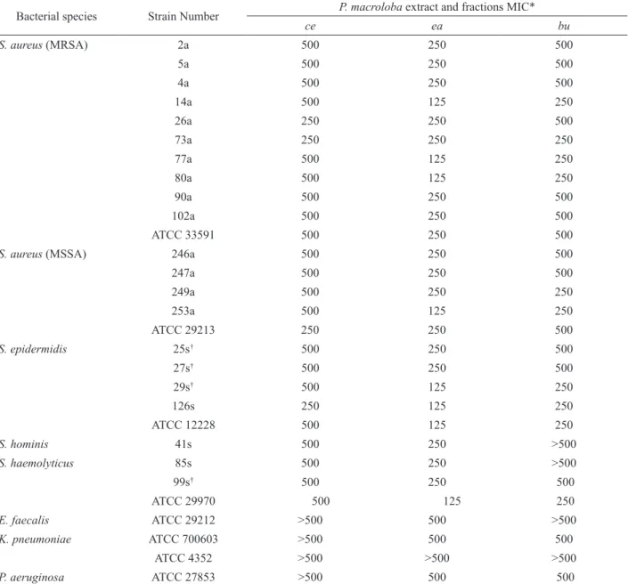
The ce, ea and bu fractions were primarily assayed against the strains described in the Table 1. At this assay
the following concentrations: 500, 250, 125 and 62.5 μg/ mL were tested. At 125 μg/mL the ea fraction inhibited
around 50% of the coagulase-negative staphylococci (CNS) as well as four clinical isolates of S. aureus, three MRSA and one MSSA. The other fractions did not show activity at this concentration.
At 250 μg/mL, all MRSA, MSSA and CNS
strains were susceptible to the ea fraction. At this concentration, the inhibition by the bu fraction was less pronounced since just six from the sixteen strains of the
S. aureus and four from the nine strains of CNS were inhibited. The ce fraction presented an unsatisfactory result once only three from the sixteen strains of S. aureus tested and one from the nine strains of CNS were inhibited.
At the major concentration investigated (500
μg/mL) both, ce and ea, were able to inhibit all MRSA,
MSSA and CNS strains assayed. However, only the last one was capable to promote inhibitory activity against the Gram-negative reference strains of K. pneumoniae (ATCC 700603) and of P. aeruginosa (ATCC 27853). So, beyond the primary results obtained it was possible to observe that the ea fraction, wealthy in tannins, exhibited the best antibacterial performance.
The extract and fractions were also investigated against one ATCC and 29 hospital strains of P. aeruginosa (fourteen resistant and ifteen sensitive to carbapenem). The results showed that just the ea fraction, at 500 μg/ mL, was active against P. aeruginosa clinical isolates. The inhibition percentage observed was around 70% of thirty strains evaluated, considering that nine out of fourteen resistant to the carbapenems were inhibited.
The fractions ce, ea and bu from P. macroloba
were also investigated against 26 clinical strains of
Acinetobacter spp. and one strain of A. lwoffii (sensitivity to carbapenem) in two different concentrations (250
and 500 μg/mL). At 250 μg/mL, just one strain of A. baumanni (resistant to carbapenem) and the strain of A. lwoffii were inhibited by the ea fraction, nevertheless, at
Table 1. Minimun inhibitory concentration (MIC) results obtained for Pentaclethra macroloba extracts against 29 bacterial strains Bacterial species Strain Number P. macroloba extract and fractions MIC*
ce ea bu
S. aureus (MRSA) 2a 500 250 500
5a 500 250 500
4a 500 250 500
14a 500 125 250
26a 250 250 500
73a 250 250 250
77a 500 125 250
80a 500 125 250
90a 500 250 500
102a 500 250 500
ATCC 33591 500 250 500
S. aureus (MSSA) 246a 500 250 500
247a 500 250 500
249a 500 250 250
253a 500 125 250
ATCC 29213 250 250 500
S. epidermidis 25s† 500 250 500
27s† 500 250 500
29s† 500 125 250
126s 250 125 250
ATCC 12228 500 125 250
S. hominis 41s 500 250 >500
S. haemolyticus 85s 500 250 >500
99s† 500 250 500
ATCC 29970 500 125 250
E. faecalis ATCC 29212 >500 500 >500
K. pneumoniae ATCC 700603 >500 500 500
ATCC 4352 >500 >500 >500
P. aeruginosa ATCC 27853 >500 500 500
*MIC-Minimum inhibitory concentration in µg/mL; ce: crude extract; ea: ethyl acetate and bu: buthanol; MSSA-Methicillin-susceptible S.aureus; MRSA-methicillin-resistant S.aureus; †methicillin-resistant coagulase-negative staphylococci.
susceptibility to this fraction. Considering a total inhibition percentage, around 96.2% of the A. baumannii
strains were inhibited by the ea fraction. The other fractions did not present activity, in exception, against the strain of A. lwoffii, that displayed susceptibility.
From the results obtained it was possible to observe, therefore, that the fraction ea was the single one capable to inhibit the major part of the Gram-negative strains. However, it is important to highlight that none of the fractions analyzed, even the ea fraction, was active against the beta-lactamase producer K. pneumoniae strain (ATCC 4352).
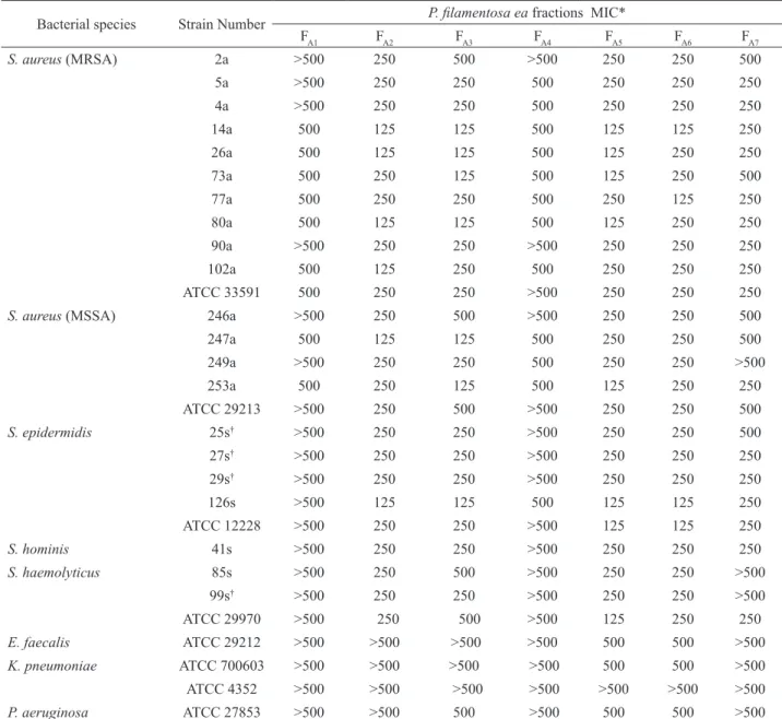
Antibacterial analysis of the subfractions obtained from the ea fraction
The main subfractions obtained from the Sephadex column chromatography of the active ea
fraction were investigated for its antibacterial properties against the same clinical and ATCC reference strains mentioned in section 2.5. The Table 2 presents the MIC for each subfraction evaluated. Four different
concentrations (500, 250, 125 and 62.5 μg/mL) were
adopted for this analysis.
Table 2. Minimun inhibitory concentration (MIC) values attributed for the fractions obtained from the ea extracts of Pentaclethra macroloba against 29 bacterial strains.
Bacterial species Strain Number P. ilamentosa ea fractions MIC*
FA1 FA2 FA3 FA4 FA5 FA6 FA7
S. aureus (MRSA) 2a >500 250 500 >500 250 250 500
5a >500 250 250 500 250 250 250
4a >500 250 250 500 250 250 250
14a 500 125 125 500 125 125 250
26a 500 125 125 500 125 250 250
73a 500 250 125 500 125 250 500
77a 500 250 250 500 250 125 250
80a 500 125 125 500 125 250 250
90a >500 250 250 >500 250 250 250
102a 500 125 250 500 250 250 250
ATCC 33591 500 250 250 >500 250 250 250
S. aureus (MSSA) 246a >500 250 500 >500 250 250 500
247a 500 125 125 500 250 250 500
249a >500 250 250 500 250 250 >500
253a 500 250 125 500 125 250 250
ATCC 29213 >500 250 500 >500 250 250 500
S. epidermidis 25s† >500 250 250 >500 250 250 500
27s† >500 250 250 >500 250 250 250
29s† >500 250 250 >500 250 250 250
126s >500 125 125 500 125 125 250
ATCC 12228 >500 250 250 >500 125 125 250
S. hominis 41s >500 250 250 >500 250 250 250
S. haemolyticus 85s >500 250 500 >500 250 250 >500
99s† >500 250 250 >500 250 250 >500
ATCC 29970 >500 250 500 >500 125 250 250
E. faecalis ATCC 29212 >500 >500 >500 >500 500 500 >500
K. pneumoniae ATCC 700603 >500 >500 >500 >500 500 500 >500
ATCC 4352 >500 >500 >500 >500 >500 >500 >500
P. aeruginosa ATCC 27853 >500 >500 500 >500 500 500 >500
*MIC: Minimum inhibitory concentration in μg/mL; MSSA-methicillin sensitive S.aureus; MRSA-methicillin resistant S.aureus; †methicillin-resistant
coagulase-negative staphylococci.
nine CNS strains. Among the subfractions analyzed, FA2
showed the most similar proile compared to FA5. The
reminiscent subfractions presented moderate activity (FA3 and FA6) or any inhibitory activity (FA1, FA4 and FA7). The other bacterial species were not inhibited at this concentration.
At 250 μg/mL, the CNS and S. aureus strains were completely inhibited by the subfractions FA2, FA5 and FA6 while the Gram-negative bacteria and the E. faecalis
strain showed to be resistant. At the highest concentration investigated the Gram-negative bacteria, in exception the ATCC 4352 of K. pneumoniae, were inhibited by FA5 and FA6.
Staphylococcus (Machado et al., 2002; Machado et al.,
2005; Pereira et al., 2006; Leal et al., 2010), investigations
on the inhibition of Gram-negative bacteria by these products are poorly found in the literature, awakening the interest for this investigation. So, the search for new strategies for the treatment of infections caused by these pathogens stimulates the investigation of alternative
sources (Machado et al., 2005). Accordingly, our indings
in respect of the tannins subfraction as an antibiotic agent
are of great relevance once this is the irst report correlating
the vegetal species studied with this property. We could realize that grand part of the clinical strains evaluated was inhibited by the phenolic fraction, considering both, Gram positive and negative strains.
Evaluation of the minimum bactericidal concentration of the subfraction FA5
A drug can be considered bactericidal in concentrations until two logs over that previously stipulated by the MIC. By the broth dilution method it
was assigned a MIC at 256 μg/mL for the subfraction FA5
against the reference strain ATCC 29213 of S. aureus, so,
higher concentrations up to 1024 µg/mL were evaluated. The results showed that at 256 μg/mL FA5 was not able
to promote a percentage of inhibition equivalent or higher
than 99.9%. Nevertheless, at 512 and 1024 μg/mL it was
not observed any bacterial growth suggesting a bactericidal mechanism of action for the subfraction FA5.
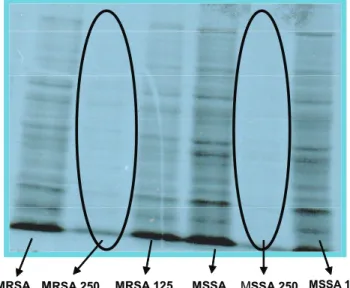
Proposal of the antibacterial mechanism of action for the tannin subfraction FA5
Analysis of the bacterial protein synthesis in the presence of FA5
Based on the fact that tannins can act beyond the bacterial metabolic system, the interference of the tannin subfraction FA5 in the protein synthesis of the reference strains ATCC 29213 (MSSA) and ATCC 33591 (MRSA) of S. aureus was investigated. In this way, our group could propose a feasible antibacterial mechanism of action for the subfraction FA5.
For this analysis were selected two different concentrations of FA5, the MIC 250 µg/mL and the
sub-MIC 125 µg/mL ones, established previously by the
broth dilution method. The aiming was to correlate the inhibitory effect observed in the antibacterial test with the potential toxic effect in the protein synthesis. The sub-MIC concentration is important to appraise once it can
show possible initial modiications in the protein synthesis proile when compared to the standard in the absence of
FA5.
The autoradiogram (Figure 4) showed that in the presence of the subfraction FA5 (250 μg/mL) the bacteria protein synthesis of the sensitive strain, as well as of
the resistant strain ATCC 33591, was inhibited. On the
contrary, it was possible to observe the incorporation of
35S-Met in the protein synthesized by the control strains
(in the absence of fraction FA5) as well as on that in the
presence of the subfraction at 125 µg/mL.
Figure 4. Analysis of staphylococcal protein synthesis.
Autoradiogram of a SDS-PAGE protein proile of the strains
ATCC 29213 (MSSA) and ATCC 33591(MRSA) of S. aureus
labeled in the presence of [35S] Met (200 μCi/mL) at two
concentrations of fraction FA5; MRSA: methicillin-resistant S. aureus; MRSA 250: strain in the presence of FA5 at 250 μg/mL;
MRSA 125: strain in the presence of FA5 at 125 μg/mL; MSSA:
Methicillin-susceptible S.aureus; MSSA 250: strain in the presence of FA5 at 250 μg/mL; MSSA 125: strain in the presence of FA5 at 125 μg/mL.
Our data illustrate the fact that in sub-MIC
dosage is still not possible to verify, apparently, an inhibitory effect in the protein synthesis of the strains analyzed. These data correlate satisfactorily with the MIC values previously assigned by the broth dilution technique. Tannins are known by linking strongly to proteins, in vitro, and by forming a complex called “tannin-protein complex” (T-PC) considerably resistant
to the degradation by digestive enzymes (Osawa,
1996). Therefore, one of the antimicrobial mechanisms of action of the tannins can be explained by the inhibition of the bacterial and fungi enzymes and/or by the formation of complexes with the substrates of them
(Scalbert, 1991). Our results could presuppose that the
ellagitannins, assigned for the first time as the major constituents in the active subfraction, would be acting by a protein synthesis inhibition mechanism probably beyond a tannin-protein complex as mentioned.
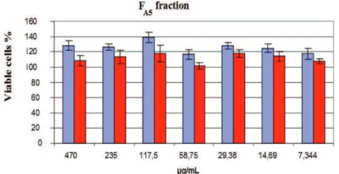
Cytotoxicity
In order to evaluate if there was speciicity of the
M
toxicological effect caused by FA5 in prokaryotic cells our
group investigated the inluence of the presence of this
subfraction in different concentrations in the eukaryotic cell growth. The cytotoxicity assay was performed aiming to determine the subfraction concentration able to cause toxic effects in eukaryotic cells, comparing it with the bacterial MIC previously established.
The Figure 5 presents the correlation between the viable cells percentages of FA5 in relation to the control (absence of the fraction) when there were incorporated different concentrations of the referred subfraction. The results were assigned after 24 and 48 h of incubation. The measurements are presented as medium of the replicates. The cells were further observed by microscopy and it was possible to detect a slightly change in the cellular morphology at the two highest concentrations investigated
(470 and 235 μg/mL) of the subfraction FA5. However, it
was not observed cellular killing. The data showed that in any concentration evaluated the subfraction investigated did not present cytotoxicity.
The data presented indicated that, at the MIC (250
μg/mL) against the clinical strains (MRSA and MSSA) as
well as against the reference strains (ATCC 29213 and ATCC 33591) of S. aureus it was not observed toxicity for the cell culture analyzed. Even at a concentration one log over the MIC it was still not observed toxicity.
Figure 5. Cytotoxicity assay by the neutral red incorporation method. Eukaryotic cells (BSC-40) were grown as adherent culture in a 96-well microplate. The fraction FA5 was added at
concentrations showed in the graphics (470 to 7.344 μg/mL)
for 24 h. The relation between sample absorbance and control absorbance calculated the relative absorbance. The measurements are expressed as average of replicates.
Our results showed that in the MIC value the
most active subfraction is not toxic for eukaryotic cells. This assay is extremely important once one of our future objectives is to suggest the use of the vegetal species P. macroloba as a constituent for a phytopharmaceutical to be used in the therapy against infections. So, our present results proved that the subfraction FA5 (ellagitannins) presented selective-toxicity for prokaryotic cells, as well as bactericidal effect against multiresistant bacterial of clinical importance.
Conclusion
The present study reports the irst in vitro
investigation about the antibacterial properties of tannin subfractions obtained from an ethyl acetate fraction of P. macroloba, enriching the research for natural products with relevant activity against clinical importance bacteria. This vegetal species has never been analyzed under this aspect. The data described in this work are in accordance with the literature which recognizes the tannins as healings. So, our results suggest the use of P. macroloba extract as a future phytopharmaceutical for the treatment of infectious diseases, however, further research is required to evaluate the practical value of this therapeutic application.
Acknowledgement
We are grateful to Marlei Silva for his assistance in the antibacterial experiment and to Professor Walter Baptista Mors for furnishing the exemplar of the plant. We thank Dr. Clarissa R Damaso and Desyree M Jesus (Instituto de Biofísica, UFRJ) for the cytotoxicity assay support. This study was supported by grants from Fundação Carlos Chagas Filho de Amparo à Pesquisa do Estado do Rio de Janeiro, Conselho Nacional de Desenvolvimento
Cientíico e Tecnológico, Coordenação de Aperfeiçoamento
Pessoal de Nível Superior and Programa de Núcleos de Excelência.
References
Clinical and Laboratory Standards Institute (CLSI) 2003.
Methods for dilution antimicrobial susceptibility tests for bacteria that grow aerobically. Wayne, Pennsylvania, USA: Approved Standards:M7-A6.
DeLeo FR, Chambers HF 2009. Reemergence of
antibiotic-resistant Staphylococcus aureus in the genomics era. J Clin Invest 119: 2464-74.
Feng Y, Chen CJ, Su LH, Hu S, Yu J, Chiu CH 2008. Evolution
and pathogenesis of Staphylococcus aureus: lessons learned from genotyping and comparative genomics.
FEMS Microbiol Rev 32: 23-37.
Fey PD, Saïd-Salim B, Rupp ME, Hinrichs SH, Boxrud DJ, Davis CC, Kreiswirth BN, Schlievert PM 2003. Comparative molecular analysis of community- or hospital-acquired methicillin-resistant Staphylococcus aureus. Antimicob Agents Chemother 47: 196-203.
Hsueh PR, Chen ML, Sun CC, Chen WH, Pan HJ, Yang LS, Chang SC, Ho SW, Lee CY, Hsieh WC, Luh KT 2002.
Antimicrobial drug resistance in pathogens causing nosocomial infections at a university hospital in Taiwan, 1981-1999. Emerg Infect Dis 8: 63-68.
Isenberg HD 1992. Antimicrobial susceptibility testing. Tests to assess bactericidal activity. In: Clinical Microbiology Procedures Handbook. Washington, D.C., USA: ASM Press.
imipenem-resistant Pseudomonas aeruginosa clone among patients in a hospital in Rio de Janeiro. J Hosp Infect 60: 19-26.
Leal ICR, Dos Santos KRN, Júnior II, Antunes OAC, Porzel A, Wessjohann L, Kuster RM 2010. Ceanothane and
lupane type triterpenes from Zizyphus joazeiro - an anti-Staphylococcal evaluation. Planta Med 76: 47-52.
Machado TB, Leal ICR, Kuster RM, Santos KRN, Silva MG,
Amaral ACF 2002. Antimicrobial ellagitannin of Punica granatum fruits. J Braz Chem Soc 13: 606-610.
Machado TB, Leal ICR, Kuster RM, Amaral ACF, Kokis V, Silva
MG, Santos KRN 2005. Brazilian phytopharmaceuticals - evaluation against hospital bacteria. Phytother Res 19: 519-525.
Nascimento-Carvalho CM, Lyra TG, Alves NN, Caldas RM,
Barberino MG 2008. Resistance to methicillin and other antimicrobials among community-acquired and nosocomial Staphylococcus aureus strains in a pediatric teaching hospital in Salvador, northeast Brazil. Microb Drug Resist 14: 129-131.
Nogueira K da S, Higuti IH, Do NAscimento AJ, Terasawa
LB, De Oliveira S, Matos AP, Souza HAPM, Cogo LL, Dalla-Costa LM 2006. Occurrence of extended-spectrum
beta-lactamases in Enterobacteriaceae isolated from hospitalized patients in Curitiba, southern Brazil. Braz J Infect Dis 10: 390-395.
Nunes APF, Schuenck RP, Bastos CCR, Magnanini MMF, Long JB, Iorio NLP, Dos Santos KRN 2007. Heterogeneous
resistance to vancomycin and teicoplanin among
Staphylococcus spp. solated from bacteremia. Braz J Infect Dis 11: 345-350.
Osawa RO 1996. Tannin-protein complex-degrading
Enterobacteria isolated from the alimentary tracts of koalas and a selective medium for their enumeration.
Appl Envir Microbiol 58: 1754-1759.
Pereira EM, Machado TB, Leal ICR, Jesus DM, Damaso CRA,
Pinto AV, Giambiagi-de-Marval M, Kuster RM, Santos KRN 2006. Tabebuia avellanedae naphthoquinones: activity against methicillin-resistant staphylococcal strains, cytotoxic activity and in vivo dermal irritability analysis. Ann Clin Microbiol Antimicrob 5:5.
Sader HS, Jones RN, Gales AC, Silva JB, Pignatari AC, The
Sentry Participants Group (Latin America) 2004. Sentry antimicrobial surveillance program report: Latin
American and Brazilian results for 1997 through 2001.
Braz J Infect Dis 8: 25-79.
Scalbert A 1991. Antimicrobial properties of tannins.
Phytochemistry 30: 3875-3883.
Schedlbauer JL, Kavanagh KL 2008. δC in Pentaclethra
macroloba trees growing at forest edges in north-eastern Costa Rica. J Trop Ecol 24: 39-47.
Silva JO, Coppede JS, Fernandes VC, Sant’ana CD, Ticli FK,
Mazzi MV, Giglio JR, Pereira PS, Soares AM, Sampaio SV 2005. Antihemorragic, antinucleolytic and other antiophidian properties of the aqueous extract from
Pentaclethra macroloba. J Ethnopharmacol 100: 145-152.
Tanaka T, Fukumori M, Ochi T, Kouno I 2003. Paeonianins A-E,
new dimeric and monomeric ellagitannins from the fruits of Paeonia lactilora. J Nat Prod 66: 759-763.
Touati A, Achour W, Cherif A, Hmida HB, Afif FB,
Jabnoun S, Khrouf N, Hassen AB 2009. Outbreak
of Acinetobacter baumannii in a neonatal intensive care unit: antimicrobial susceptibility and genotyping analysis. Ann Epidemiol 19: 372-378.
Uçkay I, Pittet D, Vaudaux P, Sax H, Lew D, Waldvogel F
2009. Foreign body infections due to Staphylococcus epidermidis. Ann Med 41: 109-119.
Viana FA, Pouliquen YBM, Andrade-Neto M, Santiago
GMP, Pessoa ODL, Rodrigues-Filho E, Braz-Filho R
2004a. Complete 1H and 13C assignments for two new
monodesmoside saponins from Pentaclethra macroloba
(Wild.) Kuntze. Magn Reson Chem 42: 695-699. Viana FA, Braz-Filho R, Pouliquen YBM, Neto MA, Santiago
GMP, Rodrigues-Filho E 2004b. Triterpenoids saponins from stem bark of Pentaclethra macroloba. J Braz Chem Soc 15: 595-602.
*Correspondence
Ivana Correa Ramos Leal
Faculdade de Farmácia, Pólo Universitário, Universidade Federal
do Rio de Janeiro, Campus Macaé
Rua Aluisio da Silva Gomes, 50, Granja dos Cavaleiros, 27930-560 Macaé-RJ, Brazil