HAND-ASSISTED RIGHT LAPAROSCOPIC LIVE DONOR
NEPHRECTOMY
ANIBAL W. BRANCO, ALCIDES J. BRANCO FILHO, WILLIAM KONDO, MARCO A.
GEORGE, RAFAEL F. MACIEL, MARIANA J. GARCIA
Department of Urology and General Surgery, Cruz Vermelha Hospital, Curitiba, Parana, and Department of Urology, Sao Jose Municipal Hospital, Joinville, Santa Catarina, Brazil
ABSTRACT
Purpose: Laparoscopic live donor nephrectomy has acquired an important role in the era of minimally invasive surgery. Laparoscopic harvesting of the right kidney is technically more challeng-ing than that of the left kidney because of the short right renal vein and the need to retract the liver away from the right kidney. The aim of this article is to report our experience with right laparoscopic live donor nephrectomies.
Materials and Methods: We performed a retrospective review of 28 patients who underwent right laparoscopic donor nephrectomies at our service. Operative data and postoperative outcomes were collected, including surgical time, estimated blood loss, warm ischemia time, length of hospital stay, conversion to laparotomy and complications.
Results: The procedure was performed successfully in all 28 patients. The mean operative time was 83.8 minutes (range 45 to 180 minutes), with an estimated blood loss of 111.4 mL (range 40 to 350 mL) and warm ischemia time of 3 minutes (range 1.5 to 8 minutes). No donor needed conver-sion to open surgery and all kidneys showed immediate function after implantation. The average time to initial fluid intake was 12 hours (range 8 to 24 hours). Two cases of postoperative ileus and a case of hematoma on the hand-port site were observed. The mean postoperative hospital stay was 3 days (range 1 to 7 days).
Conclusions: Our data confirm the safety and feasibility of right laparoscopic donor nephre-ctomy and we believe that the right kidney should not be avoided for laparoscopic donor nephrenephre-ctomy when indicated.
Key words: laparoscopy; living donors; nephrectomy; transplantation
Int Braz J Urol. 2005; 31: 421-30
INTRODUCTION
The advent of laparoscopic donor nephrec-tomy has resulted in decreased donor morbidity with less pain, shorter hospital stays, earlier return to work and regular activity, and improved cosmetics com-pared with the conventional open donor nephrectomy approach (1,2). These benefits of the minimally
in-vasive approach to kidney donation are reflected by studies that demonstrate an increased willingness to donate when the laparoscopic technique is available (3-5).
liver does not need to be retracted when left nephre-ctomy is performed (8). However, because the “bet-ter” kidney should always remain with the donor (9), occasionally the right kidney must be transplanted.
Early experience with right laparoscopic do-nor nephrectomy was marked by a high incidence of venous thrombosis and graft loss (8), which has since improved with experience and with technical modi-fications of the procedure (8,10). Recently, many ar-ticles have been published demonstrating the safety and feasibility of right donor nephrectomies (6,7,11-14), which has allowed transplant centers to main-tain the benefits of the laparoscopic era while adher-ing to the fundamental principles of patient selection established during the open surgery era (12). The purpose of this study is to report our initial experi-ence with right laparoscopic live donor nephrectomy.
MATERIALS AND METHODS
All potential living kidney donors presented to our department from May 2002 to August 2004 were considered for laparoscopic nephrectomy. Each potential donor underwent a standard preoperative immunologic and medical evaluation to confirm his/ her suitability. The exams requested to delineate re-nal vascular anatomy preoperatively were those usu-ally performed for conventional renal donors, includ-ing digital angiography and intravenous pyelogram.
The rationale for donor kidney selection for laparoscopic donor nephrectomy was identical to the standard principles used for open donor nephrectomy. In the setting of “all things being equal,” the left kid-ney was selected because of the longer left renal vein. However, if the left renal vascular anatomy was un-favorable compared with that of the right or if a right renal condition was identified, the right kidney was selected (12). We have always preserved the basic tenet that “the better kidney should remain in situ for the donor” when we have chosen which kidney would be removed (9).
Operative data and postoperative courses were reviewed. Information on donor age, sex, and previous medical history were collected. Surgical demographics included operative time, warm is-chemia time, estimated blood loss, and
intraopera-tive complications. Operaintraopera-tive time was defined as the time from the initial skin incision to closure of the external oblique fascia of the HandPort incision (7). Warm ischemia time was defined as time elapsed from the application of haemostatic clips to the renal ar-tery until the kidney was perfused with cold preser-vation fluid (7). The measured postoperative param-eters included the time to first oral intake and hospi-tal stay.
Surgical Technique
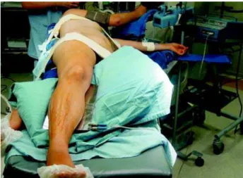
The donor is positioned in a traditional left lateral decubitus position on the operating room table with the kidney rest fully elevated and the bed in a flexed position. An axillary roll is placed beneath the donor’s arm and the right arm is maintained on an armrest in a flexed functional position (Figure-1).
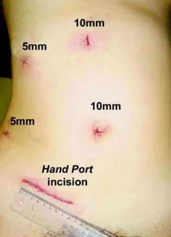
A 6 to 8-centimeter skin incision is performed in the right iliac fossa. The abdominal cavity is care-fully inspected and a wet surgical towel is placed to mobilize the colon and to assist with any bleeding. After reflecting the colon medially by incising the lateral peritoneal reflection, the ureter is identified and isolated with a Penrose drain. The HandPort (Lap Disc - Ethicon Endo-Surgery, Cincinnati, Ohio,
USA) is placed through the incision (Figure-2) and the abdomen is inflated with carbon dioxide to an intra-abdominal pressure of 12 to 14 mmHg.
Subsequently, a 10 mm trocar is placed in the periumbilical area for the 30-degree laparoscope; 2 additional 10 mm trocars are placed, one approxi-mately halfway between the xiphoid and the umbili-cus, and the other in the right middle axillary line at the umbilical level. A 5 mm trocar is then placed in the right side to retract the liver (Figure-3).
Dissection continues at the lower renal pole, posterior renal portion and superior renal pole. This can be conducted easier since the intra-abdominal hand facilitates control of the kidney and prevents rotation and potential damage to the renal hilum. The ureter isolated by the Penrose drain is dissected in a
superior and inferior direction up to the crossing of the iliac vessels, taking care with the periureteral tis-sue (between the lower pole and ureter), which must be left intact to prevent devascularization of the ure-ter. The renal vessels are dissected and freed of sur-rounding tissues (Figure-4). All side branches are clipped using LT-300 titanium clips (Ethicon Endo-Surgery) and divided. It is imperative to clear the adi-pose/lymphatic tissues off the artery and vein com-pletely so that the hemostatic clips may hold the ves-sel walls securely without risk of dislodgement. The renal artery and vein are dissected free to the level of the aorta and inferior vena cava, respectively. Throughout the operation, urine output is monitored and maintained with fluids and mannitol.
Several methods have been introduced to ligate the renal vessels. In our series, renal artery liga-ture was performed using LT-300 titanium clips or Hem-o-lok clips (Weck Closure Systems, Research Triangle Park, North Carolina, USA) and, in most cases, the technique of venous control with cotton suture and LT-300 titanium clips was used. We also tried the Hem-o-lok clips to right renal vein ligature and the caval suture after division of the renal vein with a cuff of the vena cava.
After completely isolating the renal artery and vein, the kidney was retracted laterally with the as-sistant hand. A number “0” cotton suture was passed laparoscopically around the vein before the renal
ar-Figure 2 – Placement of the HandPort in the right iliac fossa.
tery ligature, leaving the loose knot to be tightened just after sectioning the artery. The artery was ligated on the aorta side with 2 titanium clips (or Hem-o-lok clips when available) and divided sharply distal to the 2 clips. The renal vein ligature was performed by tightening the knot and placing 2 titanium clips next to it on the lateral edge of the vena cava (Figure-5). (When we perform this ligature using Hem-o-lok clips, the cotton suture is not necessary because the Hem-o-lok clip is longer than the LT-300 titanium clip, and this allows the ligature of the whole renal vein circumference). The vein was cut distal with laparoscopic scissors (Figure-6). As with the renal artery, no clips or staples were left on the vein of the graft.
When a caval suture is performed, a Satinsky clamp is introduced into the abdominal cavity through the HandPort incision after the renal artery ligature, and the clamp is placed on the inferior vena cava. Renal vessels are divided, thereby allowing the divi-sion of the renal vein with a cuff of the vena cava (Figure-7) and maximizing the renal vein length. The cavotomy is sutured laparoscopically with 4-0 Prolene and afterwards the Satinsky clamp is released from the vena cava (15).
The kidney is removed through the hand-as-sisted device and the ureter is sectioned under direct vision. The kidney is then placed in an iced preserva-tive and delivered to the recipient team for grafting. After the abdomen is checked for bleeding, the tro-car sites are closed under direct vision and the pneu-moperitoneum is evacuated (Figure-8).
Some cases were performed by retro-peritoneoscopic approach with four ports. This pro-cedure starts with a skin incision of 1 to 2 cm just below the tip of the twelfth rib. The flank muscle fi-bers are separated by blunt dissection. After sharp incision of the anterior thoracolumbar fascia, an ini-tial retroperitoneal space is created by index finger dissection. A 10 mm trocar is placed through the in-cision for the 30-degree laparoscope, and the retro-peritoneal working space is created using the laparo-scope and gas insufflation. Two additional 10 mm trocars and one 5 mm trocar (for upper renal pole exposure) are inserted in a typical diamond
arrange-Figure 5 – Reducing the caliber of the renal vein and tightening the knot (arrow).
Figure 6 – Two titanium clips are placed and the renal vein is sectioned close to the vena cava.
ment. After identification of the psoas muscle, the Gerota’s fascia is incised laterally and the ureter as well as the renal vessels are dissected (Figure-9). The kidney is then completely freed of covering fatty tis-sue. A 6-centimeter skin incision is performed over the iliac crest for the assistant’s hand. The ureter is divided under direct vision and the pneumoperito-neum is again insufflated. The renal vessels ligature is performed as previously mentioned – the vein with LT-300 titanium clips and cotton suture (Figure-10), and the artery with LT-300 titanium clips. The kid-ney is then removed from the retroperitoneal cavity through the iliac crest incision.
RESULTS
Between May 2002 and September 2004, a total of 70 healthy donors underwent laparoscopic nephrectomy for kidney transplantation in our unit,
of which 28 (40%) were on the right side. Indications for selecting the right side were multiple left renal vessels (n = 18), right renal artery fibromuscular dysplasia (n = 2), right renal cyst (n = 2), early branching of the left renal artery (n = 2), right ure-terocele (n = 1), right renal ptosis (n = 1), right renal artery aneurysm (n = 1) and left ureteral duplicity (n = 1). The procedure was performed successfully in all cases, and no patients required conversion to laparotomy. There were 13 male and 15 female pa-tients, and the mean age was 34.8 ±8 years (range 21
to 52 years). The surgery was performed by hand-assisted transperitoneal approach in 24 patients (85.7%) and by pure retroperitoneoscopic approach in the remaining 4 cases (14.3%).
Figure 8 – Final aspect of the surgery.
Figure 9 – Dissection of the renal vessels by retroperitoneoscopy.
Anatomically, 24 patients had single right renal artery (85.7%), 3 patients had double right re-nal arteries (10.7%) and another one had triple right renal arteries (3.6%). Twenty-six patients had single right renal vein (92.9%), and 2 donors had double right renal veins (7.1%).
We used LT-300 titanium clips and cotton suture for venous control in 24 patients (85.8%), Hem-o-lok clip in 2 patients (7.1%) and caval sutures in 2 patients (7.1%). The right renal artery was ligated using LT-300 titanium clip in 26 patients (92.9%) and Hem-o-lok clip in 2 patients (7.1%).
The mean operative time in our series was 83.8±37.2 minutes (range 45 to 180 minutes).
Esti-mated blood loss was 111.4 ±61.9 mL (range 40 to
350 mL) per patient and none required transfusion. Warm ischemia time was 3.0 ± 1.4 minutes (range
1.5 to 8 minutes). We had no intraoperative compli-cations and all kidneys showed immediate function after implantation.
All patients were allowed a liquid diet as soon as they were fully awake. The average time to initial fluid intake was 12.0 ±3.9 hours (range 8 to 24 hours).
The 3 postoperative donor complications in our se-ries consisted of 2 cases of postoperative ileus (7.1%), which resolved spontaneously in 7 days, as well as a case of hematoma (3.6%) at the HandPort site, which was opened and drained. The mean postoperative hospital stay was 3.0 ±1.5 days (range 1 to 7 days).
COMMENTS
Since the first successful case of laparoscopic live-donor nephrectomy reported by Ratner et al. in 1995 (16), laparoscopy has emerged as an alternative to open surgery in donor nephrectomy for transplan-tation (1,17-21), and recently it has become the stan-dard of care at increasing numbers of renal transplant programs worldwide (22).
Advantages to the laparoscopic approach are self-evident and well described, and include a reduc-tion in postoperative discomfort, narcotic require-ments and hospital stays, shortened recoveries, im-proved cosmetic results and a more rapid return to regular activities and work (1-4,21-27). All these ben-efits are obtained using laparoscopic technique with
an equivalent renal graft outcome compared with open surgery (1,28). Moreover, some centers have docu-mented an increase in the number of donations and have attributed this increase to a less invasive surgi-cal procedure that is more acceptable to the donor (5,21,25).
Several reports have confirmed some advan-tages of the hand-assisted technique described in 1998 by Wolf et al. (29). By using the hand, the surgeon can facilitate complete mobilization of the colon, easily exposing the kidney and the aorta. In addition, tissue planes are more easily defined using the intraperito-neal hand for retraction (26). Surgical time is compa-rable to open nephrectomy and tends to decrease with the learning curve. It can be performed with safety and without harm to the donor and the graft function (30). Comparisons of hand-assisted laparoscopic with stan-dard laparoscopic donor nephrectomies have not dem-onstrated any statistically significant differences in analgesic requirements, hospital stays, or allograft func-tion. Significantly shorter operative and warm ischemia times have been demonstrated in the hand-assisted laparoscopic groups compared with the standard laparoscopic groups (1,11,24,27).
group (8) shifted the Endo-GIA stapler port to the right lower quadrant. In this manner, the stapler was placed across the renal vein in a plane parallel to the vena cava in an effort to maximize the vein length. In the presence of a right renal vein shorter than 3 cm, this group also suggested a subcostal incision for open placement of a Satinsky clamp on the inferior vena cava and for graft extraction. This allowed division of the renal vein with a cuff of the vena cava and closure of the cavotomy through the incision. Re-cently, Turk et al. (11) described the use of a laparoscopic Satinsky clamp for side clamping of the vena cava to obtain a caval cuff at right renal vein transection, followed by laparoscopic suturing of the vena cava in 4 patients. The largest single-center ex-perience is from Rotterdam where the right kidney is preferred (33). They reported an unusually high rate of 73% of right laparoscopic donor nephrectomies with no differences in thrombosis, graft loss or com-plications compared to the left kidney.
Several groups currently advocate the retro-peritoneal approach, especially for right donor nephre-ctomies (11,34,35). This approach allows direct visu-alization of the aortocaval junction ensuring the maxi-mal renal vein length possible. However, the retroperi-toneal working space is smaller (13), and prior exper-tise with this approach is necessary before attempting retroperitoneal laparoscopic donor nephrectomy (12). We performed more than 40 laparoscopic transperitoneal donor nephrectomies before using the retroperitoneal approach. The choice of laparoscopic approach (whether trans- or retro-peritoneal) was at the discretion of the surgeon. The advantages of retroperitoneoscopic surgery over transperitoneal ac-cess are that bowel mobilization is not required, re-traction of the solid viscera is not needed, the risk of inadvertent gut injury and ileus is minimized, contami-nation of the peritoneal cavity is avoided, previous abdominal surgery does not preclude this approach and there is a lower incidence of long-term complications, such as port site hernia and bowel obstruction.
In many respects, the right kidney is easier to remove, with less extensive colonic dissection and ab-sence of splenic/pancreatic attachments. The typical absence of gonadal, adrenal and lumbar branches makes control of the renal vein more straightforward (13).
When evaluating our results and comparing them with those of the major university transplant centers, we found that our data compare favorably with the results of their reports. Overall, mean opera-tive time from skin incision to closure was 83.8 min-utes and this was shorter than reported in almost all other studies (115 to 218 minutes) (6,7,11-14,33,36). Our average estimated blood loss of 111.4 mL com-pares well with the reported average blood loss of 71 and 302 mL (6,7,11,12,14,33,36). Mean warm is-chemia time was 3 minutes, which is comparable with other reported studies (6,14,33,36), and superior to the experiences of Ng et al. (12) and Boorjian et al. (7), which reported 1.7 and 1.9 minutes, respectively. Considering the complications in donors, we had 2 cases of postoperative ileus (7.1%) with spon-taneous resolution in seven days. Actually, the major postoperative problem in laparoscopic donors is bowel function. Ileus prolongs hospitalization and causes readmissions. Donors report that bowel func-tion is not really normal for 7 to 10 days. Some pro-longed bowel recovery may be due to unrecognized pancreatitis (36). The two cases of prolonged ileus occurred at the beginning of our series and we ob-served that by placing the HandPort again after kid-ney removal and aspirating blood and blood clots, we did not have any other case of postoperative bowel functioning disorder. Another complication observed in our series was a case of hematoma (3.6%) at the HandPort site, which was opened and drained.
Up to 1.6% of patients undergoing right laparoscopic nephrectomies need conversion to an open method (36). Fortunately, as seen in other se-ries (7,12,13), we had no conversions.
Our mean hospital stay of 3 days is a little longer than that reported recently by other major cen-ters, which have an average hospital stay of between 1.8 and 2.6 days (6,7,12,13,36). With the increasing experience in laparoscopic nephrectomies, we started discharging our patients sooner (24 to 48 hours after surgery) in the last nine cases.
CONCLUSIONS
these findings, we state that the right kidney should not be avoided for laparoscopic donor nephrectomy when indicated.
REFERENCES
1. Jacobs SC, Cho E, Dunkin BJ, Flowers JL, Schweitzer E, Cangro C, et al.: Laparoscopic live donor nephrec-tomy: the university of Maryland 3-year experience. J Urol. 2000; 164: 1494-9.
2. Kim FJ, Ratner LE, Kavoussi LR: Renal transplanta-tion: laparoscopic live donor nephrectomy. Urol Clin North Am. 2000; 27: 777-85.
3. Cadeddu JA, Ratner L, Kavoussi LR: Laparoscopic donor nephrectomy. Semin Laparosc Surg. 2000; 7: 195-9.
4. Schweitzer EJ, Wilson J, Jacobs S, Machan CH, Philosophe B, Farney A, et al.: Increased rates of do-nation with laparoscopic donor nephrectomy. Ann Surg. 2000; 232: 392-400.
5. Finelli FC, Gongora E, Sasaki TM, Light JA: A sur-vey: the prevalence of laparoscopic donor nephrec-tomy at large U.S. transplant centers. Transplantation. 2001; 71: 1862-4.
6. Buell JF, Abreu SC, Hanaway MJ, Ng CS, Kaouk JH, Clippard M, et al.: Right donor nephrectomy: a com-parison of hand-assisted transperitoneal and retroperi-toneal laparoscopic approaches. Transplantation. 2004; 77: 521-5.
7. Boorjian S, Munver R, Sosa RE, Del Pizzo JJ: Right laparoscopic live donor nephrectomy: a single institu-tion experience. Transplantainstitu-tion. 2004; 77: 437-40. 8. Mandal AK, Cohen C, Montgomery RA, Kavoussi LR,
Ratner LE: Should the indications for laparascopic live donor nephrectomy of the right kidney be the same as for the open procedure? Anomalous left renal vascu-lature is not a contraindiction to laparoscopic left do-nor nephrectomy. Transplantation. 2001; 71: 660-4. 9. Murray JE, Harrison JH: Surgical management of fifty
patients with kidney transplants including eighteen pairs of twins. Am J Surg. 1963; 105: 205-18. 10. Buell JF, Edye M, Johnson M, Li C, Koffron A, Cho
E, et al.: Are concerns over right laparoscopic donor nephrectomy unwarranted? Ann Surg. 2001; 233: 645-51.
11. Turk IA, Deger S, Davis JW, Giesing M, Fabrizio MD, Schonberger B, et al.: Laparoscopic live donor right nephrectomy: a new technique with preservation of vascular length. J Urol. 2002; 167: 630-3.
12. Ng CS, Abreu SC, Abou El-Fettouh HI, Kaouk JH, Desai MM, Goldfarb DA, et al.: Right retroperitoneal versus left transperitoneal laparoscopic live donor ne-phrectomy. Urology. 2004; 63: 857-61.
13. Buell JF, Hanaway MJ, Potter SR, Koffron A, Kuo PC, Leventhal J, et al.: Surgical techniques in right laparoscopic donor nephrectomy. J Am Coll Surg. 2002; 195: 131-7.
14. Abrahams HM, Freise CE, Kang SM, Stoller ML, Meng MV: Technique, indications and outcomes of pure laparoscopic right donor nephrectomy. J Urol. 2004; 171: 1793-6.
15. Branco AW, Branco Filho AJ, Kondo W, George MA, Carvalho RM, Maciel RF: Maximizing the right renal vein length in laparoscopic live donor nephrectomy. Int Braz J Urol. 2004; 30: 416-9.
16. Ratner LE, Ciseck LJ, Moore RG, Cigarroa FG, Kaufman HS, Kavoussi LR: Laparoscopic live donor nephrectomy. Transplantation. 1995; 60: 1047-9. 17. Velidedeoglu E, Williams N, Brayman KL, Desai NM,
Campos L, Palanjian M, et al.: Comparison of open, laparoscopic, and hand-assisted approaches to live-do-nor nephrectomy. Transplantation. 2002; 74: 169-72. 18. Hawasli A, Boutt A, Cousins G, Schervish E, Oh H:
Laparoscopic versus conventional live donor nephre-ctomy: experience in a community transplant program. Am Surg. 2001; 67: 342-5.
19. Montgomery RA, Kavoussi LR, Su L, Sinkov V, Cohen C, Maley WR, et al.: Improved recipient results after 5 years of performing laparoscopic donor nephrectomy. Transplant Proc. 2001; 33: 1108-10.
20. Brown SL, Biehl TR, Rawlins MC, Hefty TR: Laparoscopic living donor nephrectomy: a compari-son with the conventional open approach. J Urol. 2001; 165: 766-9.
21. Ratner LE, Montgomery RA, Kavoussi LR: Laparoscopic live donor nephrectomy: the four year Johns Hopkins University experience. Nephrol Dial Transplant. 1999; 14: 2090-3.
22. Buell JF, Hanaway MJ, Woodle ES: Maximizing renal artery length in right laparoscopic donor nephrectomy by retrocaval exposure of the aortorenal junction. Transplantation. 2003; 75: 83-5.
23. Siqueira TM Jr, Gardner TA, Kuo RL, Paterson RF, Stevens LH, Lingeman JE, et al.: One versus two pro-ficient laparoscopic surgeons for laparoscopic live donor nephrectomy. Urology. 2002; 60: 406-9; discus-sion 409-10.
laparoscopic live donor nephrectomy. Ann Surg. 1997; 226: 483-9; discussion 489-90.
25. Odland MD, Ney AL, Jacobs DM, Larkin JA, Steffens EK, Kraatz JJ, et al.: Initial experience with laparoscopic live donor nephrectomy. Surgery. 1999; 126: 603-6; discussion 606-7.
26. Slakey DP, Wood JC, Hender D, Thomas R, Cheng S: Laparoscopic living donor nephrectomy: advantages of the hand-assisted method. Transplantation. 1999; 68: 581-3.
27. Buell JF, Hanaway MJ, Potter SR, Cronin DC, Yoshida A, Munda R, et al.: Hand-assisted laparoscopic liv-ing-donor nephrectomy as an alternative to traditional laparoscopic living-donor nephrectomy. Am J Trans-plant. 2002; 2: 983-8.
28. Ratner LE, Hiller J, Sroka M, Weber R, Sikorsky I, Montgomery RA, et al.: Laparoscopic live donor ne-phrectomy removes disincentives to live donation. Transplant Proc. 1997; 29: 3402-3.
29. Wolf JS Jr, Tchetgen MB, Merion RM: Hand-assisted laparoscopic living donor nephrectomy. Urology. 1998; 52: 885-7.
30. Stifelman MD, Hull D, Sosa RE, Su LM, Hyman M, Stubenbord W, et al.: Hand assisted laparoscopic
do-nor nephrectomy: a comparison with the open ap-proach. J Urol. 2001; 166: 444-8.
31. Wang DS, Bird VG, Winfield HN, Rayhill S: Hand-assisted laparoscopic right donor nephrectomy: surgi-cal technique. J Endourol. 2004; 18:205-9; discussion 209-10.
32. Barry JM: Editorial comment. Laparoscopic live do-nor nephrectomy: the university of Maryland 3-year experience. J Urol. 2000; 164: 1498-9.
33. Lind MY, Hazebroek EJ, Hop WC, Weimar W, Jaap Bonjer H, IJzermans JN: Right-sided laparoscopic live-donor nephrectomy: is reluctance still justified? Trans-plantation. 2002; 74: 1045-8.
34. Hoznek A, Olsson LE, Salomon L, Saint F, Cicco A, Chopin D, et al.: Retroperitoneal laparoscopic living-donor nephrectomy. Preliminary results. Eur Urol. 2001; 40: 614-8.
35. Gill IS, Uzzo RG, Hobart MG, Streem SB, Goldfarb DA, Noble MJ: Laparoscopic retroperitoneal live donor right nephrectomy for purposes of allotransplantation and au-totransplantation. J Urol. 2000; 164: 1500-4.
36. Jacobs SC, Cho E, Foster C, Liao P, Bartlett ST: Laparoscopic donor nephrectomy: the University of Maryland 6-year experience. J Urol. 2004; 171: 47-51.
Received: October 10, 2004 Accepted after revision: June 20, 2005
Correspondence address:
Dr. Anibal Wood Branco Rua das Palmeiras, 170 / 201 Curitiba, PR, 80620-210, Brazil Phone: + 55 41 242-6543 E-mail: [email protected]
EDITORIAL COMMENT
Laparoscopic nephrectomy has gained in-creasing popularity since the early experimental works by Gill et al. in 1994 (1) at Washington Uni-versity, and the first surgery performed in humans by Ratner et al. (2) at Johns Hopkins University. Currently, the leading institutions in USA and Eu-rope have been preferentially using the laparoscopic
In most case of live donors for kidney trans-plantation, the performance of laparoscopic nephrec-tomy has led to improved technique, allowing some contraindications to be abandoned and turning the mini-mally invasive technique into the preferential approach. In this issue of the Int Braz J Urol, the au-thors report a significant series of 28 cases of laparoscopic nephrectomy in kidney donors in most cases performed with the “hand-assisted” technique for removal of the right kidney. In many institutions, right nephrectomy is considered to be a contraindi-cation for laparoscopy due to the usually shorter length of the renal vein on this side. The authors reported good results with minor complication (7% of ileus, and one case of hematoma on the incision), thus cor-roborating the safe indication for laparoscopic nephre-ctomy on the right side as well.
On the other hand, some conventional sur-geons have tested the surgery with “mini-incision” in order to minimize the effects and sequelae of the clas-sic incision so that conventional surgery could be superposed to the laparoscopic technique. In 2003, Perry et al. (5) published a prospective analysis of patients undergoing laparoscopic nephrectomy with another group undergoing nephrectomy with “mini-incision” and still found statistically significant ad-vantages for the laparoscopic surgery in terms of post-operative pain throughout the first month, introduc-tion of oral diet, return to usual activities, esthetic satisfaction and a better emotional role as verified by validated quality of life questionnaires. Such evidence could be due to the fact that, despite incisions per-formed for removal of the kidney in both groups, the abdominal incision in the lower quadrant is more be-nign, thus providing a better postoperative period.
Despite much recent scientific evidence, in-creasingly more surgeons performing surgery
world-wide, continuous technological advancements, the decrease in limitations and the increase in donations at institutions that offer laparoscopy, many institu-tions still don’t offer this technique, for reasons that are unclear. For example, at Washington University where it all started, the transplantation team does not yet perform laparoscopic nephrectomy, in spite of the early contribution and constant advancements in the field provided by their neighbors, who are trying more and more to explore the maze of scientific advances and progress. I often prefer to think and act like Spen-cer Johnson, M.D. (6) in his book, “Who moved my cheese?”: it is safer to search in the maze than remain without the cheese.
REFERENCES
1. Gill IS, Carbone JM, Clayman RV, Fadden PA, Stone MA, Lucas BA, et al.: Laparoscopic live-donor ne-phrectomy. J Endourol. 1994; 8: 143-8.
2. Ratner LE, Ciseck LJ, Moore RG, Cigarroa FG, Kaufman HS, Kavoussi LR: Laparoscopic live donor nephrectomy. Transplantation. 1995; 60: 1047-9. 3. El-Galley R, Hood N, Young CJ, Deierhoi M, Urban
DA: Donor nephrectomy: A comparison of techniques and results of open, hand assisted and full laparoscopic nephrectomy. J Urol. 2004; 171: 40-3.
4. Lind MY, Liem YS, Bemelman WA, Dooper PM, Hop WC, Weimar W, et al.: Live donor nephrectomy and return to work: does the operative technique matter? Surg Endosc. 2003; 17: 591-5.
5. Perry KT, Freedland SJ, Hu JC, Phelan MW, Kristo B, Gritsch AH, et al.: Quality of life, pain and return to normal activities following laparoscopic donor nephre-ctomy versus open mini-incision donor nephrenephre-ctomy. J Urol. 2003; 169: 2018-21.
6. Johnson S: Who Moved My Cheese? An Amazing Way to Deal with Change. New York, G.T. Putnam’s Sons. 1998