Spontaneous Resolution Rates of Vesicoureteral Reflux in
Brazilian Children: A 30-Year Experience
Miguel Zerati Filho, Adriano A. Calado, Ubirajara Barroso Jr, Joao L. Amaro
Division of Urology, Sao Paulo State University, UNESP, Botucatu, Sao Paulo, Brazil
ABSTRACT
Objective:We evaluated clinical characteristics of primary vesicoureteral reflux (VUR) in infants in a 30-year period in Brazil with special reference to the relation of renal parenchymal damage to urinary tract infection and gender.
Materials and Methods: From 1975 through 2005, 417 girls (81.6%) and 94 boys (18.4%) with all grades of reflux were retrospectively reviewed. Patients were categorized by the worst grade of reflux, maintained on antibiotic prophylaxis and underwent yearly voiding cystourethrography until the reflux was resolved. VUR was considered resolved when a follow-up cystogram demonstrated no reflux. Surgical correction was recommended for those who fail medical therapy, severe renal scarring or persistent VUR.
Results:Grades I to V VUR resolved in 87.5%, 77.6%, 52.8%, 12.2% and 4.3%, respectively. Renal scars were present at presentation in 98 patients (19.2%). Neither gender nor bilaterality versus unilaterality was a helpful predictor of resolution. The significant difference was found among the curves using the log rank (p < 0.001) or Wilcoxon (p < 0.001) test.
Conclusion:Despite the current use of screening prenatal ultrasound, many infants are still diagnosed as having vesicoureteral reflux only after the occurrence of urinary tract infection in our country. Scarring may be associated to any reflux grade and it may be initially diagnosed at any age but half of the scars are noted with higher grades of reflux (IV and V). The incidence of reflux related morbidity in children has significantly diminished over the last three decades.
Key words: ureter; vesico-ureteral reflux; reconstructive surgical procedures
Int Braz J Urol. 2007; 33: 204-15
INTRODUCTION
Vesicoureteral reflux (VUR) is a common pediatric problem. The possibility of renal damage due to VUR and consequently renal function impairment and hypertension gives the necessary impetus for the diagnosis or exclusion of this problem (1). In general, it was thought that medical and surgical management represented equally efficacious treatment options (1,2). Both treatment forms have advantages and
disadvan-tages that must be taken into account and shared with the parents when choosing the best approach. If, on one hand, clinical treatment requires chronic antibi-otic prophylaxis, periodic surveillance of urine cultures and periodic voiding cystourethrograms (VCUGs), which despite the low morbidity may cause discom-fort, on the other hand, surgery is related to the risks of a surgical-anesthetic procedure.
spon-taneous resolution for patients with VUR undergoing medical management. Due to the lack of similar data in our country, specialists have used these data for management of the patients.
The primary endpoint of this study was de-fining the rates of spontaneous resolution for patients with VUR in our country. These data may be helpful to determine the need for surgical intervention and the proper follow-up schedule in patients on antibiotic prophylaxis.
On the other hand, the group selected in this report, which was conservatively managed over a 30-year period, is of particular interest in understand-ing the changes in the approach of VUR in this pe-riod of time. Besides finding out the overall resolu-tion rate, we saw if we could find differences be-tween the groups according to the period diagnosis was carried out.
MATERIALS AND METHODS
Of the 813 consecutive patients with VUR treated at our institution from January 1975 to De-cember 2005, 302 were excluded because in 121 a VCUG was not repeated, in 105 follow-up were in-sufficient (< 12 months) and 76 children had sec-ondary VUR. 511 children were retrospectively evaluated and met the inclusion criteria: a) age < 10 years at the time of diagnosis; b) radiological tests confirming VUR and VUR grade; c) minimum fol-low-up time of 12 months; d) no voiding dysfunction, neuropathic bladder, duplicated systems, ectopic ure-ters, ureterocele, and other associated pathologies. VUR was detected by VCUG and the grade classi-fied according to the International Reflux Study Com-mittee (3).
The renal parenchyma was evaluated using renal cortical scintigraphy (99mTc-DMSA), intravenous
urography or both (4-6). Patients were kept under con-tinuous prophylaxis and had urine culture collected on a monthly basis, upper urinary tract assessment with renal ultrasonography (RUS) every six months and intravenous urography or scintigraphy and VCUG once a year. Reflux was considered resolved when absent on a single cystogram.
Patients with any of the following parameters underwent surgery: failure of medical management (breakthrough urinary tract infections and patient non-compliance), increase or development of new renal scars and reflux that fails to resolve (mainly grade IV or V).
After obtaining demographic data, patients were divided into three groups according to the pe-riod diagnosis was carried out: Group A (n = 80) from 1975 to 1985; Group B (n = 205) from 1986 to 1995; Group C (n = 226) from 1996 to 2005.
The results were statistically analyzed using the Minitab® statistical computing software, with p <
0.05 considered to indicate statistical significance. Kaplan-Meier curves were constructed to estimate the reflux resolution rate per grade and the rates were compared using the log rank and Wilcox on tests. Data shown in the tables were analyzed by the chi-square test. The study protocol was reviewed and approved by the local ethics committee.
RESULTS
The initial clinical manifestation leading to the diagnosis of VUR in 85% of the cases was urinary tract infection, confirmed by urinary culture, followed by fever of indeterminate cause (5.4%), lumbar pain (3.5%), antenatal hydronephrosis (2.9%), hematuria (2.5%) and others. Mean age at presentation was 3.1 years and median follow-up was 2.7 years (1.2 - 12.4 years). VUR was unilateral in 250 (48.9%) and bilat-eral in 261 (51.1%) cases. Grade II VUR was de-tected more frequently (192 cases – 37.6%), followed by grade III (174 cases - 34%), grade IV (74 cases – 14.5%), grade I (48 cases – 9.4%) and grade V (23 cases – 4.5%).
The large majority of diagnosis, 344 (67.3%), was made before the 3rd year of life, of which 170
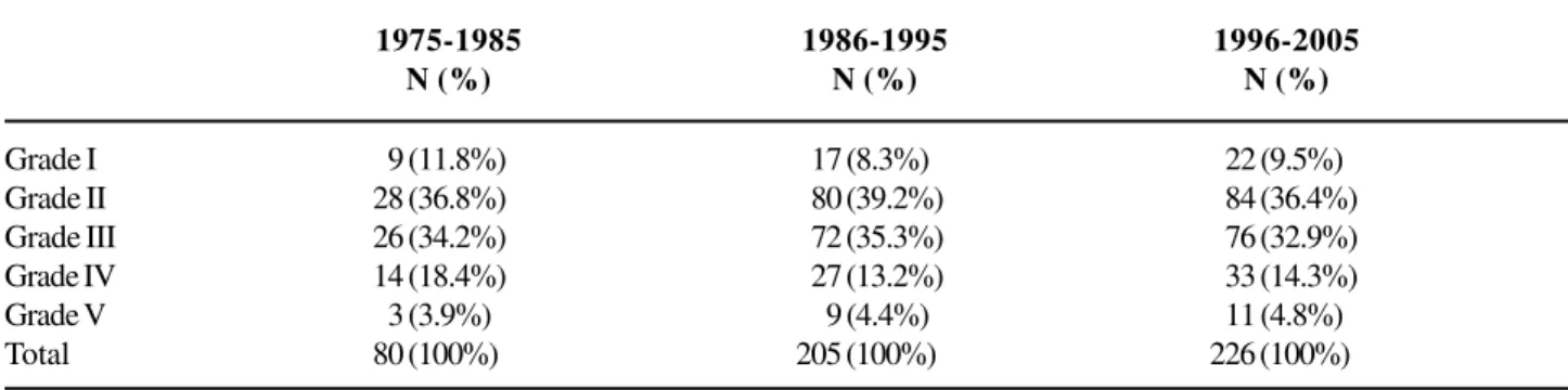
(33.2%) before the first year (Figure-1). The stratifi-cation of patients according to VUR grade shows a similar distribution among the three groups (Table-1). There was no significant difference among the study groups (A, B and C) for the severity of VUR. Table-2 shows the tests carried out in the initial evaluation of the patients in the study groups. For the statistical analysis, groups A and B were analyzed together due to the small sample of patients who carried out RUS and DMSA scan in group A. In the first period, intra-venous urography was carried out in 93.7% of the cases and RUS and DMSA scan in 1.2% and 13.7% respectively. In the last decade there is an important change, and intravenous urography is used in only 21.2% of the children and RUS and scintigraphy is used in 88% and 23.4% respectively. There is a sig-nificant difference between groups (A+B) and C for the use of RUS, scintigraphy and intravenous urogra-phy (p < 0.001).
Patients were also evaluated for the pres-ence of renal scars at the time of diagnosis. In group A, 31.2% (25/80) had renal scars, in group B, 20.5% (42/205); and in group C, 13.7% (31/226) (Figure-2). There was a significant difference between groups for the presence of renal scars in the initial evalua-tion (p = 0.02), with a clear decrease of renal scars in the last period, from 1996 to 2005. Table-3 strati-fies patients with renal scars according to age at the presentation. There is a clear correlation between age and renal scars, which are significantly more frequent in the group of patients over 5 years of age (p < 0.001).
Of the 125 patients requiring surgery, 30% (24/80) were from Group A, 30.2% (62/205) from Group B and 17.2% (39/226) from Group C. When the number of surgical procedures is compared, a sig-nificant difference is observed between Groups A and B compared to Group C (p = 0.003), with a clear de-crease of surgical indications over time. In the overall characteristics of patients (Table-4), severe cases of
reflux (IV and V) are observed in 19% of the chil-dren and the incidence of renal injury is proportional do VUR grade, i.e., the more intensive the VUR, the
greater is the probability of developing scars. How-ever, even low grade VUR (I and II) was associated with renal scarring in this cohort.
Table 1 – The distribution of reflux grades according to the study groups.
Grade I 09 (11.8%) 017 (8.3%) 022 (9.5%)
Grade II 28 (36.8%) 080 (39.2%) 084 (36.4%)
Grade III 26 (34.2%) 072 (35.3%) 076 (32.9%)
Grade IV 14 (18.4%) 027 (13.2%) 033 (14.3%)
Grade V 03 (3.9%) 009 (4.4%) 011 (4.8%)
Total 80 (100%) 205 (100%) 226 (100%)
1975-1985 N (%)
1986-1995 N (%)
1996-2005 N (%)
p > 0.05
Table 2 – Tests carried out in the initial evaluation of the patients in the study groups.
Renal ultrasound 01 (1.2%) 061 (29.7%) 199 (88%)
Renal scintigraphy (DMSA) 11 (13.7%) 030 (14.6%) 053 (23.4%)
Intravenous urography 75 (93.7%) 163 (79.5%) 048 (21.2%)
Total 87 (n = 80) 254 (n = 205) 300 (n = 226)
p < 0.001
1975-1985 N (%)
1986-1995 N (%)
1996-2005 N (%)
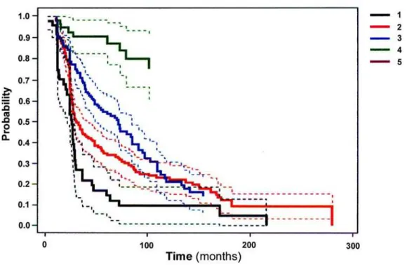
When the rates of spontaneous resolution of reflux were analyzed for different grades of VUR throughout the medical management, 87.5% (42/48) of the patients with VUR grade I; 77.6% (149/192) of patients with VUR grade II and 52.8% (92/174) of
patients with VUR grade III had spontaneous resolu-tion (Figure-3). Of the 74 patients with VUR grade IV, spontaneous resolution was observed in 9 (12.2%) patients. Only 1 patient with VUR grade V had spon-taneous resolution during follow-up.
There is no significant difference of sponta-neous resolution with clinical treatment when boys and girls are compared. The resolution of VUR was not affected by age at entry. As to the laterality of VUR, there was no significant difference of cure for unilat-eral or bilatunilat-eral VUR for grades I, II and III (p = 0.731; p = 0.344; p = 0.204, respectively). However, for patients with VUR grade IV, there was spontane-ous resolution in 8 patients with unilateral (19%) dis-ease and in only 1 case of bilateral VUR (3.1%) (p = 0.068).
Age (years) Patients(N) Renal Scars – N (%)
< 1 170 20 (11.8%)
1-3 years 174 20 (11.5%)
3-5 years 089 16 (18%)
> 5 years 078 42 (53.8%)
Table 3 – Patients with renal scars according to age at the presentation.
p < 0.001
Table 4 – Characteristics of patients studied (n = 511).
N Mean age
Girl : Boy Renal scars* UTI
UTI with fever Surgery
Grade I
48 2.3 years (30d - 10a)
42 : 6 3 (6.2%) 18 (37.5%)
3 (6.2%) 3 (6.2%)
Grade II
192 1.7 years (44d - 9.3a)
163 : 29 17 (8.8%) 44 (23%) 33 (17.2%) 20 (10.4%)
Grade III
174 1.5 years (27 d - 8,5a)
138 : 36 22 (12.6%) 50 (28.7%) 37 (21.2%) 35 (20.1%)
Grade IV
74 2.7 years (0 d - 10a)
58 : 16 36 (48.6%) 24 (32.4%) 20 (27%) 45 (60.8%)
Grade V
23 1.3 years (0 d - 8a) 12 : 11 20 (87%)
** ** 22 (95.6%)
* Renal scars detected through DMSA or intravenous urography at the moment of the diagnosis; ** Anti-reflux surgery was performed in almost all patients with grade V.
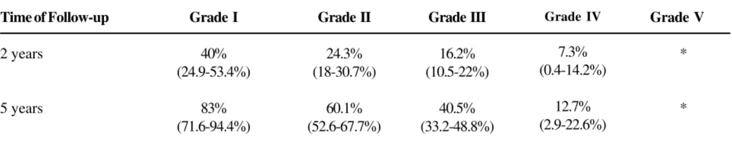
Table 5 – The probability of reflux resolution with continuous antibiotics prophylaxis has been documented at 2 and 5 years after diagnosis.
Time of Follow-up
2 years
5 years
Grade I
40% (24.9-53.4%)
83% (71.6-94.4%)
Grade II
24.3% (18-30.7%)
60.1% (52.6-67.7%)
Grade III
16.2% (10.5-22%)
40.5% (33.2-48.8%)
Grade IV
7.3% (0.4-14.2%)
12.7% (2.9-22.6%)
Grade V
*
*
The analysis of Kaplan-Meier curves (Fig-ure-3) showed a significant difference (p < 0.0001). Table-5 shows the probability of spontaneous cure of VUR at 2 and 5 years of follow-up based on a trans-versal section of the Kaplan-Meier curves.
COMMENTS
Despite the large number of children with reflux, management among urologists is still contro-versial. One of the most debated aspects is the choice between observation or surgical treatment. Although there is no consensus for the treatment of VUR in childhood, several principles based on literature data and corroborated by this study may be used as pa-rameters when choosing optimal therapy for each case.
Chronic reflux nephropathy is a well known and feared disease and is the cause of chronic renal
failure in up to 25% of children undergoing dialysis and in 10 to 15% of adults waiting for renal transplan-tation (7).
Several factors are taken into consideration when choosing therapy for VUR, such as: age, VUR grade, presence of renal injury, history of urinary in-fections and tolerance to antibiotics. The prevention of irreversible renal injury is the final goal of any of the therapeutic modalities, and so far there is no evi-dence of the superiority of clinical treatment over sur-gical therapy (8).
The incidence of renal scarring at the time of VUR diagnosis was 31.2% in the first period (Group A) against 13.7% in the last group (Group C). A prob-able explanation for the high incidence of renal scars in the first period (group A) is a combination of many fac-tors. First of all in the 1970s and early 1980s It was not a common practice for infants following their first di-agnosed UTI to be referred to routine diagnostic imag-ing and we know that The likelihood of developimag-ing re-nal scars depends on factors such as the number of symptomatic UTI and the delay in their treatment. Ac-tually the importance of prompt treatment of urinary infection or symptomatic recurrence of UTI was em-phasized to local physicians. A second explanation is that patients admitted until the end of the 80’s had a greater risk of UTI than children with a diagnosis of VUR after 1990. Possibly, this fact reflects an improve-ment in our understanding of the risk factors for UTI in this population, especially the recognition and treatment of voiding dysfunction and constipation.
In 24.5% (125/511) of the cases, surgical pro-cedures were carried out to repair VUR. Skoog et al. (13) and Greenfield et al. (12), using similar indication criteria, reported 14% to 20% of surgical treatment. The surgical indications decreased from 30%, in the first period, to 17.2% in the last, following the cur-rently used criteria. This decrease is probably related to a better understanding of the disease and more adequate follow-up of these children. Despite the ex-cellent success rates following antireflux surgery one has to bear in mind that surgery only corrects the ana-tomical abnormality. The long-term outcome with re-gard to renal function, urinary tract infections and ar-terial hypertension does not differ significantly from the medication group.
In our study, the Kaplan-Meier curves of VUR resolution show a clear relationship between the possibility of cure and the follow-up time, showing that VUR grades I and II were cured significantly faster than grades III and IV. Other authors (8,14) have mentioned similar results. The information re-garding the probability of resolution is essential when passing information onto the parents and choosing the best therapy to be adopted. Our spontaneous cure results are in agreement with those of other studies (15,16). Schawab et al. (8) have reported 83% and with primary vesicoureteral reflux does more good
than harm. Assuming intervention is beneficial, it is not clear whether antibiotics alone or reimplantation surgery alone are most effective in reducing the risk of urinary tract infection and renal parenchymal ab-normality. Furthermore, the trials, which have been undertaken comparing surgery and antibiotics with antibiotics alone, have not shown any additional ben-efit of surgery except for a reduction in risk of febrile urinary tract infections.
Because of the good results obtained with clinical therapy using low dose antibiotics, in addition to the fact that there are few prospective, random-ized, studies comparing clinical treatment to surgery (9-11), the large majority of children with VUR is clini-cally treated. Thus, high rates of spontaneous resolu-tion are obtained, which depend especially on VUR grade and age at the time of diagnosis.
The most frequent clinical abnormality lead-ing to the diagnosis of VUR is UTI. In our series, 85% of the children had UTI as an early manifesta-tion. Recent studies have shown that other causes, such as antenatal hydronephrosis and voiding dysfunc-tion have played a larger role in the diagnosis of VUR (12). In the evaluation of International Reflux Study in Children (IRSC) (10), UTI was observed in 89% to 92% of children with VUR, and in approximately 30% of the cases there was some degree of renal injury at the time of diagnosis.
76% of spontaneous regression for grades I and II re-spectively. Tamminen-Mobius et al. (17) obtained reso-lution in 63% of VUR grade II and 50% in grade III. In our casuistic we obtained 87.5% of spontaneous cure for grade I, 77.6% for grade II and 52.8% for grade III, when the follow-up period is taken into consider-ation. The spontaneous resolution of VUR grade IV with clinical therapy is known to be low. In our study, the expected probability of cure at 5 years is only 12.2%. Results have shown a spontaneous cure ranging from 9 to 16% in patients with VUR grade IV (13,16).
The well-known association between vesicoureteral reflux and urinary tract infection is the basis for pathophysiological and therapeutic implica-tions, which have dominated the literature on the sub-ject for the last 3 decades. The pathogenesis of renal damage in children with VUR is controversial, as the exact role of UTI and pressure effects of sterile re-flux on the developing kidneys is still unknown.
Primary VUR diagnosed after prenatal hy-dronephrosis is usually severe and is often associated to a congenital renal scars, which is primary and not the result of UTI. In our study diagnosis of VUR re-sulting from antenatal hydronephrosis were observed only in 15 (3%) children. This type of VUR affects male more than female infants and the male predomi-nance is thought to be secondary to a transient infravesical obstruction during pregnancy. This spe-cific group of children achieves up to 43% of sponta-neous cure for grades IV and V (18-20). Although in the first type of reflux renal scarring is often present at diagnosis, then probably congenital, it may always progress after UTI; hence the importance of early diagnosis and careful follow-up in each infant with primary VUR.
Our results showed that the incidence of re-flux-related morbidity in children has significantly di-minished over the last three decades. In our country, the rates of reflux-related nephropathy are much less than reported historically and are consistent with the most recently reported international data.
In our experience, and based on current knowledge and guidelines, which derive from a thor-ough review of the literature the recommendation of a treatment strategy for VUR is not, however, straight-forward. In deciding whether to recommend surgical
correction of VUR, factors that should be considered to include the previous and potential future morbidity of VUR in that individual, the risk of uncorrected VUR, the likelihood of spontaneous resolution or significant reduction in VUR, the efficacy and complications of medical therapy, the morbidity and discomfort associ-ated to serial screening for VUR, the benefits and risks of surgical therapy, and economic factors.
The probability of spontaneous resolution of VUR forms the basis of the decision to treat using antibiotic prophylaxis. Undoubtedly, the most relevant finding in our clinical study is the rates of spontaneous resolution in a large Brazilian cohort.
Because the chance of spontaneous resolu-tion is as high as 87.5%, 77.6% and 52.8% in VUR grades I, II and III, respectively, only exceptional cases should undergo primary surgical correction.
The different procedures should be analyzed for their costs in order to optimize the therapeutic egy in different health systems. An individualized strat-egy seems to be the ultimate goal and should take into consideration the large number of issues.
CONCLUSION
In conclusion, urinary tract infection is still the major cause of VUR diagnosis. However, early diag-nosis and adequate treatment allowed a significantly decrease of morbidity caused by renal injury. The currently employed clinical treatment might be safely used in most of the cases. Spontaneous resolution is directly related to VUR severity and is less likely to occur in grades IV and V for which surgery is a more adequate alternative.
CONFLICT OF INTEREST
None declared.
REFERENCES
vesicoureteral reflux randomly allocated to operation or long-term antibacterial prophylaxis. The International Reflux Study in Children. J Urol. 1992; 148: 1650-2. 2. Wingen AM, Koskimies O, Olbing H, Seppanen J,
Tamminen-Mobius T: Growth and weight gain in chil-dren with vesicoureteral reflux receiving medical ver-sus surgical treatment: 10-year results of a prospec-tive, randomized study. International Reflux Study in Children (European Branch). Acta Paediatr. 1999; 88: 56-61.
3. [No authors listed]: Medical versus surgical treatment of primary vesicoureteral reflux: report of the Interna-tional Reflux Study Committee. Pediatrics. 1981; 67: 392-400.
4. Smellie JM: The intravenous urogram in the detection and evaluation of renal damage following urinary tract infection. Pediatr Nephrol. 1995; 9: 213-9; discussion 219-20.
5. Goldraich NP, Ramos OL, Goldraich IH: Urography versus DMSA scan in children with vesicoureteric re-flux. Pediatr Nephrol. 1989; 3: 1-5.
6. Merguerian PA, Jamal MA, Agarwal SK, McLorie GA, Bagli DJ, Shuckett B, et al.: Utility of SPECT DMSA renal scanning in the evaluation of children with pri-mary vesicoureteral reflux. Urology. 1999; 53: 1024-8. 7. Bailey RR, Mailing TMJ, Swainson CP: Vesicoureteric
reflux and reflux nephropathy. In: Scherier RW, Gottschalk CW (eds.), Disease of the Kidney, Fifth edition. Boston, Litthe, Brown & Co. 1993; pp. 689-727.
8. Wheeler D, Vimalachandra D, Hodson EM, Roy LP, Smith G, Craig JC: Antibiotics and surgery for vesicoureteric reflux: a meta-analysis of randomised controlled trials. Arch Dis Child. 2003; 88: 688-94. 9. Birminghan Reflux Study Group: Prospective trial of
operative versus non operative traitement of severe vesico-ureteric reflux in children: five years observa-tion. Brit Med J. 1987; 295: 237-41.
10. Weiss R, Tamminen-Mobius T, Koskimies O, Olbing H, Smellie JM, Hirche H, et al.: Characteristics at entry of children with severe primary vesicoureteral reflux
recruited for a multicenter, international therapeutic trial comparing medical and surgical management. The In-ternational Reflux Study in Children. J Urol. 1992; 148: 1644-9.
11. Scholtmeijer RJ: Treatment of vesicoureteric reflux: re-sults after 3 years in a prospective study. Child Nephrol Urol. 1991; 11: 29-32.
12. Greenfield SP, Ng M, Wan J: Experience with vesicoureteral reflux in children: clinical characteris-tics. J Urol. 1997; 158: 574-7.
13. Chand DH, Rhoades T, Poe SA, Kraus S, Strife CF: Incidence and severity of vesicoureteral reflux in chil-dren related to age, gender, race and diagnosis. J Urol. 2003; 170: 1548-50.
14. Skoog SJ, Belman AB, Majd M: A nonsurgical approach to the management of primary vesicoureteral reflux. J Urol. 1987; 138: 941-6.
15. Elder JS, Peters CA, Arant BS Jr, Ewalt DH, Hawtrey CE, Hurwitz RS, et al.: Pediatric Vesicoureteral Reflux Guidelines Panel summary report on the management of primary vesicoureteral reflux in children. J Urol. 1997; 157: 1846-51.
16. Arant BS Jr: Medical management of mild and moder-ate vesicoureteral reflux: followup studies of infants and young children. A preliminary report of the South-west Pediatric Nephrology Study Group. J Urol. 1992; 148: 1683-7.
17. Tamminen-Mobius T, Brunier E, Ebel KD, Lebowitz R, Olbing H, Seppanen U, et al.: Cessation of vesicoureteral reflux for 5 years in infants and children allocated to medical treatment. The International Re-flux Study in Children. J Urol. 1992; 148: 1662-6. 18. Duckett JW: Vesicoureteral reflux: a ‘conservative’
analysis. Am J Kidney Dis. 1983; 3: 139-44.
19. Yeung CK, Godley ML, Dhillon HK, Gordon I, Duffy PG, Ransley PG: The characteristics of primary vesico-ureteric reflux in male and female infants with pre-natal hydronephrosis. Br J Urol. 1997; 80: 319-27.
20. Sillen U, Hjalmas K, Aili M, Bjure J, Hanson E, Hansson S: Pronounced detrusor hypercontractility in infants with gross bilateral reflux. J Urol. 1992; 148: 598-9.
Accepted after revision: October 20, 2006
Correspondence address: Dr. Adriano Almeida Calado Rua Conselheiro Portela, 285 / 602 Recife, PE, 52020-030, Brazil
EDITORIAL COMMENT
Vesicoureteral reflux (VUR) was originally described in medieval times; the first ureteral reimplantation to correct VUR was performed in the late 19th century. Despite the intervening centuries and their numerous accompanying medical advances, reflux remains in the 21st century a vexing manage-ment problem for the urologist. The traditional thera-peutic options of antibiotic chemoprophylaxis and open ureteroneocystostomy are increasingly being chal-lenged in the United States and Europe by newer tech-nologies such as endoscopic injection of bulking agents and laparoscopic or robotic ureteroneocystostomy. It remains unclear which of these techniques, if any, will ultimately become the worldwide standard of care. Diverse issues such as cost, cosmesis, and efficacy have yet to be fully determined on all of these treat-ment modalities, and will ultimately need to be bal-anced against one another. Against this backdrop of uncertainty, it is critical that the urologist treating VUR
understand the natural history of the disease. The authors are to be credited for their efforts with this study, a large, retrospective, population-based cohort of over 500 Brazilian children. As the study spans a 30-year period, the authors are in a unique position to comment on the evolution of VUR evaluation and management. It is noteworthy that both surgical in-tervention and renal scarring were significantly re-duced in the most recent cohort, particularly in light of the fact that the proportion of high-grade VUR re-mained relatively constant. I agree with the authors that this is likely due to earlier diagnosis. Although the urologic community may argue about what exactly the optimal treatment modality for VUR is in the 21st century, these data would seem to imply that we are nonetheless able to agree that some treatment is re-quired to prevent long-term renal dysfunction. This study would appear to support the notion that we are succeeding in that regard.
Dr. Jonathan C. Routh
Department of Urology Mayo Clinic Rochester, Minnesota, USA E-mail: [email protected]
EDITORIAL COMMENT
I congratulate the authors on a detailed study of children born with vesicoureteral reflux (VUR). Despite the retrospective nature, the thorough review of data produced useful information that is extremely relevant today when endoscopic therapy is being popularized. The authors point out that the goal of treating this disorder is prevention of infections and scarring. As I teach our residents, there is a difference in treating “children with reflux” and treating “reflux.”
This manuscript reiterates known rates of spontaneous resolution. These high rates of resolution
As exemplified in this article and others, most scarring is congenital and few children develop cortical loss while under medical therapy. Knowing this, any surgical therapy will likely only achieve a lower incidence of infection and add little benefit over antibiotics to prevention of scarring. This has been demonstrated in several studies (1). Since long-term, low dose antibiotics are known to be safe (2), one has to question the determination to treat younger and younger patients surgically (3), removing any hope of a natural cure. The advent of endoscopic treatment appears to have clouded our goals in treating this disorder. There is no better repair than spontaneous resolution. In the 1990’s we were discussing observation off antibiotics. Somehow, in the 21st century we have regressed to discussing surgical treatment of non-refluxing ureters and orifices that
dilate during cystoscopy. Dr. Filho’s manuscript reminds us that spontaneous resolution is likely with patience.
REFERENCES
1. Wheeler D, Vimalachandra D, Hodson EM, Roy LP, Smith G, Craig JC: Antibiotics and surgery for vesicoureteric reflux: a meta-analysis of randomised controlled trials. Arch Dis Child. 2003; 88: 688-94. 2. Karpman E, Kurzrock EA: Adverse reactions of
nitro-furantoin, trimethoprim and sulfamethoxazole in chil-dren. J Urol. 2004; 172: 448-53.
3. Puri P, Pirker M, Mohanan N, Dawrant M, Dass L, Colhoun E: Subureteral dextranomer/hyaluronic acid injection as first line treatment in the management of high grade vesicoureteral reflux. J Urol. 2006; 176: 1856-9; discussion 1859-60.
Dr. Eric A. Kurzrock
Chief, Pediatric Urology University of California, Davis Children’s Hospital Sacramento, California, USA E-mail: [email protected]
EDITORIAL COMMENT
The authors detail their 30 year experience in 511 children with reflux; the majority of patients presenting with urinary infection. These children, with no confounding anatomic or neurological abnormalities presented at a mean age of 3.1 years and were followed for a median of 2.7 years. The distribution of grades and spontaneous resolution rates were congruent with those reported in previous studies. Surgical correction was reserved for approximately 25% of the original cohort.
This study reminds us that the majority of cases of mild to moderate reflux will resolve with time. It also underscores the evolution in our management
(3-6). Surgery is then reserved for those who experience pyelonephritis off of medical treatment. Concomitant with this approach, a competing philosophy has arisen: immediate injection therapy shortly after initial diagnosis of reflux (7). This latter approach has been gaining popularity owing in no small way to the development of the first FDA approved injectable agent in 2001. Preliminary data suggest that injection rates for vesicoureteral reflux have risen sharply in the United States in the last few years (8). In addition, there are increasing concerns over the safety and lack of evidence regarding the efficacy of long-term low dose antibiotic use in children (9-11). Despite 55 years since Hutch first described the relationship between pyelonephritis and reflux in paraplegics (12) we still require well designed prospective randomized controlled trials comparing long-term renal outcomes in patients undergoing conventional medical and surgical management versus observation without antibiotic suppression. Until such time that this information is available, we need to be cautious regarding the enthusiastic adoption of new surgical interventions just because they offer relatively simple and quick closure.
REFERENCES
1. Wan J: Editorial comment. J Urol. 2002; 168: 707. 2. Edwards D, Normand IC, Prescod N, Smellie JM:
Disappearance of vesicoureteric reflux during long term prophylaxis of urinary tract infection in children. Br Med J. 1977; 2 (6082): 285-8.
3. Cooper CS, Chung BI, Kirsch AJ, Canning DA, Snyder HM: The outcome of stopping antibiotics in older
children with vesicoureteral reflux. J Urol. 2000; 163: 269-72, discussion 272-3.
4. Thompson RH, Chen JJ, Pugach J, Naseer S, Steinhardt GF: Cessation of prophylactic antibiotics for management of persistent vesicoureteral reflux. J Urol. 2001; 166: 1465-9.
5. Al- Sayyad AJ, Pike JG, Leonard MP: Can prophylactic antibiotics safely be discontinued in children with vesicoureteral reflux? J Urol. 2005; 174 (4 pt 2): 1587-9. 6. Georgaki-Angelaki H, Kostaridou S, Daikos GL, Kapoyiannis A, Veletzas Z, Michos AG, et al.: Long-term follow-up of children with vesicoureteral reflux with and without antibiotic prophylaxis. Scan J Infect Dis. 2005; 37: 842-5.
7. Puri P, Priker M, Mohanan N, Dawrant M, Dass L, Colhoun E: Subureteral Dextranomer / Hyaluronic acid injection as first line treatment in the management of high grade vesicoureteral reflux. J Urol. 2006; 176: 1856-60.
8. Lendvay TS, Sorensen M, Cowan CA, Joyner BD, Mitchell MM, Grady RW: The evolution of vesicoureteral reflux management in the era of Dextranomer / Hyaluronic acid copolymer: A pediatric health information system database study. J Urol. 2006; 176: 1864-7.
9. Le Saux N, Pham B, Moher D: Evaluating the benefits of antimicrobial prophylaxis to prevent urinary tract infections in children. A systematic review. CMAJ 2000; 163: 523-9.
10. Velicer CM, Heckbert SR, Lampe JW, Potter JD, Robertson CA, Taplin SH: Antibiotic use in relation to the risk of breast cancer. JAMA. 2004; 291: 827-35. 11. Allen UD, MacDonald N, Fuite L, Chan F, Stephens D:
Risk factors for resistance to “first-line” antimicrobials among urinary tract isolates of Escherichia coli in children. CMAJ. 1999; 160: 1436-40.
12. Hutch JA: Vesicoureteral reflux in the paraplegic: cause and correction. J Urol. 1952; 68: 457-69.
Dr. Andrew E. MacNeily