INTRODUCTION
In 2002, there were approximately 60 mil-lion women at reproductive age in the United States; about 1.2 million, or 2% of these women made a medical visit related to infertility during that year and roughly 10% underwent some type of treatment for infertility at some time of their lives. Additionally, 7% of the couples with the woman at reproductive age (2.1 million couples) reported not having reached pregnancy after 12 months of frequent sexual intercourse with no use of contraceptive methods (1).
Among the techniques of assisted repro-duction currently available for treatment of these couples, in vitro fertilization (IVF) is the one that offers the best pregnancy rates (1,2). This tech-nique has been used successfully since the late 1970s, and its results show a signifi cant improve-ment with experience, nevertheless patients with severe spermogram alterations still had little al-ternative, since the results of IVF for these cases continued poor.
The development of gamete microma-nipulation techniques with subsequent intracyto-plasmatic sperm injection (ICSI), fi rst performed Objective: To determine the infl uence of the concentration of oval spermatozoa
ac-cording to the strict morphology criterion in men with normal sperm concentration following the World Health Organization criteria on the results of classic IVF. Materials and Methods: Based on review of patient charts, this study included infer-tile couples presenting with female causes for infertility or unexplained infertility, in whom men presented with normal spermogram values for sperm concentration, sperm motility, volume of ejaculate and total sperm count after semen processing greater than 20 million. Based on the value obtained in strict sperm morphology, patients were divided into three groups: in Group A, patients with values between 0% and 4%; in group B, between 5% and 14%, and in group C, patients with sperm morphology greater than 14%. The outcomes analyzed were oocyte fertilization rate, biochemical pregnancy rate, clinical pregnancy rate and rate of liveborns. Results: A total of 244 cases met the inclusion criteria, 27 of them in group A, 165 in group B, and 52 in group C. The mean fertilization rate and the rate of liveborns were, respectively: 71.9% and 33.3% in group A; 80.9% and 24.2% in group B, and 78.8% and 28.8% in group C. There was no statistical difference among the groups in any of the outcomes analyzed.
Conclusion: The values of strict sperm morphology, as proposed by Kruger and ad-opted by the World Health Organization, had no infl uence on the results of classic in vitro fertilization in the studied sample.
Infl uence of strict sperm morphology on the results of
classic in vitro fertilization
_______________________________________________
Milton Ghirelli-Filho, Françoise Elia Mizrahi, Antonio Carlos Lima Pompeo, Sidney Glina
Projeto Alfa Clinic, Sao Paulo and Faculdade de Medicina do ABC, Santo Andre, SP, Brazil
ABSTRACT
ARTICLE
INFO
_______________________________________________________________ _____________________
Key words:
Fertilization; In Vitro; Infertility; Spermatozoa
Int Braz J Urol. 2012; 38: 519-28
________________
Submitted for publication: February, 02, 2012
________________
in Brussels, in 1992 (3), brought new horizons to the field of human reproduction and allowed men with minimal sperm production to produce preg-nancies, even through alternative techniques for sperm recovery, such as epididymal puncture and testicular biopsy, decreasing the impact of seminal parameters on IVF results.
Unfortunately, not all assisted reproduc-tion procedures result in pregnancy, and among these pregnancies, not all result in the birth of a child. In 2006, of the 99199 cycles of IVF carried out in human reproduction clinics in the United States, with the use of fresh semen, 34719 (35%) led to a pregnancy, but only 28404 (29%) resulted in a birth. In other words, 18% of the gestations conceived by IVF did not result in a birth (1).
Among the factors that influenced the re-sults of IVF, semen quality parameters are accept-ed as a good indicator of the capacity for oocyte fertilization and attainment of pregnancy, along with the quality of the oocyte (4). The semen qual-ity parameters recognized as vital for assessment of fertility are sperm concentration, motility and morphology. Various authors consistently corre-late the values of sperm evaluation criteria with oocyte fertilization rates in IVF (5-7).
Sperm morphology is among the spermo-gram factors that displays a potential impact on male fertility. Among the various sperm morpholo-gy classification systems, the Tygerberg strict crite-rion, originally described by Kruger et al., in 1987 (8), which uses morphometric analysis to determine if the spermatozoon fits within a strict range con-sidered normal, where normal is established as the value of more than 14% of the spermatozoa within the criteria established in a sample.
The currently recommended method for sperm morphology assessment is a classification of normal/abnormal spermatozoa, with an op-tional report of the site of abnormalities in abnor-mal sperm (8,9). With the goal of performing this evaluation, the spermatozoa, which anatomically comprise head, neck, middle piece, main piece, and end piece, are divided only into head (and neck) and tail (middle piece and main piece). For a spermatozoon to be considered normal, both its head and tail must be normal. All the borderline forms should be considered abnormal (10).
Nevertheless, technical procedures for as-sessing sperm morphology are associated with a series of difficulties related to lack of objectivity, variation in interpretation, and poor performance in external evaluations of quality control (10).
In 1988, Kruger et al. demonstrated the importance of sperm morphology as a significant prognostic factor in IVF (11); currently however, strict morphology lacks reproducibility and clin-ical applicability as there is still doubt in more recent studies about the true implications of IVF results, particularly in reference to the classic technique (12).
Sperm morphology has been questioned by some authors about its real value as a prognostic factor for IVF, and most studies use the ICSI tech-nique as standard for evaluation (13,14). In a study performed in 2010, French et al. suggested that the prognostic value of strict morphology should be reconsidered in cycles of assisted reproduction in-volving IVF with ICSI, since the sperm morpholo-gy evaluated by Kruger strict morpholomorpholo-gy showed little prognostic value in IVF cycles with ICSI and did not seem to influence the development or the morphology of the blastocyst (15).
In studies carried out with the ICSI tech-nique, the correlation between the two factors does not seem to exist, but when the technique used for IVF is the classic technique, the question remains about the true prognostic role of sperm morphology, even after three decades of experi-ence with IVF (12,15).
Considering latest data about sperm mor-phology, the World Health Organization changed the normal values for strict sperm morphology from 14% to 4% in order to fit the normality cut-off point to new evidence (16).
The objective of the present study is to de-termine the influence of the values of strict sperm morphology in men with normal sperm concentra-tions, according to the World Health Organization criteria, on the results of classic IVF.
MATERIALS AND METHODS
Patients
who sought the assisted reproduction department of the Projeto Alfa clinic for pregnancy, during the years 2007 and 2010, due to conjugal infertility for at least 12 months.
This study included couples with female causes for infertility or couples with unexplained infertility at the clinical assesment. Prior participa-tion in an IVF cycle or in any other assisted repro-duction technique was not considered an exclusion factor for the study.
The male factor was assessed by means of a spermogram, and as an additional inclusion crite-rion, only men who presented with a spermogram within the range of normality for the parameters sperm concentration, sperm motility, volume of ejaculate, and total sperm count after semen pro-cessing by discontinuous gradient technique were included.
The following normality values were ad-opted: sperm concentration greater than 20 mil-lion per milliliter, sperm motility greater than 50% of directional motility in the sample, volume of ejaculate greater than 1.5 milliliter, and total sperm count after semen processing greater than 20 mil-lion. All spermograms were evaluated by the same biologist using the manual technique.
This study was submitted to the Ethics and Research Committee of the Faculdade de Medicina do ABC and approved under Protocol # 054/2010.
Study design
Based on the sperm morphology of the se-men sample collected for the initial evaluation, pa-tients were divided into three groups: in group A, patients with sperm morphology values between 0% and 4%; in group B, between 5% and 14%, and in group C, patients with sperm morphology great-er than 14%. Group C was considgreat-ered the control group, and groups A and B were compared to the control group regarding parameters of female age, number of oocytes retrieved, number of embryos ob-tained, number of embryos transferred and male age. The outcomes analyzed were oocyte fertil-ization rate, biochemical pregnancy rate, clinical pregnancy rate, and rate of liveborns. Comparison among the groups as to the outcomes was per-formed in a manner similar to that previously de-scribed for the other parameters.
The oocyte fertilization rate was defined by the number of zygotes with two pronucleii and two polar bodies when assessed between 16 and 18 hours after insemination, divided by the num-ber of oocytes retrieved. Biochemical pregnancy was defined as patients with beta-HCG dosing greater than 20.0 mIU/dL 14 days after the em-bryo transfer. Clinical pregnancy was defined as the presence of at least one gestational sac with fetal heartbeats on ultrasonography, one month after embryo transfer.
Seminal Preparation and Evaluation of Sperm Morphology
Semen samples were collected by mastur-bation two to five days after the last ejaculation, both for initial seminal analysis and for IVF. The initial seminal analysis of sperm concentration, sperm morphology and volume of ejaculate was performed according to the World Health Organi-zation criteria (10).
Seminal processing was performed using the discontinuous gradient technique with the use of the culture medium Isolate (Irvine Scientific) and Spermgrad (Vitrolife), followed by evaluation of total seminal count in the sample retrieved.
Sperm morphology was evaluated in a single sample for the initial assessment of patients, count-ing 100 cells, accordcount-ing to the strict criteria proposed by Kruger/Tygerberg and adopted by the World Health Organization (10), using Papanicolaou stain.
IVF Protocols
Ovarian stimulation of multiple follicles was done with purified or recombinant gonado-trophines; GnRH agonists or antagonists were used to suppress endogenous secretion of go-nadotrophines, with medication doses adjusted for each individual case, taking into consideration the woman’s age and ancillary tests. Patients who presented with at least three follicles with at least 17 millimeters diameter on ultrasonography after ovarian stimulation received injections of hCG. Follicular puncture and aspiration was guided by transvaginal ultrasound and was performed 36 hours after the injection of hCG.
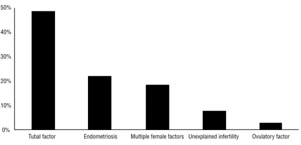
tech-Figure 1 - Proporcion of causes that led to IVF.
nique using semen obtained by masturbation (17). Each oocyte was inseminated with approximately 200.000 sperm cells.
Statistical Analysis
Groups A and B were individually com-pared to group C, used as the control group due to better sperm morphology. The variables of fe-male age, fe-male age, number of oocytes retrieved, number of embryos transferred, number of em-bryos obtained, and oocyte fertilization rate were analyzed using Student’s two-tailed t test. The variables biochemical pregnancy rate, clinical pregnancy rate, and rate of liveborns were evalu-ated using the two-tailed chi-squared test. In both evaluations, a 5% level of significance was ad-opted, with 80% statistical power.
Additionaly, the same statistical analysis above described was performed to compare group A to groups B and C together, according to the new World Health Organization criteria (16).
The sample size calculated to obtain a sta-tistical power of 80% and a significance level of 5% for the variable clinical pregnancy was 27 cases in each group, based on analyses of the data from studies on the topic (18). The fertilization rate vari-able needs a smaller sample size in order to obtain
the same statistical power with only 19 cases in each group (11).
RESULTS
Patients
Analysis was made of 1721 clinical charts of couples who underwent IVF cycles during the stated period, in which 244 met the inclusion cri-teria for this study. Based on review of the clini-cal charts, the three proposed groups were formed; group A included 27 cases; group B included 165 cases, and group C included 52 cases.
Among the causes of infertility that jus-tified the use of IVF, the most common was the tubal factor with 119 cases, representing 48.8% of the total (Figure-1).
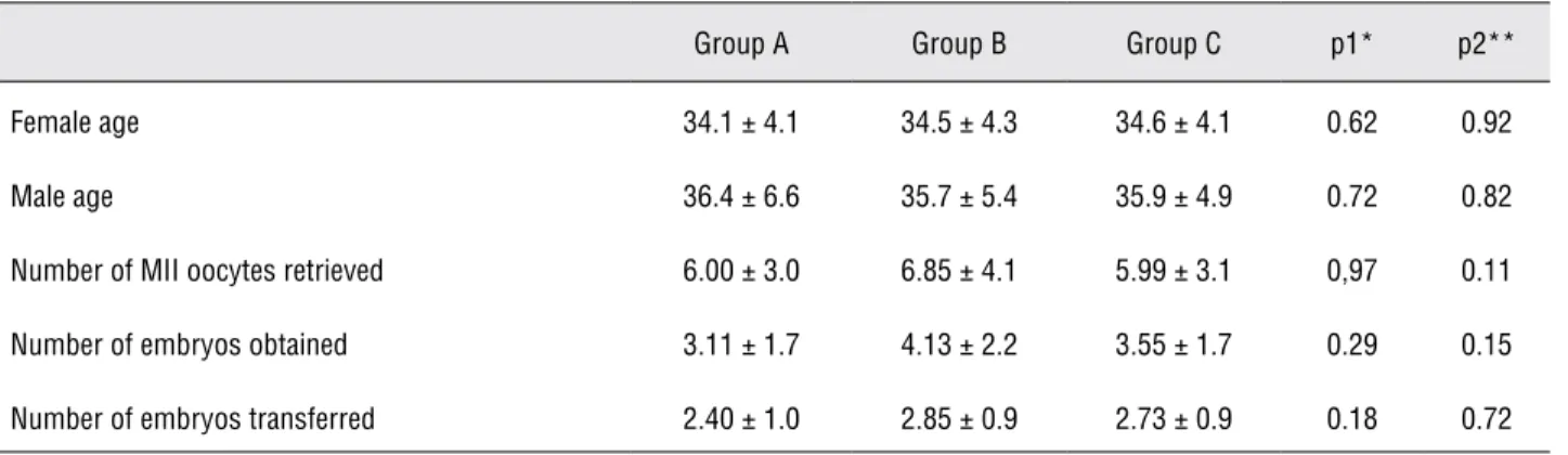
Groups A and B were compared isolatedly to group C in an initial evaluation as to the vari-ables female age, male age, number of oocytes re-trieved in metaphase II (MII), number of embryos obtained and number of embryos transferred, and no statistical differences were found in any pa-rameter among the groups (Table-1).
The overall mean of all 244 cases for fe-male age were 34.5 ± 4.26 years; for fe-male age it was 35.8 ± 5.44 years; for the number of oocytes retrieved in metaphase II it was 6.57 ± 3.84
oo-Tubal factor Endometriosis Multiple female factors Unexplained infertility Ovulatory factor 50%
40%
30%
20%
10%
523
cytes; for the number de embryos obtained it was 3.80 ± 2.11 embryos; and for the number of em-bryos transferred it was 2.77 ± 0.96 emem-bryos.IVF Results
the results of IVF were compared in a simi-lar manner to the parameters previously described in Table-1. In the variables analyzed: oocyte fertil-ization rate, biochemical pregnancy rate, clinical pregnancy rate, and rate of liveborn, no statistical difference was seen among the groups (Table-2).
The overall mean of the 244 cases for oocyte fertilization rate was 79.4% ± 22.1%; biochemical pregnancy rate was 37.2%, clinical pregnancy rate was 27.8%, and the rate of liveborns was 26.2%.
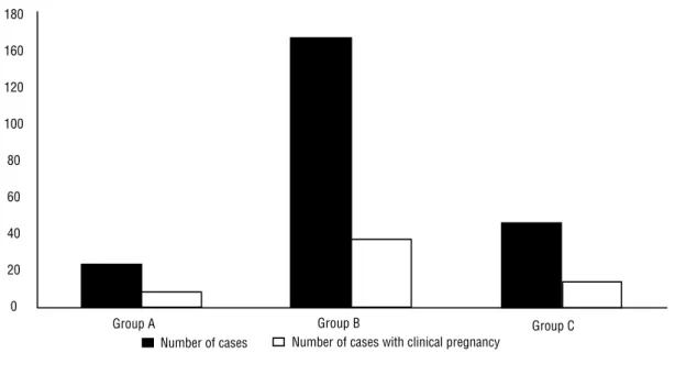
The distribution of the total number of cases in each group and the respective number of gestations are represented in the graphic below (Figure-2). The mean oocyte fertilization rates are shown for each group (Figure-3).
Comparing group A to groups B and C to-gether, no statistical difference on the variables female age, male age, number of oocytes re-trieved in metaphase II (MII), number of embry-os obtained and number of embryembry-os transferred was found. Similarly to the analysis performed to groups A, B and C isolated, the comparison between group A to groups B and C together showed no statistical difference in the variables of IVF results (Table-3).
Table 1 - Means of groups and p value for statistical comparison of groups A and B with group C.
Group A Group B Group C p1* p2**
Female age 34.1 ± 4.1 34.5 ± 4.3 34.6 ± 4.1 0.62 0.92
Male age 36.4 ± 6.6 35.7 ± 5.4 35.9 ± 4.9 0.72 0.82
Number of MII oocytes retrieved 6.00 ± 3.0 6.85 ± 4.1 5.99 ± 3.1 0,97 0.11
Number of embryos obtained 3.11 ± 1.7 4.13 ± 2.2 3.55 ± 1.7 0.29 0.15
Number of embryos transferred 2.40 ± 1.0 2.85 ± 0.9 2.73 ± 0.9 0.18 0.72
*p1 = group A x C ** p2 = group B x C Key: MII: Metaphase II
Table 2 - Fertilization and pregnancy rates in the groups and p value for statistical comparison of groups A and B to group C.
Group A Group B Group C p1* p2**
Fertilization rate (Mean) 71.9% 80.9% 78.8% 0.21 0.51
Biochemical pregnancy rate 44.4% 33.3% 36.5% 0.49 0.97
Clinical pregnancy rate 37.0% 25.4% 30.8% 0.57 0.45
Liveborn rate 33.3% 24.2% 28.8% 0.68 0.50
Figure 2 - Number of cases and number of cases with clinical pregnancy in the groups presented.
Figure 3 - Oocyte fertilization rate in the groups presented.
180
160
120
100
80
60
40
20
0
Group A Group B Group C
Number of cases Number of cases with clinical pregnancy
100%
80%
60%
40%
20%
0%
Group A Group B Group C
DISCUSSION
The present study is one of the few per-formed correlating sperm morphology exclusive-ly with classic technique IVF cycles (4,6,11,18-20), not including cycles carried out using the ICSI technique. Its results demonstrated that the values of sperm morphology showed no correla-tion with pregnancy rates in IVF cycles.
Another positive characteristic of this study was sperm morphology evaluation as an isolated variable in spermogram, since the oth-er seminal parametoth-ers woth-ere within normal val-ues. Some similar studies performed previously showed other spermogram variables, such as sperm concentration or motility, variables that may have an influence on the results of IVF re-gardless of the values of sperm morphology (13). The values of sperm morphology proposed to form the groups in this study were based on prior studies demonstrating that men with sper-mograms showing sperm morphology values of 14% or more would produce better results for IVF. In this way, the group with the morphology of 14% or more was established as the control group for comparison to the other two groups, which were divided using the cut-off value of 4% proposed in prior studies (8,9,11).
The sperm morphology cut-off values also follow the new classification proposed by the World Health Organization. The sperm mor-phology assessment proposed in World Helth Organization´s latest manual intends to limit what is identified as normal to the potentially
fertilizing spermatozoa. Using these guidelines, the range of percentage normal values for both fertile and infertile men is likely to be 0-30%, with few samples exceeding 25% of normal spermatozoa. This low value will inevitably pro-duce low thresholds; indeed reference limits and thresholds of 3-5% normal forms (16).
The new normality cut-off point proposed in the referred manual is 4% and this informa-tion led to another statistical analysis between the group A and the groups B and C together representing the samples considered as morpho-logically normal.
The three groups formed showed no statis-tical differences when compared to possible con-founding factors, such as female age, number of oocytes retrieved, number of embryos obtained, and the number of embryos transferred, allowing sperm morphology to remain as the only spermo-gram variable among the groups, which proved to be comparable.
Other factors with possible influence on the results were not statistically analyzed, such as the cause for female infertility that indicated IVF and the medication used for ovarian stimula-tion as well as its doses, due to the high number of variables within each factor, which would not allow sufficient numbers in each group for ad-equate statistical analysis.
One potential factor of influence in the study results was that the morphology reading was performed on a spermogram prior to the per-formance of IVF and not on the sample used for the procedure itself. Even considering that the
Table 3 - Fertilization and pregnancy rates in the groups and p value for statistical comparison of group A to groups B and C together.
Group A Group B + C p
Fertilization rate (Mean) 71.9% 80.4% 0.30
Biochemical pregnancy rate 44.4% 34.1% 0.29
Clinical pregnancy rate 37.0% 26.7% 0.26
time elapsed between the sample for the reading and the performance of IVF was no more than two months, changes in the values of morphol-ogy between the two readings could influence the study results. Nevertheless, the authors did not consider this factor relevant, in general, to the sample.
Keegan et al. carried out a study with 495 infertile couples in which men presented with spermograms containing more than two million mobile spermatozoa after seminal processing, and compared patients with strict morphology greater than or equal to five with patients who had strict morphology smaller than five using classic IVF and ICSI cycles; no statistical differ-ence was noted between the oocyte fertilization rates and liveborn rates with either one of the techniques (12). In a similar study, French et al. analyzed 1074 IVF cycles done exclusively with the ICSI technique, and compared the results of oocyte fertilization rates and rates of liveborns in patients with strict morphology values that var-ied between zero and seven, without finding any statistical difference between the groups, how-ever in this study the patients with other altered spermogram parameters were not excluded from the study (15).
The results of the present study go against those reached in primary studies on the topic (4,8,9,11,21). Among these studies, one of the most significant is the one by Kruger et al., who first compared results of oocyte fertilization rates and pregnancy rates after IVF cycles in 96 cou-ples, in which men presented with a spermogram with strict morphology greater than or equal to 14% and other seminal parameters within nor-mality, noting a statistical difference in the given variables in favor of the group with strict mor-phology greater than 14% (8).
One possible explanation for the result herein presented having been different from ini-tial studies correlating sperm morphology and IVF results may be attributed to the sperm mor-phology assessment technique used, hence the significant variability and inconsistency in the results when comparing different institutions, different professionals, and even among obser-vations made by the same professional (22).
In this study, the spermograms were all interpreted by the same professional with the in-tention of decreasing error and variability noted among different professionals and following the criteria for morphological sperm evaluation ad-opted by the World Health Organization (10) in the strictest way possible.
The prognostic evaluation for IVF found-ed on spermogram parameters is basfound-ed on sperm concentration and motility and lacks instruments for sperm morphology evaluation with precise and reproducible results. Perhaps new morpho-logical classifications are needed to improve ac-curacy of the evaluation.
One of the techniques proposed for mor-phological sperm assessment is the motile sperm organelle morphology examination (MSOME), which evaluates the sperm under the microscope with a magnification of at least 1000 times; this technique positively correlated sperm nuclear morphology with oocyte fertilization and preg-nancy rates, although experience with this tech-nique is still too limited to enable definitive con-clusions on the topic (23).
Use of the electronic microscope to evalu-ate ultrastructural spermatic morphology of the components of the sperm head was used in a study by Mashiasch et al., in 1992, and correlated with the capacity for fertilization of the sperm in in vitro assessments (7).
Other forms of evaluating the sperm to determine the prognosis of IVF have been pro-posed, basing the tests on the functional evalua-tion of the sperm, such as, for example, acrosome reaction test and hemizona assay, although these tests still lack clinical applicability, and despite promising results presented, remain as tools for the experimental laboratory.
The conclusion of this study was that the values of strict sperm morphology, as proposed by Kruger and adopted by the World Health Or-ganization, had no influence on the results of classic in vitro fertilization in the sample studied.
CONFLICT OF INTEREST
REFERENCES
1. Sunderam S, Chang J, Flowers L, Kulkarni A, Sentelle G, Jeng G, et al.: Assisted reproductive technology surveil-lance--United States, 2006. MMWR Surveill Summ. 2009; 58: 1-25.
2. ESHRE Capri Workshop Group: Intrauterine insemination. Hum Reprod Update. 2009; 15: 265-77.
3. Palermo G, Joris H, Devroey P, Van Steirteghem AC: Preg-nancies after intracytoplasmic injection of single spermato-zoon into an oocyte. Lancet. 1992; 340: 17-8.
4. Kruger TF, Menkveld R, Stander FS, Lombard CJ, Van der Merwe JP, van Zyl JA, et al.: Sperm morphologic features as a prognostic factor in in vitro fertilization. Fertil Steril. 1986; 46: 1118-23.
5. Oehninger S, Kruger T: The diagnosis of male infertility by semen quality. Clinical significance of sperm morphology as-sessment. Hum Reprod. 1995; 10: 1037-8.
6. Enginsu ME, Dumoulin JC, Pieters MH, Evers JL, Geraedts JP: Predictive value of morphologically normal sperm con-centration in the medium for in-vitro fertilization. Int J An-drol. 1993; 16: 113-20.
7. Mashiach R, Fisch B, Eltes F, Tadir Y, Ovadia J, Bartoov B: The relationship between sperm ultrastructural features and fertilizing capacity in vitro. Fertil Steril. 1992; 57: 1052-7. 8. Kruger TF, Acosta AA, Simmons KF, Swanson RJ, Matta JF,
Veeck LL, et al.: New method of evaluating sperm morphol-ogy with predictive value for human in vitro fertilization. Urology. 1987; 30: 248-51.
9. Coetzee K, Kruge TF, Lombard CJ: Predictive value of normal sperm morphology: a structured literature review. Hum Re-prod Update. 1998; 4: 73-82.
10. World Health Organization. WHO laboratory manual for the examination and processing of human semen. 4th ed. Ge-neva: World Health Organization. 1999.
11. Kruger TF, Acosta AA, Simmons KF, Swanson RJ, Matta JF, Oehninger S: Predictive value of abnormal sperm morphol-ogy in in vitro fertilization. Fertil Steril. 1988; 49: 112-7. 12. Keegan BR, Barton S, Sanchez X, Berkeley AS, Krey LC, Grifo
J: Isolated teratozoospermia does not affect in vitro fertiliza-tion outcome and is not an indicafertiliza-tion for intracytoplasmic sperm injection. Fertil Steril. 2007; 88: 1583-8.
13. McKenzie LJ, Kovanci E, Amato P, Cisneros P, Lamb D, Car-son SA: Pregnancy outcome of in vitro fertilization/intracyto-plasmic sperm injection with profound teratospermia. Fertil Steril. 2004; 82: 847-9.
14. Høst E, Ernst E, Lindenberg S, Smidt-Jensen S: Morphology of spermatozoa used in IVF and ICSI from oligozoospermic men. Reprod Biomed Online. 2001; 3: 212-5.
15. French DB, Sabanegh ES Jr, Goldfarb J, Desai N: Does se-vere teratozoospermia affect blastocyst formation, live birth rate, and other clinical outcome parameters in ICSI cycles? Fertil Steril. 2010; 93: 1097-103.
16. World Health Organization. WHO laboratory manual for the examination and processing of human semen. 5th ed. Ge-neva: World Health Organization. 2010.
17. Gianaroli L, Plachot M, van Kooij R, Al-Hasani S, Dawson K, DeVos A, et al.: ESHRE guidelines for good practice in IVF laboratories. Committee of the Special Interest Group on Embryology of the European Society of Human Reproduc-tion and Embryology. Hum Reprod. 2000; 15: 2241-6. 18. Ombelet W, Fourie FL, Vandeput H, Bosmans E, Cox A,
Jans-sen M, Kruger T: Teratozoospermia and in-vitro fertilization: a randomized prospective study. Hum Reprod. 1994; 9: 1479-84.
19. Grow DR, Oehninger S, Seltman HJ, Toner JP, Swanson RJ, Kruger TF, et al.: Sperm morphology as diagnosed by strict criteria: probing the impact of teratozoospermia on tion rate and pregnancy outcome in a large in vitro fertiliza-tion populafertiliza-tion. Fertil Steril. 1994; 62: 559-67.
20. Robinson JN, Lockwood GM, Dokras A, Egan DM, Nicholson SC, Ross C, et al.: Does isolated teratozoospermia affect per-formance in in-vitro fertilization and embryo transfer? Hum Reprod. 1994; 9: 870-4.
21. Kobayashi T, Jinno M, Sugimura K, Nozawa S, Sugiyama T, Iida E: Sperm morphological assessment based on strict cri-teria and in-vitro fertilization outcome. Hum Reprod. 1991; 6: 983-6.
22. Ombelet W, Bosmans E, Janssen M, Cox A, Maes M, Punjabi U, et al.: Multicenter study on reproducibility of sperm mor-phology assessments. Arch Androl. 1998; 41: 103-14. 23. Bartoov B, Berkovitz A, Eltes F, Kogosowski A, Menezo Y, Barak
Y.: Real-time fine morphology of motile human sperm cells is associated with IVF-ICSI outcome. J Androl. 2002; 23: 1-8.
______________________ Correspondence address:
REFERENCES
1. Esteves SC, Zini A, Aziz N, Alvarez JG, Sabanegh ES Jr, Agar-wal A: Critical appraisal of World Health Organization’s new reference values for human semen characteristics and ef-fect on diagnosis and treatment of subfertile men. Urology. 2012; 79: 16-22.
EDITORIAL COMMENT
This is an interesting retrospective series demonstrating the lack of influence of isolated low morphology values in biochemical and clini-cal pregnancy rates following classic IVF treat-ment. Care was taken only to include couples with an unknown or female factor cause of in-fertility.
Assessing morphology is not a very pre-cise laboratory task as it may vary according to subjective biologist evaluation and even from same patient’s sample collection at different
oc-casions. Use of semen reference values provided by 2010 WHO latest publication must also be looked at with care (1).
This paper conclusion makes us wonder if there has been overtreatment of isolate terato-zoospermia patients with IVF/ ICSI as pregnancy rates seem equivalent throughout low and higher values. Prospective studies might help answering this question as would analysis of low morpholo-gy-associated functional variables such as sperm DNA integrity and chromatin condensation (2).