Sarcoidosis in the south of Brazil: a study of 92 patients*
LUIZ CARLOS CORRÊA DA SILVA1, FELIPE TEIXEIRA HERTZ2, DENNIS BARONI CRUZ2, FERNANDA CARAVER2, JULIANA CARDOZO FERNANDES3, FABRÍCIO PICOLI FORTUNA4, KLAUS IRION5, NELSON DA SILVA PORTO6
Keywords: Sarcoidosis, pulmonary/diagnosis; Sarcoidosis, pulmonary/pathology; Lung diseases/pathology; Prognosis; Retrospective studies
*Study conducted in the Department of Pulmonology, Thoracic Surgery and Radiological Diagnosis at the Pavilhão Pereira Filho (PPF, Pereira Filho Hospital) of the Complexo Hospitalar da Santa Casa de Porto Alegre (Santa Casa Hospital Complex of Porto Alegre), in the Pulmonology Department of the Fundação Faculdade Federal de Ciências Médicas de Porto Alegre (FFFCMPA, Foundation of the Porto Alegre Federal School of Medical Sciences) and as part of the Programa de Pós-Graduação (PPG, Postgraduate Program) in Respiratory Medicine at the Universidade Federal do Rio Grande do Sul (UFRGS, Federal University of Rio Grande do Sul)
Research financed in part by the Fundação de Assistência à Pesquisa do Rio Grande do Sul (FAPERGS, Foundation for the Support of Research in [the state of] Rio Grande do Sul)
1. Head Professor in the Pulmonology Department of the Fundação Faculdade Federal de Ciências Médicas de Porto Alegre (FFFCMPA, Foundation of the Porto Alegre Federal School of Medical Sciences)
2. Medical student at the Fundação Faculdade Federal de Ciências Médicas de Porto Alegre (FFFCMPA, Foundation of the Porto Alegre Federal School of Medical Sciences)
3. Masters student in Respiratory Medicine, Pavilhão Pereira Filho (PPF, Pereira Filho Hospital), Universidade Federal do Rio Grande do Sul (UFRGS, Federal University of Rio Grande do Sul
4. Pulmonologist, former Resident, Pavilhão Pereira Filho (PPF, Pereira Filho Hospital), Porto Alegre, Rio Grande do Sul, Brazil 5. Radiologist, Pavilhão Pereira Filho (PPF, Pereira Filho Hospital), Porto Alegre, Rio Grande do Sul, Brazil
6. Director of the Radiological Diagnosis sector of the Pavilhão Pereira Filho (PPF, Pereira Filho Hospital), Porto Alegre, Rio Grande do Sul, Brazil
Correspondence to: Luiz Carlos Corrêa da Silva. Rua Pedro Ivo 532/302, Bela Vista, Porto Alegre-RS - Brasil. CEP: 90450-210; Tel/Fax: 55 51 3221.8522. E-mail: [email protected]
Submitted: 21 July 2004. Accepted, after review: 2 May 2005.
ABSTRACT
Objective: This case study, conducted in the state of Rio Grande do Sul (RS), Brazil, aims to determine the local profile of sarcoidosis, describing patient characteristics, clinical presentation and pulmonary function, as well as analyzing the results of radiological, histopathological and biochemical tests, at the time of diagnosis in a series of sarcoidosis patients.
INTRODUCTION
Sarcoidosis is a multisystemic granulomatous disease of unknown cause, presenting an increased cellular immune response, particularly by CD4 cells and macrophages.(1-6) Despite the fact that sarcoidosis may affect any compartment of the organism, the lungs and intrathoracic ganglia are the preferred locations and are affected in 90% of cases.(7-8) Immune mechanisms are important in the pathogenesis, assuming that various factors can trigger the cascade of immunological and inflammatory events that characterize the disease.(9) Genetic and racial factors can also increase susceptibility to sarcoidosis.
Epidemiological studies conducted in various r e g i o n s h a v e d e m o n s t r a t e d c o n s i d e r a b l e differences in the prevalence, seasonality and clinical presentation of sarcoidosis.(10-19) The disease typically affects young people between 20 and 40 years of age, occurring with equal frequency in men and women.(20)
In sarcoidosis, systemic involvement and the alterations seen in the various organs vary depending on the geographic region and on the characteristics of the referral centers.
A diagnosis of sarcoidosis is made by exclusion and depends upon a set of clinical findings, radiological findings and laboratory test results. A radiological finding of hilar bilateral adenopathies in an asymptomatic patient is highly suggestive of sarcoidosis.(21)
The basic histopathological finding is well-formed noncaseating granuloma, without necrosis, with abundant epithelioid and multinucleated giant cells, surrounded by a ring of lymphocytes. In the differential diagnosis, granulomatous diseases of known causes and high prevalence in our region, such as tuberculosis and systemic mycosis, should be considered.(20, 22-24)
The treatment of sarcoidosis is aimed at control of the symptoms, recovery of function and regression of the lesions. As a rule, asymptomatic patients should be monitored but do not require treatment. In the last few decades, the use of systemic corticosteroids has gained increasing support as the therapy of choice for patients with the chronic form of the disease or for those suffering periods of exacerbation.(6,25-26)
In general, sarcoidosis resolves spontaneously.
Indicators favoring a cure are type I radiological findings, or type I and type II radiological findings accompanied by clinical findings consistent with the acute or subacute form of the disease. Type III radiological findings and clinical findings consistent with the chronic form of the disease, as well as spirometric abnormalities, especially forced vital capacity below 1.5 liters, are indicators of a worse prognosis. Death from sarcoidosis is a rare event, occurring only in cases of multiple organ failure caused by irreversible fibrosis.(27-28)
In 1999, several international societies (jointly) published a set of guidelines that serve as an aid to those interested in this disease.(1-2)
There have been no recent publications reporting large case series of Brazilian patients with sarcoidosis. This was yet another motivation for the present study, which had the objective of evaluating the data obtained from a group of patients diagnosed with sarcoidosis and establishing a profile of the disease in our region. To that end, data that characterize patients with sarcoidosis were collected. In addition, clinical manifestations, diagnostic procedures and treatment were evaluated.
METHODS
This was a transversal, retrospective study that was conducted in the Department of Pulmonology, Thoracic Surgery and Radiological Diagnosis at the Pavilhão Pereira Filho (Pereira Filho Hospital) of the Santa Casa Hospital Complex of Porto Alegre, a university hospital. The local Ethics in Research Committee approved the study design.
The hospital records of 123 patients diagnosed with sarcoidosis between 1990 and 2003 were reviewed. A total of 92 cases were selected based on the following inclusion criteria: clinical and radiological findings suggestive of, or at least consistent with, sarcoidosis; and biopsy sample demonstrating granuloma consistent with the disease. All cases in which there were data missing from the patient chart were excluded.
facility in question, an initial sarcoidosis evaluation protocol is used, with mandatory collection of the above mentioned data and, when possible, ophthalmologic evaluation (including examination of the anterior uvea using a slit lamp), electrocardiogram (or, if necessary, echocardiogram), computed tomography of the chest, fiberoptic bronchoscopy (with transbronchial biopsy and bronchoalveolar lavage), and complete abdominal ultrasound. In rare cases, Gallium-67 scintigraphy is performed. The Kveim test is not performed, nor are serum levels of angiotensin-converting enzyme determined. In addition, no molecular biology assays of interleukins, interferon, tumor necrosis factor, etc. were performed. Since the Thoracic Surgery Department is well integrated with the Clinical Pulmonology Department, patients are frequently sent directly for open-lung biopsy of the mediastinal or pulmonary ganglia and are not seen by a pulmonologist until after the procedure.
Conventional chest X-ray exams included standard frontal and lateral views, with the esophagus opacified, as well as an X-ray giving an overpenetrated view of the mediastinum.
The evaluation of pulmonary function consisted of spirometry, in the majority of the cases including determination of forced vital capacity, forced expiratory volume in one second, and the relationship between the two. In most cases, the pulmonary volumes and the diffusing capacity were not measured. The equation and the normality criteria used were those established by Knudson in 1983.
Ruling out other causes, the clinical manifestations were attributed to sarcoidosis and were classified as follows: absence of symptoms (patient presented no signs or symptoms, or those found could not be attributed to sarcoidosis); pulmonary symptoms (cough, dyspnea and chest pain); skin manifestations (skin alteration probably associated with sarcoidosis, especially erythema nodosum); manifestations in the joints (arthralgia and swelling); systemic manifestations (fever, weight loss and night sweats); other manifestations (cardiological, ocular or central nervous system).
Using the routine radiological classification recommended in the guidelines established jointly by the American Thoracic Society, European Respiratory Society and World Association of Sarcoidosis and Other Granulomatous Disorders,(1) which include the radiological types: 0 (normal radiological findings); I
(bilateral mediastinal hilar adenopathies); II (adenopathies and pulmonary infiltrates); III (pulmonary infiltrates only); and IV (pulmonary fibrosis). All cases were re-evaluated by department radiologists. The authors of the present study opted to use the term "type" rather than "stage" because sarcoidosis is a systemic disease in which the findings produced in the imaging of the chest often do not reflect the disease phase or overall severity. Although high-resolution computed tomography usually shows aspects typical of the disease, economic restraints precluded its use in the protocol.
The specimens collected for histopathological analysis were obtained through endobronchial or transbronchial biopsy (using fiberoptic bronchoscopy), mediastinoscopy or open-lung biopsy. In a few cases, histopathological confirmation was achieved through biopsy of the superficial structure (ganglia or skin).
The histopathological profile associated with sarcoidosis includes findings such as well-formed noncaseating granuloma, as well as abundant epithelioid and multinucleated giant cells, surrounded by a ring of lymphocytes.
RESULTS
The group consisted of 92 patients, 53 females (58%) and 39 males (42%), with a mean age of 41.8 ± 14.1 years (range, 11 to 78 years). There was no significant gender-based difference in age: men, 40.1 ± 14.4 years; women, 43.1 ± 13.9 years (p = 0.32). As for ethnicity, there were 77 Caucasians (84%) and 15 non-Caucasians (16%). There were 8 smokers (10%), 24 former smokers (29%) and 51 nonsmokers (61%) (Figure 1).
In the over-30 age bracket, there was a predominance of women diagnosed with the disease, (46 cases; 87% vs. 27 cases; 69% among the men), a slightly significant difference (p = 0.04) (Figure 2).
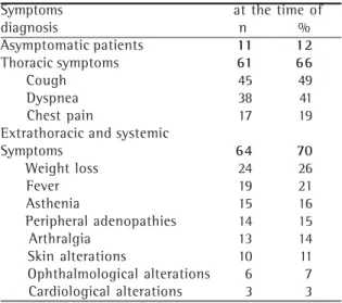
At the time of diagnosis of sarcoidosis, only 11 individuals (12%) were asymptomatic. The frequency of symptoms was comparable between men and women (87% and 89%, respectively; p = 0.83). Nor was there any difference between men and women in terms of the presentation of thoracic symptoms (79% vs. 92%; p = 0.47) and extrathoracic symptoms (82% vs. 77%; p = 0.53).
Among the symptomatic patients, 61 (66%) presented thoracic symptoms, and 64 (70%) presented extrathoracic and systemic symptoms. Exclusively thoracic symptoms were seen in 17 cases (18%) and exclusively extrathoracic symptoms were seen in 20 cases (22%). There were 44 patients (48%) who presented thoracic and extrathoracic symptoms concomitant to systemic symptoms. Among the thoracic symptoms, cough was the most frequent, being reported by 45 patients (49%), followed by dyspnea (38 patients; 41%), and chest pain (17 patients; 19%). Among the extrathoracic and systemic manifestations, the most frequent were
weight loss in (24 patients; 26%); fever (19 patients; 21%); asthenia (15 patients; 16%); peripheral adenopathies (14 patients; 15%) and arthralgia in (13 patients; 14%). Skin alterations were seen in 10 patients (11%), 6 (7%) being diagnosed with erythema nodosum. Ophthalmological alterations occurred in 6 patients (7%), and cardiological alterations were seen in 3 (3%). Episodes of urinary lithiasis occurred in 5 patients (5%) (Table 1).
In the group of symptomatic patients, the mean interval between the onset of symptoms and the first examination was 6.1 ± 7 months, 59% of the
10
29
61
0 10 20 30 40 50 60 70
Tabagistas Ex-tabagistas Nunca fumaram
%
dos
P
a
c
ie
nt
e
s
Figure 1 - Smoking in patients with sarcoidosis (n = 83)
0 5 10 15 20
<20 21-30 31-40 41-50 51-60 61-70 >70
Faixa etária (anos)
%
d
o
s
ca
so
s
Masculino Feminino
Figure 2 - Distribution of 92 patients with sarcoidosis by age and gender. Data for men and women are shown separately.
TABLE 1
Most common sarcoidosis-related symptoms and clinical findings reported at the time of
diagnosis (n = 92)
smokers former smokers nonsmokers
%
of
the
pa
ti
en
ts
Male
Female
Age bracket (years)
Symptoms at the time of
diagnosis n %
Asymptomatic patients 11 12
Thoracic symptoms 61 66
Cough 45 49
Dyspnea 38 41
Chest pain 17 19
Extrathoracic and systemic
Symptoms 64 70
Weight loss 24 26
Fever 19 21
Asthenia 15 16
Peripheral adenopathies 14 15
Arthralgia 13 14
Skin alterations 10 11
Ophthalmological alterations 6 7
patients seeking medical attention within 3 months. The spirometric evaluation (simple spirometry) was performed at the time of diagnosis in 73 patients (79%) and the results were normal in 40 (55%) of those 73.
Restrictive ventilatory disorders were seen in 17 patients (23%), obstructive ventilatory disorders in 13 (18%), and mixed ventilatory disorders in 3 (4%). In the cases in which there were spirometric alterations (33 cases), the ventilatory disorders were mild in 20 cases (28% of 73 patients evaluated) and of moderate intensity in 13 (18% of 73 patients evaluated). No severe ventilatory disorders were found in this group (Figure 3).
Among the patients submitted to spirometry, alterations in pulmonary function were found in 58% of the men (18 patients) and in 36% of the women (15 patients), a less than significant difference (p = 0.058).
In 49 patients (53%), 24-hour calciuria was measured. Calciuria greater than 300 mg/24 h was
found in 12 cases (24% of the patients evaluated), with a mean of 225.4 ± 145.8 mg/24 h.
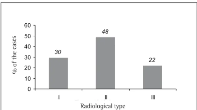
Chest X-rays were available for 78 patients, all having been taken at the time of diagnosis. Among these 78 patients, 23 (30%) presented radiological type I, 38 (48%) presented radiological type II, and 17 (22%) presented radiological type III (22%). None were classified as type IV (pulmonary fibrosis), and none of the patients presented normal chest X-rays at the time of diagnosis (Figure 4).
The specimens for anatomopathological examination were of intrathoracic origin in 84 patients (91%) and were obtained through various techniques: mediastinoscopy in 46 (50%); transbronchial biopsy in 19 (21%); and open-lung biopsy in 19 (21%). Peripheral lymph node biopsy was performed in 5 patients (6%), and integumentary biopsy was performed in 3 (3%).
As the principal therapeutic measure, systemic corticosteroid therapy was prescribed in 55 cases (60%), including all of those presenting radiological type III, the rest presenting type II. No adverse effects that would have justified the discontinuation of the corticosteroid therapy were observed.
Correlations were drawn between various variables in order to determine their significance and practical importance.
Patients that presented episodes of fever as the initial symptom of sarcoidosis sought medical treatment in significantly less time than did those that presented no such fever (2.6 ± 2.5 months vs. 7.4 ± 7.7 months; p = 0.009).
Male patients presented levels of 24-hour urinary calciuria significantly higher than those observed among female patients (288.5 ± 171.0 mg/dL vs. 184.1 ± 111.5 mg/dL; p = 0.014).
When the radiological type was compared with mean urinary calciuria, the group of patients presenting radiological type III had a mean calciuria that was significantly greater that that of the patients presenting radiological types I or II: type I, 198.6 ± 107.3 mg/24 h; type II, 213.2 ± 112.2 mg/24 h; and type III, 349.6 ± 181.3 mg/24 h. The analysis of variance showed a significant difference between type III and types I and II (p = 0.011).
To date, on the Pereira Filho Hospital, none of the patients with sarcoidosis were submitted to lung transplant, yet another indication that, in our region, the disease does not affect the lungs in a severe 22
48
30
0 10 20 30 40 50 60
I II III
%
dos
C
a
s
os
Tipo radiológico
Figure 4 - Distribution of 78 patients with sarcoidosis by radiological classification.
Figure 3 - Result of the spirometric evaluation performed at the time of diagnosis of sarcoidosis in 73 patients. RVD: restrictive ventilatory disorder; OVD: obstructive ventilatory disorder; MVD: mixed ventilatory disorder
%
of
the
c
as
es
Normal RVD OVD MVD
Disorder severe
moderate
mild
%
of
the
c
as
es
fashion, i.e. does not lead to respiratory failure.
DISCUSSION
In the present study, patients diagnosed with sarcoidosis and treated in a hospital specializing in respiratory diseases were evaluated retrospectively. Based on the characteristics of the samples, the results obtained cannot necessarily be extrapolated to the general population of patients with sarcoidosis, although they might present a profile that can be used as a reference for medical practices in our region.
Some correlations found between the data observed in this study and those seen in the literature are hereafter presented.
Initially, some data collected in a previous study conducted on the Pereira Filho Hospital should be analyzed. That study involved 138 patients diagnosed with sarcoidosis between 1965 and 1989 and presenting the following characteristics: mean age, 30.7 years - 21 patients (15%) below the age of 20, 91 patients (66%) between 20 and 40 years of age, and 26 patients (19%) over the age of 40; 79 female patients (57%) and 59 male patients (43%); 106 Caucasian patients (77%) and 32 black patients (23%).(29)
The current study represents the temporal sequence of this previous large study of sarcoidosis conducted the Pereira Filho Hospital. In analyzing the characteristics of the patients, similarities were found, with the exception of the age bracket since, in the last two decades, the mean age of patients with sarcoidosis has increased, from 30.7 years to 41.8 years. This increase in the age bracket has also been observed in other studies such as that designated A Case Control Etiologic Study of Sarcoidosis, conducted in the USA, in which the mean age was over 40 years(30), and another, carried out in the Netherlands, in which the mean age was 46.7 years.(11) In some countries, such as Sweden and Japan, a second peak has been observed in the sixth decade of life, especially among women.
The previously mentioned American study (A Case Control Etiologic Study of Sarcoidosis) was a multicenter project carried out by a group of researchers from ten large clinical facilities. One of the objectives of the study was to identify the sites (organs) involved in the group of patients as a whole and in subgroups divided by gender, race and age.(30-31) This population was heterogeneous
in terms of race (53% white, 44% black), gender (64% women, 36% men) and age (46% below the age of 40, 54% of 40 years of age or older). Female patients present greater ocular involvement, neurological involvement, erythema nodosum and age above 40, whereas male patients presented greater greater hypercalcemia. Black patients presented greater involvement of the skin (excluding erythema nodosum), eyes, liver, bone marrow and extrathoracic lymph nodes. The authors concluded that the initial presentation of sarcoidosis was dependent upon gender, age and, in particular, black race.
In the USA, sarcoidosis is more common among African-Americans than among Caucasians, with adjusted annual incidences of 35.5/100,000 inhabitants and 10.9/100,000 inhabitants, respectively.(1) Worldwide, however, approximately 80% of sarcoidosis patients are white. In the present study, we also observed this tendency, despite the lower (16%) proportion of black patients. According to the Instituto Brasileiro de Geografia e Estatística (Brazilian Institute of Geography and Statistics) , only 4% of the population of Rio Grande do Sul is black.(32) In our study sample, Caucasian patients predominated (84%), which is in contrast with the findings of other studies in the literature. A potential socioeconomic bias can be observed in the sample since a large number of the patients were referred by private health insurance companies. Similar findings have been observed in studies of populations presenting the same characteristics.(5)
The slight predominance of women over men in our sample (58% vs. 42%) is similar to that observed in other studies.(11,33) In a populational study, the incidence of sarcoidosis reported by one health organization was 21.6 women/100,000 inhabitants/ year and 15.3 men/100,000 inhabitants/year.(34) The mean age at the time of diagnosis was 40.9 years in both genders. In relation to female patients, these data are in agreement with those of other studies.(5) In the Case Control Etiologic Study of Sarcoidosis, the age at the time of diagnosis among men was predominantly below 40.(30)
Sarcoidosis frequently presents with no clinical manifestations or with symptoms of mild intensity, which, on one hand, can delay the diagnosis and, on the other, can aid the differential diagnosis greatly. This is a characteristic found in the majority of studies in the literature. In other studies, delayed diagnosis of sarcoidosis has also been reported, apparently occurring more due to the characteristics of the presentation of the disease than to the characteristics of the patient or physician.(36)
The organ most affected in isolation was the lung, as evidenced by the finding that 75% of the symptoms were thoracic, in keeping with those of other studies, in which the incidence ranged from 50% to 95%. Pulmonary symptoms in isolation may explain delays in diagnosis, principally caused by the lack of specificity of such symptoms.(6) This variable was not evaluated in the present study. The mean interval between the onset of symptoms and the initial examination was six months, the great majority of the patients being diagnosed within three months after the first appearance of symptoms (59%). Among the extrathoracic symptoms, the most common were weight loss, fever and asthenia, together characterizing the systemic component of the disease. In one study, persistent fatigue was reported as the principal presentation symptom of sarcoidosis (71% of the cases).(11) In the present study, persistent fatigue was reported by 16% of the patients. In that same study, a questionnaire was sent to the members of the Dutch Sarcoidosis Society. A total of 1026 completed questionnaires were completed and returned (58% of the total number of questionnaires distributed). The mean age was 46.7 + 11.6 years, and 63% of the patients were women. There were 176 cases of familiar sarcoidosis. The symptoms most frequently seen were fatigue (71%), dyspnea (70%), arthralgia (52%), myalgia (39%) and chest pain (27%). Systemic corticosteroid therapy was used in 565 patients (55.1%).
Although the radiological presentations seen in the present were not significantly different than those reported in the literature, there were some noticeable differences. For example: 30% of the cases evaluated in the current study presented radiological type I, compared with the 40% to 50% reported in the literature; radiological type II was seen in 48% of our patients, whereas other authors have reported this type in only 30%; and
radiological type III was observed in 22% of our patients, which is quite comparable to the value found in the literature.(3-4,29,33) The fact that our sample included no cases presenting radiological type 0 (normal X-ray) can be explained, in part, by the fact that our facility specializes in pulmonology, which limits the entrance of patients presenting no abnormalities on chest X-rays. In our facility, we rarely classify patients as type IV at the time of diagnosis since it is not possible to verify the exclusivity of the fibrotic component at that time. Cases can only be classified as type IV when there are abnormalities suggestive of fibrosis, lesions that persist throughout a long follow-up period, and no other lesions that might prove to be reversible.
The results of the spirometry indicate that sarcoidosis typically has little impact on pulmonary function since more than half of all patients present normal exam results, and, when alterations are seen, the dysfunction is of mild intensity. Ventilatory disorders can be obstructive or restrictive, depending on the distribution of the lesions (in the lung parenchyma vs. in the airways) and on the presence of comorbidities.
C o r t i c o s t e r o i d t h e r a p y w a s p r e s c r i b e d according to the criteria recommended in the literature, recently standardized into international guidelines, and was used by approximately half of the patients in this study, similar to the proportions reported in other studies. There are no reliable data in the literature to support our choice to administer the corticosteroids on alternate days, in the same manner as for daily use. However, this technique has proven efficacious in our facility.
Since this study was retrospective and involved a limited number of cases, various other correlations cited in the results of this study, such as those related to age, gender, ethnicity, symptoms, radiological type, calciuria and extrathoracic symptoms, merit further evaluation.
Taken together, the characteristics of the patients with sarcoidosis in this study, the majority of whom came from the state of Rio Grande do Sul, did not differ significantly from the findings in the literature.(37) The fact that we found no patients that had been treated for tuberculosis prior to being diagnosed with sarcoidosis can be explained, at least in part, by the strict standards of the Rio Grande do Sul State Tuberculosis Control Program and by the considerable accuracy of the radiological records kept by our facility (as well as by others) regarding the suspicion of sarcoidosis.
The possibility that many patients will go undiagnosed, the lack of reporting of known cases, the inability to identify the etiology, diagnostic difficulties (in making the diagnosis a n d i n r u l i n g o u t o t h e r c a u s e s ) a n d t h e impossibility of specific therapeutic intervention make sarcoidosis a disease presenting many unknown factors and one whose etiopathogenesis is not well understood. However, sarcoidosis presents some clinical patterns that facilitate its recognition, and, as a rule, medical practices can be carried out based on the guidelines.(1-2) Therefore, the pulmonologist or other specialist working in the sector can, in the majority of cases, use accessible criteria to diagnose and treat their patients.
In view of the fact that comparative epidemiological studies have demonstrated that geographic, ethnic and genetic factors are correlated with specific characteristics of patients with sarcoidosis, further case series studies should be conducted in other regions.
REFERENCES
1. American Thoracic Society (ATS), European Respiratory Society (ERS) and World Association of Sarcoidosis and Other Granulomatous Disorders (WASOG): Statement on sarcoidosis. Joint Statement of the American Thoracic Society, ERS and WASOG. Am J Respir Crit Care Med. 1999;160(2):736-55.
2. Costabel U, Hunninghake GW. ATS/ERS/WASOG statement on sarcoidosis. Sarcoidosis Statement Committee. American Thoracic Society. European Respiratory Society. World Association for Sarcoidosis and Other Granulomatous Disorders . Eur Respir J. 1999;14(4):735-7.
3. Crofton J, Douglas AC. Sarcoidosis. In: Crofton J, Doublas AC. Respiratory diseases. 3rd ed. Oxford: Blackwell; 1981. 4. James DG. Clinical concepts of sarcoidosis. Am Rev
Respir Dis. 1961;84(Pt 2):66-70.
5. Judson MA, Baughman RP, Teirstein AS, Terrin ML, Yeager H Jr. Defining organ involvement in sarcoidosis: the ACCESS proposed instrument. Sarcoidosis Vasc Diffuse Lung Dis. 1999;16(1):75-86.
6. Johns, CJ, Michele, TM. The clinical management of sarcoidosis: a 50-year experience at the Johns Hopkins Hospital. Medicine. 1999;78:65-111.
7. DeRemee RA. Sarcoidosis. Mayo Clin Proc. 1995;70(2): 177-81.
8. Tarantino AB, Corrêa da Silva LC. Sarcoidose. In: Tarantino AB. Doenças pulmonares. 5a ed. Rio de Janeiro: Guanabara Koogan; 2002. p.814-26.
9. Reich JM. What is Sarcoidosis? Chest. 2003;124(1):367-71. 10. Wurm K, Rosner R. Prognosis of chronic sarcoidosis. Ann
NY Acad Sci. 1976;278:732-5.
11. Wirnsberger RM, de Vries J, Wouters EF, Drent M. Clinical presentation of sarcoidosis in the Netherlands an epidemiological study. Neth J Med. 1998;53(2):53-60. 1 2 . H i l l s S E , P a r k e s S A , B a k e r S B . E p i d e m i o l o g y o f sarcoidosis in the Isle of man-2: evidence for space-time clustering. Thorax. 1987;42(6):427-30. 1 3 . Hiraga Y. An epidemiological study of clustering of
sarcoidosis cases. Nippon Rinsho. 1994;52(6):1438-42. Japanese.
14. Panayeas S, Theodorakopoulos P, Bouras, Constantopoulos S. Seasonal occurrence of sarcoidosis in Greece. Lancet. 1991;338(8765):510-1.
15. Bardinas F, Morera J, Fite E, Plasencia A. Seasonal clustering of sarcoidosis. Lancet. 1989;2(8660):455-6. 1 6 . Hosoda Y, Yamaguchi M, Hiraga Y. Global epidemiology of sarcoidosis. What story do prevalence and incidence tell us? Clin Chest Med. 1997;18(4):681-94.
17. Loddenkemper R, Kloppenborg A, Schoenfeld N, Grosser H, Costabel U. Clinical findings in 715 patients with newly detected pulmonary sarcoidosis - Results of a cooperative study in former West Germany and Switzerland. Sarcoidosis Vasc Diffuse Lung Dis. 1998;15(2):178-82.
2 0 . Corrêa da Silva LC. Sarcoidose. In: Corrêa da Silva LC. Condutas em pneumologia. Rio de Janeiro: Revinter; 2001. p.494-505.
2 1 . Winterbauer RH, Belie N, Moores KN. A clinical interpretation of bilateral hilar adenopathy. Ann Intern Med. 1973;78:65-71.
22. Bethlem N, Bethlem EP, Lemle A, Capone D, Souza GRM, Corrêa JC et al. Revisão e sugestões de normas para o diagnóstico, tratamento e acompanhamento da sarcoidose gangliopulmonar. J Pneumol. 1995;21(3):123-31. 2 3 . Mitchell DN, Scadding JG, Heard BE, Hinson KF.
Sarcoidosis: histopathological definition and clinical diagnosis. J Clin Pathol. 1977;30(5):395-8.
24. Baughman RP, Ianuzzi MC. Editorial - Diagnosis of sarcoidosis: When is a peek good enough? Chest 2000; 117:(4)931-2. 2 5 . Judson MA. An approach to the treatment of pulmonary
sarcoidosis with corticosteroids: the six phases of treatment. Chest. 1999;115(4):1158-65.
26. Paramothayan S, Jones PW. Corticosteroid therapy in pulmonary sarcoidosis. A systematic review. JAMA. 2002;287(10):1301-7.
2 7 . Neville E, Walker NA, James DG. Prognostic factors predicting the outcome of sarcoidosis: an analysis of 818 patients. Q J Med. 1983;52(208):525-33. 2 8 . James DG. Life-threatening situations in sarcoidosis.
Sarcoidosis Vasc Diffuse Lung Dis. 1998;15(2):134-9. 2 9 . Pereira, M. Sarcoidose: análise de 138 casos [tese]. Porto Alegre:Universidade Federal do Rio Grande do Sul; 1994. 128 p.
3 0 . Baughman RP, Teirstein AS, Judson MA, Rossman MD, Yeager H, Bresnitz EA; Control Etiologic Study of Sarcoidosis (ACCESS) research group, et al. Clinical characteristics of patients in a case control study of sarcoidosis. Am J Respir Crit Care Med. 2001;164(10 Pt 1):1885-9.
31. Design of a case control etiologic study of sarcoidosis (ACCESS). ACCESS Research Group. J Clin Epidemiol. 1999;52(12):1173-86.
3 2 . IBGE. Instituto Brasileiro de Geografia e Estatística. Recenseamento geral do Brasil: Censo demográfico 2000. Rio de Janeiro; 2001.
3 3 . James DG, Neville E, Siltzbach LE. A worldwide review of sarcoidosis. Ann N Y Acad Sci. 1976;278:321-34. 3 4 . Rybicki BA, Major M, Popovich J Jr, Maliarik MJ,
Iannuzzi MC. Racial differences in sarcoidosis incidence: a 5-year study in health maintenance organization. Am J Epidemiol. 1997;145(3):234-41.
3 5 . Peros-Golubicic T, Ljubic S. Cigarette smoking and sarcoidosis. Acta Med Croatica. 1995;49(4-5):187-93. 3 6 . Judson MA, Thompson BW, Rabin DL, Steimel J, Knattereud GL, Lackland DT; ACCESS Research Group, et al. The diagnostic pathway to sarcoidosis. Chest. 2003;123(2):406-12.